*Gülhane Medical Faculty Department of Orthopedics and Traumatology. Ankara/Turkey.
**Antalya Education and Research Hospital Department of Orthopedics and
Traumatology Antalya/Turkey.
***Medipol University Department of Orthopedics and Traumatology. Istanbul/ Turkey.
****Gülhane Medical Faculty Department of Radiology. Ankara/Turkey
Ayrı Basım İsteği: Tolga Ege
Gülhane Medical Faculty Department of Orthopedics and Traumatology. Ankara/Turkey. ([email protected])
Makalenin Geliş Tarihi: Nov 22, 2014 • Kabul Tarihi: Jun 14, 2015 • Çevrim İçi Basım Tarihi: 30 Eylül 2016
Erişkin kalça displazisinin değerlendirilmesinde normal
radyolojik ölçüm değerleri; Anadolu toplumunda 1732
sağlıklı kalçanın değerlendirilmesi
Tolga Ege(*), Özkan Köse (**) , Bahtiyar Demiralp(***), Doğan Bek(*), Tuba Sanal(****)
ÖZET
Amaç: Çalışmanın amacı, sağlıklı Anadolu insanında kalça ekleminin, özellikle de kalça displazinin değerlendirilmesi amacı ile normal radyolojik parametrelerinin saptanmasıdır.
Hastalar ve metot: Prospektif olarak yapılan çalışmamıza, klinik olarak herhangi kalça rahatsızlığı bulunmayan ve merkezimizde rutin tarama amaçlı pelvik bölgeyi içeren, ön-arka direkt grafisi çekilmiş 866 erişkin hasta ( 18 yaşından büyük) dahil edilmiştir. Asetabular Sharp açısı, CE açısı, asetabular indeks (AI), ACM açısı ve Reimer’in migrasyon indeksi standart radyografiler üzerinden ölçüldü. Her iki cinsiyet ve her iki taraf kalça için ayrı olarak normal değerler saptanıp istatistiksel karşılaştırma yapılmıştır.
Sonuçlar: CE (E: 30.3±3.4 - K: 28.8±2.7) ve ACM açıları (E: 41.0±1.8 - K: 40.5±1.8) erkek hastalarda istatistiksel olarak yüksek iken; AI (E: 3.5±0.6 -K: 3.8±0.9) ve Sharp açıları (E: 37.9±2.5 -K: 38.5±2.1) bayanlarda daha fazla idi. Reimer’in migrasyon indeksi cinsiyetler arasında benzer değerlere sahipti (E: 12.6±3.5-K: 13.0±3.6). Erkek hastalardaki AI (asetabular indeks) açıları haricinde tüm parametreler her iki cinste de sağ ve sol kalça için farklı idi.
Çıkarımlar: Asetabular bölgeyi içeren ortopedik cerrahi işlemlerde kendi toplumumuzdan elde edilmiş normal asetabular açı değerlerinin kullanılmasını vurgulamaktayız.
Anahtar Kelimeler: Astebular displazi, sharp acisi, aseyabular indeks, merkez kenar
acisi, ACM acisi, Reimer’in migrasyon indeksi, Turk popülasyonu, normal data.
SUMMARY
Normal values of radiographic measurements used for the assessment of adult hip dysplasia; analysis of 1732 healthy hips in Anatolian population
Objectives: The purpose of this study is to determine the normal values of radiographic measurements used for the assessment of hip joint, particularly focusing on the hip dysplasia, on healthy young adults in Anatolian population. Materials and methods: This prospective study consisted 866 subjects (>18 years of age) without clinical evidence of hip disorder who underwent anterior-posterior (AP) x-ray of pelvic region for routine screening in our institution. Acetabular angle of Sharp, center edge angle, ACM angle, acetabular index, and Reimer’s migration index were measured. Normative data regarding radiographic parameters were presented for both gender and body side and statistical comparison was performed between gender and body sides. Results: CE angles (M: 30.3±3.4 vs F: 28.8±2.7) and ACM (M: 41.0±1.8 vs F: 40.5±1.8) angles were higher in male subjects; AI (M: 3.5±0.6 vs F: 3.8±0.9) and Sharp angle (M: 37.9±2.5 vs F: 38.5±2.1) was higher in female subjects. Reimer’s migration index was similar between genders (M: 12.6±3.5 vs F: 13.0±3.6). All measured variables were statistically different between body sides, except AI in male subjects. Conclusion: We emphasize that normal limits of acetabular angles obtained from our own population should be used as reference values in various orthopedic operations regarding acetabular region.
Key words: Acetebular dysplasia, sharp angle, acetebular index, center edge angle,
ACM angle, Reimer’s migration index, Turkish population, normative data.
Gülhane Tıp Derg 2016;58: 245-249 © Gülhane Tıp Fakültesi 2016 doi: 10.5455/Gülhane. 172969
ARAŞTIRMA/ORIGINAL ARTICLE
Introduction
Assessment of a hip joint and determining whether it is nor-mal or abnornor-mal is essential in patients undergoing hip joint surgery. In order to understand and define what is ‘abnormal’; first we need to know what is ‘normal’. Direct radiographic exa-mination of the pelvis is generally used as a first-step diagnos-tic tool for the evaluation of the hip joint. Several radiographic measurements have been described to assess and quantify the anatomic structure and morphology of the hip including acetabular index (AI), which is described by Hilgenreiner and Tönnis (1,2), acetabular angle of Sharp (3), ACM angle desc-ribed by Delberger and Frank (1), acetabular index of weight bearing zone, CE angle of Wrisberg (4) and Reimer’s migra-tion index (5). These objective measures are frequently used for the diagnosis, contemplating a prompt surgical plan and follow-up of patients with acetebular dysplasia as well as pa-tients undergoing hip joint preserving surgeries and total hip replacement.
It is well known that hip joint morphology may vary between different ethnic groups and populations (6-9). The normal va-lues of radiographic measurements used for the assessment of adult hip dysplasia in Turkish population have been previ-ously reported by Ozcelik et al (10). However, this study was performed only on patients residing in the province of Eskise-hir, and it was not clearly explained that the study population are healthy subjects or not. Therefore, this previous normative data may not be generalized to whole country, and may not be representative for healthy subjects.
We have performed this study on a larger population and included only healthy subjects from all regions of the country. The purpose of this study is to determine the normal values of radiographic measurements used for the assessment of hip jo-int, particularly focusing on the hip dysplasia, on healthy young adults in Anatolian population.
Patients and methods
This descriptive cross-sectional study comprised of 866 adults subjects (>18 years of age) without clinical evidence of hip disorder who underwent anterior-posterior (AP) x-ray of pelvic region for routine screening in Gülhane Medical Faculty during the last three-year period. Subjects with history of chro-nic hip pain, inflammatory arthritis, congenital or neuromuscu-lar disease or abnormality of the lower limp, and subjects with a history of prior hip and lower limb surgery were excluded from the study. All subjects were job applicants for Turkish Ar-med Forces and apparently healthy subjects without systemic disease. This prospective study was carried out according to Declaration of Helsinki and the Institutional Review Board at
246
•
Eylül 2016 • Gülhane Tıp Derg Ege ve ark.our institution approved the study protocol. All subjects gave their informed consent prior to their inclusion in the study.
All antero-posterior (AP) standard standing radiographs inc-luding pelvic region were taken from a film-to-focus distance of 115cm and x-ray beam was centered to the symphysis pubis. The AP view was obtained with approximately 20° of internal rotation of the lower limbs, to compensate for femoral ante version. The criteria for an acceptable pelvic radiograph inc-lude a symmetric appearance of obturator foramina and iliac crests, and a true AP view of both femoral necks. Inappropriate radiographs on which femoral head and acetabulum are not clearly displayed and anatomic landmarks cannot be clearly revealed were excluded from the study.
Radiographic measurements
Acetabular angle of Sharp, center edge angle of Wiberg, ACM angle, acetabular index of weight bearing zone, and Reimer’s migration index were measured by the senior aut-hors who was familiar with these measurements and had used them in clinical practice. All measurements were performed using the software program Clinical Workstation Simple (ver. 4.5.16, TURMAP, MedPlus, Turkey) on the digital workstation (Figure 1). Demographic data including age, sex and place of birth was also recorded.
Statistical analysis
Continuous variables were stated as mean and standard deviation and categorical variables as percentages and frequ-ency distribution. Independent sample t-test and paired samp-le t-test were used for statistical analysis. A p value samp-less than 0.05 was accepted as significant.
Results
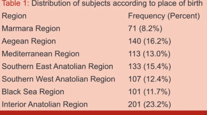
There were 482 male and 384 female subjects with a mean age of 22.7±1.6 years (range, 18-26). A total of 1732 hip joints were assessed. Distribution of subjects according to place of birth is presented in Table 1.
Table 1: Distribution of subjects according to place of birth
Region Frequency (Percent)
Marmara Region 71 (8.2%)
Aegean Region 140 (16.2%)
Mediterranean Region 113 (13.0%)
Southern East Anatolian Region 133 (15.4%)
Southern West Anatolian Region 107 (12.4%)
Black Sea Region 101 (11.7%)
Interior Anatolian Region 201 (23.2%)
Sharp’s Angle: The mean Sharp’s Angle in female and male subjects was 38.5±2.1 (range, 30-49) and 37.9±2.5 (range 29-49) respectively and statistically significant difference detected between genders (p=0.0001). When we compare the different body sites, in both gender left hips had slightly higher values compared to right hips.
Center edge angle of Wiberg: The mean CE angle was 29.7±3.2 degrees (range, 22-38) in all hips. The mean CE
ang-le in femaang-le and maang-le subjects was 28.8±2.7 (range, 23-38) and 30.9±3.4 degrees (range, 22-38) respectively (p=0.0001). Again in both gender right hips had slightly higher values com-pared to left hips.
ACM Angle: ACM angle was statistically higher in male sub-jects (M: 41.0±1.8 vs F: 40.5±1.8) (p<0.05). In both gender right hips had greater values compared to left hips.
Acetabular index of weight bearing zone: Female subjects tent to have higher values compared to male subjects (F: 3.8±0.9 M: 3.5±0.6) Again in female gender, left hips have slightly higher values compared to right side. However in male subjects AI values were similar between body sites.
Reimer’s migration index: The mean Reimer’s migrati-on index for female and male subjects was 13.0±3.6 % and 12.6±3.5 % respectively and there was no statistically signifi-cant difference between genders. However when we compare left and right hips, in both genders left hips had slightly higher values compared to right hip.
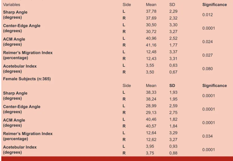
Summary of all measurements in both genders are presen-ted in Table 2. Comparison of male and female subjects accor-ding to right and left hips is presented in Table 3. There was no statistically significant difference between regions of Turkey.
Table 2: Summary of normative data
Measurement Sex Mean±SD Range 95% CI
Sharp Angle (°) Male 37.9±2.5 29-49 37.7-38.0 Female 38.5±2.1 30-49 38.3-38.6 Center-Edge Angle (°) Male 30.3±3.4 22-38 30.0-30.5 Female 28.8±2.7 23-38 28.6-28.9 ACM Angle (°) Male 41.0±1.8 36-49 41.6-41.9 Female 40.5±1.8 36-49 40.3-40.6 Migration Index (%) Male 12.6±3.5 7-26 12.3-12.8 Female 13.0±3.6 6-26 12.7-13.2 Acetebular Index (°) Male 3.5±0.6 2-5 3.4-3.5
Female 3.8±0.9 2-6 3.7-3.8
Center-edge (CE) angle less than 20° is accepted as dysplastic, a CE angle between 20° and 25° is accepted as borderline dysplasia, and CE angle greater than 25° is accepted as normal hip. Acetebular index of weight bearing zone greater than 10° is considered as abnormal; values above 10° are frequently found in acetabular dysplasia. Sharp angle greater than 42 degrees is considered as abnormal. The migration index of Reimers is considered to be abnormal if greater than 20%. An ACM angle greater than 49 degrees is considered abnormal.
Discussion
This study aimed to determine the normal ranges of plain radiographic measurements used for the assessment of adult hip dysplasia in Anatolian population. Several similar studies have been performed previously in different ethnic populations (Table 4). Results of our study showed that acetebular morp-hology and related parameters specific to Anatolian population was unique and different with some aspects from the other populations.
One of the most common used radiologic parameter in ace-tabular dysplasia is aceace-tabular index (AI). It shows the slope of the acetabular roof as previously described by Hilgenreiner (2). However reference points vary in children and adults so it is not a reliable method for long-term follow-up of dysplas-tic acetabulum. Moreover this angle is mostly affected by the
Table 3. Comparison of Right and Left Hips according to gender (Abbreviations, SD: Standard Deviation, L: Left, R: Right) Male Subjects (n:464)
Variables Side Mean SD Significance
Sharp Angle (degrees) L 37,78 2,29 0.012 R 37,69 2,32 Center-Edge Angle (degrees) L 30,50 3,30 0.0001 R 30,72 3,27 ACM Angle (degrees) L 40,96 2,52 0.024 R 41,16 1,77
Reimer’s Migration Index
(percentage) L 12,48 3,37 0.027 R 12,43 3,31 Acetebular Index (degrees) L 3,55 0,63 0.080 R 3,50 0,67 Female Subjects (n:365)
Side Mean SD Significance
Sharp Angle (degrees) L 38,33 1,93 0.0001 R 38,24 1,95 Center-Edge Angle (degrees) L 28,99 2,59 0.0001 R 29,13 2,75 ACM Angle (degrees) L 40,46 1,82 0.0001 R 40,57 1,84
Reimer’s Migration Index (percentage) L 12,64 3,29 0.034 R 12,62 3,27 Acetebular Index (degrees) L 3,95 0,93 0.0001 R 3,75 0,88
248
•
Eylül 2016 • Gülhane Tıp Derg Ege ve ark.patient’s position on plain radiographs (1,10). According to the Tönnis (1) the upper limit of normal AI value in adults is stated as 10 degrees and the method of measurement described by Tönnis found to be reliable by Nelitz et al. (11). Again Leques-ne et al. stated that values greater than 12 degrees must be accepted as dysplastic (12). In male subjects, we have found the mean AI value as 3.5±0.6, which was statistically different from the female subjects (3.8±0.9). However on a local zone in Turkey, Ozcelik et al (10) found no statistically significant difference between genders in terms of AI values. While our overall AI results were congruent with Ozcelik et all, they were significantly lower than the specified values in foreign countri-es.( 6,8,9 ).
Another common radiologic parameter used in acetabular dysplasia is Sharp angle. Sharp states that acetabular angle determines the angle of acetabulum instead of its depth (3). But, the major benefit of this angle, it remains the same throug-hout the person’s life. So it can be a diagnostic tool for determi-ning healthy hip joints. However, it can be difficult to measure especially in severely dysplastic hips as the teardrops may be deformed (1,10). In the current study, all of our subjects were otherwise healthy so we could measure this angle without any problem. Furthermore, the other major benefit of the aceta-bular angle of sharp over acetaaceta-bular index that it is minimally affected from the pelvic position during radiologic procedure as it can be major problem in busy outpatient radiology clinic environment (3,10). In his original article Sharp et al. (3) refer-red the normal acetabular angle below 420 in adults; moreover Tönnis et al. (1) reported the upper limit of normal as 43 0. In the current study we found the mean value of Sharp angle 37.9±2.5 degrees in males and 38.5±2.1 degrees in females. The difference between genders was statistically significant and the average Sharp angle values in our study were close to the normative values reported in some western studies (6,13) but significantly lower from the values reported in some eas-tern studies ( 8,9).
ACM angle is a radiologic parameter that measures the depth of acetabulum and correlates with dysplastic acetabu-lum. However it doesn’t give any information about obliquity of acetabular roof. The major advantage of the ACM angle is not affected by the patient age and position on pelvic radiographs (10). According to Tönnis et al. (14) major concern about this radiologic measurement is the patient age. It is difficult to mea-sure ACM angle in patients below 10 years old as the B point, (inferior edge of the acetabulum) is hard to be determined on pelvic radiographs. In our series we did not have any problem regarding ACM angle measurements as our subjects was in adolescent group. In the current literature values between 40-50 degrees are reported normal (1) and Tönnis et al (1,14) sta-tes that values greater than 49 degrees after 2 years old must be accepted as pathologic. We found the mean ACM angle 41.0±1.8 in males and 40.5±1.8 in females. Our results were congruous with the current literature.
The CE angle is another radiologic parameter that was first introduced in 1939 (15) and shows the relationship between the femoral head and the acetabulum. It has a physiological range of 20–40 degrees. Cut-off values below 20 degrees indicates hip dysplasia and values between 20– 25 degrees indicates borderline cases, and >25 reveals normal hips (7). However Sharp (3) stated three limitations of CE angle which were the center point of deformed femoral head, joint space
subluxation and lateral edge of acetabulum which affects CE result. In the present study we have found the mean CE angles 30.3±3.4 degrees for males and 28.8±2.7degrees for females and our results were consistent with other studies performed in different ethnic populations. (6,7,16-18),(table 4).
The femoral head extrusion index (FHEI) reveals the amount of femoral head covered by the acetabular roof. The normal range of this index was initially reported to be normal between 70–100 %, with an average of 90 % (19). It reveals the amount of femoral head covered by the acetabular roof. In the later period, Cooperman et al.(20) proposed the cut off values as 75 %. This has been supported by findings by the Danish gro-up, presented as an inverse index, called the lateral migration index, with values above 25 % being indicative of dysplasia (13). The results of the present study revealed the Reimer’s migration index 12.6±3.5 and 13.0±3.6 for males and females respectively. Our results were slightly lower compared to other studies. (7,13,18)
Our study has some strengths and limitations. Although we have large enough population from all around the country; our study group was limited to same age group. Also inter-obser-ver variability is not taken into consideration. Howeinter-obser-ver, to our knowledge this is the largest population based study, which measures the several acetabular angles in young adult Turkish population.
As a conclusion we believe that knowing the normal aceta-bular parameters as a first step before the treatment is very important and we must use our own public radiologic parame-ters in the future studies. And surgeons should be familiar with their own parameters
References
1. Tönnis D. Congenital dysplasia and dislocation of the hip in children and adults. 1st ed. Berlin: Springer-Ver-lag; 1987.
2. Thieme WT, Thiersch JB (translators). Classic. Transla-tion: Hilgenreiner on congenital hip dislocation. J Pedi-atr Orthop 1986;6:202-14
3. Sharp IK. Acetabular dysplasia. The acetabular angle. J Bone Joint Surg [Br] 1961;43:268-72
4. Wiberg G. Studies on dysplastic acetabula and conge-nital subluxation of the hip joint. Acta Chir Scand Suppl. 1939; 58:5–132
5. Heyman CH, Herndon CH. Legg-Perthes disease; a method for the measurement of the roentgenographic result. J Bone Joint Surg.1950;32(A:4):767–78
6. Jeremic D, Macuzic IZ, Vulovic M. Sex differen-ces in anatomical parameters of acetabulum among asymptomatic Serbian population. Vojnosanit Pregl. 2011;68(11):935–9.
7. Laborie LB, Engesæter I, Lehmann TG, Sera F, De-zateux C, Engesæter LB, Rosendahl K. Radiographic measurements of hip dysplasia at skeletal maturity-new reference intervals based on 2,038 19-year-old Norwe-gians Skeletal Radiol. 2013 Jul;42(7):925-35
8. Mohd Yusof Baharuddin, Ahmad Hafiz Zulkifly, Moham-med Rafiq Abdul Kadir, Azlin Saat, Azian Abdul Aziz and Muhammad Hisyam Lee, 2011. Morphometric Study of
the Acetabular in Malay Population Normal Hips and its Clinical Applications. Journal of Medical Sciences, 11: 213-219
9. Umer M, Thambyah A, Tan WT, Das De S. Acetabular morphometry for determining hip dysplasia in the Sin-gaporean population. J Orthop Surg (Hong Kong). 2006 Apr;14(1):27-31.
10. Ozçelik A, Omeroğlu H, Inan U, Ozyurt B, Seber S. Nor-mal values of several acetabular angles on hip radiog-raphs obtained from individuals living in the Eskişehir region.Acta Orthop Traumatol Turc. 2002;36(2):100-5 11. Nelitz M, Guenther KP, Gunkel S, Puhl W. Reliability
of radiological measurements in the assessment of hip dysplasia in adults. Br J Radiol 1999; 72:331-4
12. Lequesne M, Malghem J, Dion E. The normal hip jo-int space: variations in width, shape, and architectu-re on 223 pelvic radiographs. Ann Rheum Dis. 2004 Sep;63(9):1145-51
13. Jacobsen S, Sonne-Holm S, Soballe K, Gebuhr P, Lund B. Hip dysplasia and osteoarthrosis: a survey of 4,151 subjects from the Osteoarthrosis Substudy of the Copenhagen City Heart Study. Acta Orthop. 2005;76(2):149–58
14. Tönnis D. Normal values of the hip joint for the evalua-tion of X- rays in children and adults. Clin Orthop Relat Res. 1976;119:39–47
15. Wiberg G. Studies on Dysplastic Acetabula and Con-genital Subluxation of the Hip Joint. Acta Chir Scand. 1939;(83) Suppl 58:7.
16. Shi YY, Liu TJ, Zhao Q, Zhang LJ, Ji SJ, Wang EB. The normal centre-edge angle of Wiberg in the Chinese po-pulation: a population-based cross-sectional study. J Bone Joint Surg. 2010;92B(8):1144–7
17. Saikia KC, Bhuyan SK, Rongphar R.Anthropometric study of the hip joint in northeastern region population with computed tomography scan. Indian J Orthop. 2008 Jul;42(3):260-6.
18. Aly TA. Hip morphologic measurements in an Egyptian population. Orthopedics. 2011;34(4):262
19. Heyman CH, Herndon CH. Legg-Perthes disease; a method for the measurement of the roentgenographic result. J Bone Joint Surg. 1950;32(A:4):767–78
20. Cooperman DR, Wallensten R, Stulberg SD. Aceta-bular dysplasia in the adult. Clin Orthop Relat Res. 1983;175:79–85