Received Date / Geliş Tarihi: 15.04.2012 Accepted Date / Kabul Tarihi: 23.06.2012 © Telif Hakkı 2012 AVES Yayıncılık Ltd. Şti. Makale metnine www.jarem.org web sayfasından ulaşılabilir. © Copyright 2012 by AVES Yayıncılık Ltd. Available on-line at www.jarem.org
doi: 10.5152/jarem.2012.13 Address for Correspondence / Yazışma Adresi: Dr. Türker Özkan,
Department of Hand Surgery, Faculty of Medicine, Istanbul University, İstanbul, Turkey
Phone: +90 212 246 52 53 E-mail: turker.ozkan@istanbul.edu.tr
Upper Extremity Surgery in Spastic Cerebral Palsy
Serebral Paralizide Üst Ekstremite Cerrahisi
Türker Özkan
1, Serdar Tunçer
21Department of Hand Surgery, Faculty of Medicine, Istanbul University, İstanbul, Turkey
2Department of Plastic and Reconstructive Surgery, Faculty of Medicine, Istanbul Bilim University, İstanbul, Turkey
ABSTRACT
Involvement of the upper extremity in cerebral palsy often results in a typical pattern of spasticity, with elbow flexion, forearm pronation, ulnar devia-tion and flexion of the wrist, and adducdevia-tion-flexion posture of the thumb. Although only a relatively small subset of cerebral palsy patients are can-didates for surgery, properly selected patients and procedures yield a reasonable improvement. Nonsurgical treatment modalities include physical therapy, orthoses, and medications aimed at decreasing spasticity. Surgical options for the management of the spastic upper extremity vary with the specific parts, however they are focused around three basic principles: weakening the overactive muscle/tendons, strengthening the underactive muscle/tendons, and stabilizing non-stable joints. Surgical management of the spastic upper limb in cerebral palsy requires meticulous evaluation and planning. It is important to know that upper extremity deformities are secondary manifestations of the cerebral injury. Therefore, in addition to evaluation of upper extremity function, consideration should be given to the intelligence and motivation of the patient, and voluntary use of the up-per extremity. Prior to surgery, the overall level of function needs to be considered. These are also important details for the patients and their families, who should know that surgery is aimed at improving the upper extremity deformity, and not the primary disorder. Appropriately indicated surgery can significantly contribute to upper extremity function. (JAREM 2012; 2: 43-54)
Key Words: Cerebral palsy, spasticity, upper extremity surgery, tendon transfers ÖZET
Serebral paralizide üst ekstremite tutulumu, tipik bir spastisite paterni oluşturur. Bunun sonucunda da sıklıkla dirsek fleksiyonu, önkol pronasyonu, el bileğinde fleksiyon ve ulnar deviasyon, başparmakta adduksiyon-fleksiyon postürüyle karakterize bir deformite meydana gelir. Serebral paralizi hasta-larının yalnızca az bir kısmı cerrahi için uygun adaylar olsa da, uygun seçilmiş hastalarda cerrahi tedavi yüz güldürücü sonuçlar verebilmektedir. Cerrahi dışı tedaviler arasında fizik tedavi, ortezler ve spastisiteyi azaltmaya yönelik ilaçlar vardır. Spastik üst ekstremite tedavisindeki cerrahi seçenekler defor-mitenin bulunduğu bölgeye göre değişir, ancak üç temel özellik üzerinde odaklanır: aşırı aktif kas ve tendonların zayıflatılması, yetersiz aktiviteye sahip olan kas ve tendonların güçlendirilmesi ve stabil olmayan eklemlerin stabilizasyonu. Dirsek fleksiyon deformitesi serebral paralizide nadiren bir cerrahi endikasyon oluşturur, ancak ileri derecede olup işlevi engellediğinde biceps ve brachialis gevşetilmesi, fleksor pronator kas kaydırma gibi işlemler ile düzeltilebilir. Önkol pronasyon postürü, supinasyon gerektiren, ele alınan bir cismin avuç içinde tutulması veya kişisel hijyen gibi görevlerin yapılama-masına neden olabilir. Bu durumlarda ideal olan, mevcut pronasyonu bozmadan hastaya supinasyon hareketi kazandırmaktır. Fleksor karpi ulnarisin ekstansor karpi radialis brevis’e transferi, pronator teres rerouting, brachioradialis rerouting, brachialis rerouting işlemleri pronasyon deformitesinin tedavisinde kullanılabilecek işlemlerdir. El bileği fleksiyon deformitesi, ekstrensek parmak fleksorlarının tutma gücünü azaltır, bu nedenle tendon trans-ferleri ile tedavi edilebilirler. Ancak, el bileğini ekstansiyona getiren her tedavinin, parmak ekstansiyonunu azaltabileceği akılda tutulmalıdır. El bilek ekstansiyonu için en sık yapılan işlem, fleksor karpi ulnarisin ekstansor karpi radialis brevis’e transferidir. Çok ileri olgularda el bilek artrodezi yapılabilir. Parmak fleksorleri gergin ise, fraksiyone uzatma, z uzatma veya yüzeyel fleksorların derin fleksorlara verildiği superficialis to profundus işlemi uygula-nabilir. Başparmak serebral paralizili hastalarda avuç içinde kalmış olabilir. Bu durumda gevşetme ve tendon transferleri ile başparmağın abduksiyonu ve avuç içinden uzaklaştırılması amaçlanır. Serebral paralizide spastik üst ekstremitenin cerrahi tedavisi titiz bir değerlendirme ve planlama gerektirir. Üst ekstremite deformitelerinin serebral hasarın sekonder birer belirtisinin olduğu bilinmelidir. Bu nedenle, üst ekstremite işlevlerine ek olarak hastanın zeka düzeyi ve motivasyonuna, üst ekstremitenin istemli kullanımına dikkat edilmelidir. Ameliyattan önce üst ekstremitenin genel olarak kullanımına bakılmalıdır. Bunlar, ameliyatın hastalığın kendisini değil deformitelerini düzeltme amacını taşıdığını bilmesi gereken hasta ve aileleri için de önemli detaylardır. Uygun bir endikasyon ile yapılmış olan cerrahi tedavi, üst ekstremite işlevine anlamlı katkıda bulunacaktır. (JAREM 2012; 2: 43-54)
Anahtar Sözcükler: Serebral paralizi, spastisite, üst ekstremite cerrahisi, tendon transferleri
INTRODUCTION
Spasticity of the upper limb is most frequently caused by cerebral palsy, which describes a group of movement disorders attributed to nonprogressive injuries in the developing fetal or infant brain (1). Involvement of the upper extremity in cerebral palsy often results in a typical pattern of spasticity, with elbow flexion, forearm pro-nation, ulnar deviation and flexion of the wrist, and adduction-flexion posture of the thumb (2). Cerebral palsy can be classified into spastic, dyskinetic, ataxic, or mixed types. The spastic type is often the type most suitable for surgical treatment, as the results are more predictable in the spastic group compared with others. The goal of surgery in a cerebral palsy patient should not be at-taining a normal upper extremity, but rather to improve assistive
function, posture, and hygiene. Surgical procedures are selected to improve activities of daily living, to increase the speed of hand flexion and extension, and improvement in the rotational axis of the forearm. Although only a small subset of cerebral palsy pa-tients are candidates for surgery, properly selected papa-tients and procedures yield reasonable improvements. Nonsurgical treat-ment modalities include physical therapy, orthoses, and medica-tions aimed at decreasing spasticity.
Surgical options for the management of the spastic upper ex-tremity vary with the specific parts, however, they are focused around three basic principles: weakening the overactive muscle/ tendons, strengthening the underactive muscle/tendons, and stabilizing non-stable joints. Weakening spastic muscles is the
most predictable operative procedure. The muscle tendon unit is weakened by releasing its origin, detaching at the insertion, or lengthening the unit in the midportion, using Z lengthening or fractional lengthening (3). Releasing the tendon at the origin or lengthening at midportion are preferable since they preserve some function, whereas release from the insertion often elimi-nates function of the muscle. Strengthening the weak muscles is often performed using tendon transfers. Due to the involvement of the central nervous system, re-education after transfer in the cerebral palsy patient is difficult. It is therefore preferable to use a donor tendon that is working “in-phase” with the desired func-tion, which enables active function after surgery without compre-hensive reeducation (3).Due to the difficulties in application and the unpredictability of the results, surgery and especially dynamic tendon transfers, are not recommended for the dyskinetic cases. Finally, stabilization of the joints is done either by plication of the joint capsule, or joint arthrodeses.
Patient Evaluation
The examination of the patient should be carried out in a calm setting with familiar people (4). Serial examinations are preferred, as the spasticity may affect upper extremity movement. The most important observation in the evaluation of the patient is whether the child voluntarily attempts to use the hand during activities. If the child does not use the hand, surgery is not advised. Surgery cannot induce functional activity in a functionally ignored limb, and will be of little benefit to the child (3).
The examination includes documentation of passive and active ranges of motion for the shoulder, elbow, forearm, wrist and fin-gers; muscle strength of upper limb motors; patterns of defor-mity; sensibility; functional activities of pinch, grasp, and release; and size measurements of circumference and length of arm, fore-arm and hand (4).
The passive range of motion of each joint is assessed. Long standing muscle spasticity with joint imbalance can lead to joint contractures, most often seen as elbow flexion or wrist flexion contractures (4). Measurement of Volkmann’s angle is performed by extending the wrist while keeping the fingers extended. If ex-trinsic finger flexor contracture is present, it results in a wrist posi-tion less than neutral. Surgical correcposi-tion of a wrist to neutral or dorsiflexed position in such a condition will cause a clenched fist unless the extrinsic finger flexors are corrected simultaneously. Next, the patient is observed while performing certain tasks. This
will enable assessment of joint position, and the muscles respon-sible for the dynamic deformity. The authors use the House Func-tional Use Classification for overall assessment of upper limb function (Table 1).
Timing of surgical treatment
Although opinions on the subject are controversial, it is often reported that ideally the reconstructive surgery should be per-formed between the ages of 4-6. In this age range the central nervous system is mature and the deformities are easily detect-ed. Also, the child can cooperate with the rehabilitation team (5-9). Even though surgery is recommended at early ages, a rela-tively older age is not a contraindication as long as the patient tries to control the hand voluntarily and the hand functionality is improved through its restoration (10).
In time, contractures develop in the motionless joints, while the skin and the soft tissues lose elasticity. Therefore, tendon trans-fers must be made as early as possible. In the meantime, the joint range of motion and elasticity of the soft tissues must be pro-tected via passive exercises until surgery. It should be noted that no tendon transfer can activate a joint lacking passive movement and that the active movement of a joint cannot surpass its pre-transfer passive movement. If it is impossible to overcome the spasticity, joint movement limitations and soft tissue contractures via intense and continuous rehabilitation treatments, then these problems must be solved by surgical release and lengthening techniques prior to tendon transfer.
Surgical procedures according to the affected the upper extremity parts
Elbow
Spastic elbow flexion deformity is usually present in hemiplegic children. The elbow enables the upper extremity to be spatially directed towards the target. Severe elbow flexion contractures may hold the arm attached to the body. In addition to problems with hygiene and cosmesis, the functionality is impaired (11). It is secondary to spasticity of the brachioradialis, biceps, and brachialis muscles. Although elbow flexion deformity is rather common, it is rarely severe enough to create a surgical indica-tion. Generally, less than 30º of flexion contracture in the elbow does not cause a functional limitation (12). Fixed contractures of 45º or above or flexion deformities exceeding 80º during activ-ity are considered as surgical indications (6, 7). Among the most frequently used surgical techniques are loosening/lengthening
Level Designation Activity level
0 Does not use Does not use
1 Poor passive assist Uses as stabilizing weight only 2 Fair passive assist Can hold onto object placed in hand
3 Good passive assist Can hold onto object and stabilize it for use by other hand 4 Poor active assist Can actively grasp object and hold it weakly
5 Fair active assist Can actively grasp object and stabilize it well
6 Good active assist Can actively grasp object and manipulate it against other hand
7 Spontaneous use, partial Can perform bimanual activities easily and occasionally uses the hand spontaneously 8 Spontaneous use, complete Uses hand completely independently without reference to the other hand
of elbow flexor muscles/tendons, releasing the contracted soft tissues, separating the flexor-pronator muscle group from the adhesion point and shifting them distally. Overzealous release of the flexors should be avoided in order to prevent loss of elbow flexion.
Forearm pronation deformity
Forearm pronation is usually caused by spasticity of both the pro-nator teres and propro-nator quadratus muscles. Severe deformity causes a reverse-grasp posture, reaching for objects with the ul-nar side of the hand positioned superiorly and the radial side of the hand inferiorly. It significantly limits the functions of the hand (13). Interosseous membrane contracture, secondary curvatures at the radius and ulna and dislocations at the radius head -espe-cially in the posterior direction- may develop as a result of long-lasting pronation contractures (6, 14, 15).
Both pronation and supination movements are needed for ad-equate functionality of the hand (6). Therefore, the ideal surgical method for treating pronation deformity in patients with cerebral palsy must aim at restoring the active supination movement with-out compromising the existing pronation movement.
The surgical methods used to restore the pronation deformity can be classified in two groups. In the first group, the pronator contracture is released as a secondary benefit of a procedure which originally aims at restoring another deformity:
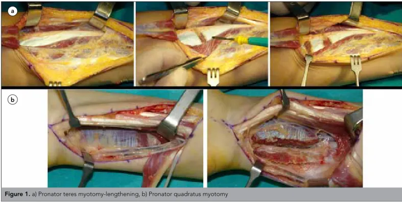
flexor/pro-nator release or transferring flexor carpi ulnaris (FCU) to the wrist extensors. In the second group are the surgical methods directly aimed at the release of pronation contracture: pronator teres my-otomy or lengthening (Figure 1a), pronator quadratus mymy-otomy (Figure 1b), transfer of pronator teres to the wrist extensors and transfer of the FCU tendon to brachioradialis tendon insertion. Pronator release only versus release with active tendon transfer in the treatment of pronation deformity is still a subject of dis-cussion. Removal of the deforming force causing pronation de-formity may imrove supination. However, it is generally reported that an active transfer is needed to further augment supination. Gschwind and Tonkin have defined four types of pronation defor-mity and recommended treatment methods for each one, shown in Table 2. In our practice, we are using a modified version of this classification and treatment algorithm having included Brachio-radialis re-routing technique for Type 3 and Type 4 cases. Taking these principles into account, the techniques used in the surgical treatment of pronation deformity can be summarized as follows:
FCU→ECRB transfer: As a classical method, defined by Green, and generally used to increase the wrist extension, this is transfer of the flexor carpi ulnaris tendon to the extensor carpi radialis
tendon. The primary target of this technique is to restore wrist
Type Treatment
Type 1. Active supination beyond neutral Surgery is unnecessary
Type 2. Active supination to less than, or to neutral position Pronator quadratus release. Flexor aponeurotic release Type 3. No active supination, free passive supination Pronator teres transfer (Brachioradialis re-routing)
Type 4. No active supination, tight passive supination Pronator quadratus and flexor aponeurotic release (Pronator teres myotomy/lengthening+Brachioradialis re-routing)
Table 2. Pronation deformity classification and treatment options by Gschwind and Tonkin (18)
a
b
extension. As a secondary benefit, the forearm gains substantial active supination. This additional advantage is documented in certain biomechanical studies (16).
Pronator Teres Rerouting: This procedure was first described by
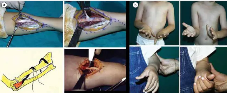
Sakellarides et al. (17). Later, Strecker et al. (14) and Gschwind et al. (18) published their results. Van Heest et al. (19) mechanically demonstrated on cadavers the improvement in supination via re-routing the pronator teres around the radius (Figure 2).
Brachioradialis re-routing: Described by Özkan et al. (20): This
technique is used to correct forearm pronation deformity and re-store active supination. Adequate active supination is achieved by altering the direction of the brachioradialis (re-routing). Since the pronator teres muscle, which is the major pronator muscle of the forearm, is not used as a motor, the forearm pronation is not disturbed. In a biomechanical study, Cheema et al. (16) have demonstrated that brachioradialis rerouting is the second best technique when compared with other methods (Figure 3).
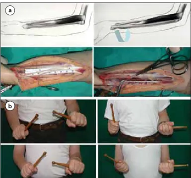
Transfer of the brachialis muscle to the re-routed biceps ten-don: This is an alternative technique which can be used in
re-storing forearm supination. It can be performed especially in the absence of a suitable motor tendon, and also is an option when there is a need to decrease the excessive flexion at the elbow (Figure 4). In a recent study, we reported on four patients who un-derwent a brachialis rerouting supinatorplasty. Active supination increased in two (60° and 50°), minimally increased in one (5°), and did not change in one patient (21).
Other surgical techniques used in repairing the pronation de-formity: Active increase in forearm supination can be expected
by combining pronator teres release with passing the flexor carpi
ulnaris around the ulnar side of the forearm and transferring it to ECBR or BR tendons. By transferring pronator teres to ECRB tendon, the pronator dominance over the forearm is broken. This force can be transferred to the weak wrist or finger extensors (22).
Wrist
Wrist and finger flexion deformities are among the most common problems in patients with spastic palsy (23). The flexion deformity of the wrist is caused by the wrist flexors, especially the flexor carpi ulnaris. Also, spastic finger flexors worsen the situation. When the wrist is flexed, the finger flexors present substantial dysfunction especially during grip. Lengthening the wrist flex-ors decreases the flexor dominance on the wrist. This provides a mechanical advantage for the extensor muscle group. If the wrist extensors are not strong enough to balance out this flexor dominance, they may be actively supported by tendon transfers. Prior to the transfer for the augmentation of the wrist extensors, the spasticity on the extrinsic finger flexors should be assessed. When the wrist is at the neutral position, the spastic finger flex-ors will be mechanically advantageous. The patient cannot loos-en the clloos-enched fist, which precludes grip. In such conditions, lengthening of the finger flexors must be included in the surgery plan. Loss of active extension requires correction with a tendon transfer.
Wrist Flexion Deformity
There are several drawbacks of having the wrist at flexion con-stantly: 1) Flexed wrist creates a mechanical disadvantage for the flexor muscles and reduces the grip power. 2) Especially in patients with deficient stereognosis, the coordination of hand movements may be maintained visually. With the flexion posture
a b
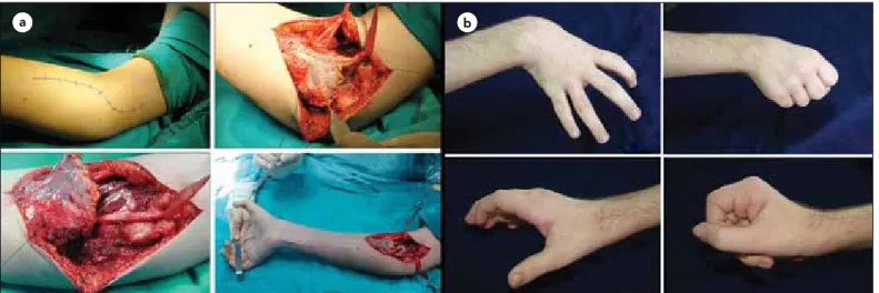
Figure 2. Pronator teres re-routing-Surgical technique: The forearm is brought to maximum supination and the pronator insertion at mid-1/3 radius
is palpated. An approximately 3 cm incision is made in this area, the pronator teres insertion is found between the brachioradialis and ECRL ten-dons. Pronator teres tendon is stripped off the radius with 2 cm of periosteum. The interosseous membrane is dissected extra-periosteally proxi-mal to the original insertion point. The pronator teres tendon is passed in a volar-dorsal direction through this window. The tendon is reinserted on the lateral aspect of the radius close to the original insertion point using classical techniques or suture anchor. Alternatively, the pronator tendon is Z lengthened, the distal tendon slip is passed from dorsal to volar and then sutured to the proximal tendon. Postoperatively, an above elbow splint is applied, keeping the elbow at 45-90 degrees flexion and the forearm in 45-60 degrees supination for 4-5 weeks.
b) Pronator teres re-routing-Sample Case : Preoperative (above) and postoperative (below) active supination and protected active pronation views of a 5-year-old patient with cerebral palsy with supination deficit on the left forearm
of the wrist, the fingers remain in the inward segment of the hand which impedes visualization. 3) The aesthetic appearance of the wrist at flexion may disturb the patient and the family members. 4) In case of severe flexion, there may be lesions on the volar side skin of the wrist.
The wrist plays a key role in movements of the hand. Even when there is no active movement on the fingers, flexion and extension can be made with the tenodesis effect. A moving wrist provides at least 25% of hand function. Therefore, wrist arthrodesis should be performed only in severe deformities, where treatment with dynamic methods fails. In cerebral palsy surgery, a mobile wrist is always among the priorities.
Wrist deformity may develop due to flexor tendon tightness, in-adequate wrist extensors, contracture of the wrist volar capsule or a combination of these factors.
I. Wrist and Finger Flexor Tightness and Treatment
Wrist flexion deformity is generally caused by the spasticity of the flexor carpi ulnaris tendon. In some patients, flexor carpi ra-dialis and Palmaris longus tendons are also spastic. Tenseness on the finger flexors, if any, impedes full extension of the wrist and conduces to deformity. Before a transfer to bring the wrist to extension, factors impeding the passive joint movement span must be eliminated.
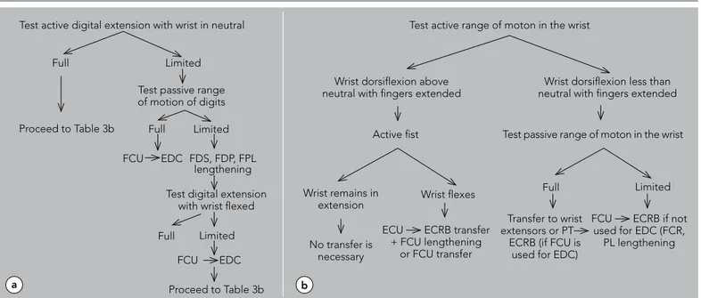
The treatment algorithm below may facilitate the treatment pref-erences for maintaining passive movement of the wrist:
Lengthening of the wrist flexor tendons: The muscle which
prevents passive extension of the wrist most is the spastic flexor carpi ulnaris. Furthermore, the spasticity of this muscle may cause ulnar deviation of the wrist. Extensor carpi ulnaris may create a strong ulnar deviation on the wrist during pronation and also a flexion deformity of the wrist when it is in volar subluxation. In the latter case, the extensor carpi ulnaris tendon can be trans-ferred to the extensor carpi radialis brevis tendon. In moderate Figure 3. a) Brachioradialisre-routing - Surgical technique: An incision is
made on the distal volar –radial aspect of the forearm, and the brachio-radialis muscle-tendon unit is exposed. The radial artery and the radial nerve branch are retracted. The brachioradialis muscle and tendon are dissected and prepared for transfer. A very important step to be taken in the meantime is to isolate the muscle from all the surrounding fasci-al connections. Otherwise, the excursion of the muscle will not be ade-quate. The free end of the tendon that is detached and separated from the insertion point is passed beneath the flexor pollicis longus muscle and taken volarly from dorsal through the interosseous space. The ten-don is re-routed extraperiostally around the radius while preserving the radial artery. The tension is adjusted while the forearm is held at neutral and the original length of the tendon is preserved. The tendon is re-inserted to the radius via classical methods or with a suture anchor. b) Brachioradialis re-routing-Sample Case: A 12-year-old cerebral palsy patient with lack of supination in the left forearm. Preoperative (above) and postoperative (below) views show active supination and preserved active pronation
a
b
Figure 4. a)Transfer of the brachialis muscle to the re-routed biceps tendon– Surgical technique: The brachialis muscle is completely separated
from its the insertion point and released proximally. The biceps tendon is split longitudinally, while the insertion is left intact. The biceps slip on the radial side is re-routed around the radius neck. By fixing it to the musculo-tendinous segment of the brachialis muscle prepared before, the supinatorplasty is completed. b)Transfer of the brachialis muscle to the re-routed biceps tendon-Sample Case: Preoperative (above) and postope-rative (below) active supination and preserved active pronation in a 10-year-old patient with cerebral palsy with lack of supination in the left forearm.
cases, only fractional or Z plasty lengthening of the flexor carpi ulnaris tendon may be sufficient. If an active wrist extension is desired, the flexor carpi ulnaris tendon can be transferred to wrist extensor tendons. The palmaris longus tendon can be separated from the distal insertion point and be used for transfer (e.g. to the thumb extensor tendon). Although the flexor carpi radialis tendon can be used in tendon transfers, only lengthening of this tendon is recommended since releasing both wrist flexors may lead to hyperextension deformity, which may create complica-tions especially when the finger extension is not adequate.
Release of finger flexor tendons: All flexor tendon lengthening
techniques result in a decrease in the power of the flexor ten-dons. However, this can be compensated by the wrist, strength-ening the grip during extension. While fractional lengthstrength-ening creates minimal strength loss, the superficialis-to-profundus (STP) tendon transfer procedure may cause severe strength loss. The effect of Z-lengthening stands between these two procedures. Other bone procedures like proximal row carpectomy, and wrist arthrodesis are not recommended on functional hands. These methods may be performed on rather severe cases for hygiene- and cosmesis-related concerns.
i) Fractional Tendon Lengthening has minimal excessive
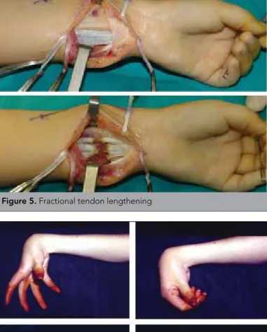
length-ening risk and minimal negative effect on the grabbing power of the fingers, therefore it is preferred in well-functioning hands. A longitunal incision on the mid 1/3 of the volar forearm is sufficient if only fractional lengthening is planned. Two transverse tenoto-mies are made on the musculotendinous region of the tendon to be lengthened. The most distal tenotomy must be at least 2 cm away from the most distal point of the musculotendinous junc-tion, and there must be a minimum 1 cm distance between the tenotomies. Only the tendinous part of the musculotendinous connection should be cut and the muscular part must be kept untouched (Figure 5).
If passive extension of the fingers is still limited despite full flex-ion of the wrist, then possibly fractflex-ional lengthening will not be adequate and one of the other two techniques will be required.
ii) Tendon Z-lengthening: This method can be used in cases where
fractional lengthening is inadequate. It is especially used in FPL lengthening. Here, the most important criterion is lengthening the tendon by 0.5 cm for each degree of joint opening desired. The tendon is cut at the radial aspect on one side and ulnar on the other. After the lengthening, the tendon ends are fixed with Pulvertaft weave or end-to-side technique. If the lengthening is performed on the thumb, fixation is performed when the wrist is at neutral and the MP and IP joints are at mild flexion. When the wrist is at extension, the thumb must be touching the second finger. Furthermore, the thumb must be out of the palm with the flexion of the wrist.
iii) Superficialis-to-profundus procedure (STP): This procedure
must be performed on dysfunctional hands with severe flexion contracture as it causes considerable loss in grip power (Figure 6).
Flexor/Pronator slide: Pronator teres can be detached from its
origin, the medial condyle, and moved distally. This procedure, called the flexor-pronator slide (Figure 7) is usually performed on
patients with CP sequela and central hemiparesis, who have se-vere flexor dominance in the elbow, wrist and fingers. The proce-dure may also be performed to involve all the flexors, originating from the pronator/flexor common origin at the medial epicon-dyle level (24).
In addition, proximal row carpectomy can shorten the forearm by approximately 1 cm and maintain a passive dorsiflexion increase of 25º on the wrist.
II. Extension Loss of the Wrist and Its Management
After elimination of the factors preventing passive extension of the wrist, active extension of the wrist must be achieved. The sur-gical classification by Zancolli (6) can serve as a reference point for the active tendon transfer:
The cases in Group 1 can have full finger extension with less than 20º wrist flexion. In this group, usually the flexor carpi ulnaris mus-cle is spastic, and lengthening of this musmus-cle solves the problem in most cases.
Figure 5. Fractional tendon lengthening
Figure 6. Superficialis to profundus tendon transfer: Preoperative
In Group 2, the active finger extension can be provided only with a wrist flexion greater than 20º. In Subgroup 2A, there is weak wrist extension when the fingers are in flexion. In Subgroup 2B, on the other hand, there is no wrist extension at all. The latter group needs supportive transfer procedures to maintain the wrist extension after flexor release.
The transfers mostly used for wrist extension are: FCU→ECRB, ECU→ECRB, PT→ECRB, BR→ECRB and FDS→ECRB. If a more radial deviation of the wrist is required, these transfers should be made to the ECRL tendon. Additionally, the FCU→ECRB transfer performed to maintain wrist extension actively adds to the fore-arm supination (Figure 8).
The cases in Group 3 do not have active finger extension. In gen-eral, there is severe involvement. Transfer to the finger extensors (e.g. FCU→EDC) is needed after release of the contractures in the flexor muscles.
Surgical techniques commonly used in wrist extension insuf-ficiency treatment:
1. Green Transfer (Flexor carpi ulnaris tendon → Extensor carpi
radialis brevis tendon transfer): Technical details of this
meth-od are summarized in Figure 9.
2. Extensor carpi ulnaris tendon →Extensor carpi radialis
bre-vis tendon transfer: This transfer is performed on patients
who have sufficient wrist extension and whose wrists become flexed during grip under the influence of finger flexors. This transfer must always be performed together with fractional lengthening of FCU muscle. It cannot be performed simul-taneously with wrist flexors→wrist extensors transfer. Other-wise, extension deformity may develop (25, 26).
3. Pronator teres tendon→Extensor carpi radialis brevis tendon
transfer: This technique is suitable for patients whose FCU
tendon is dysfunctional or used as a motor for another trans-fer. However, due to the limited excursion of pronator teres, the wrist movements are reduced. Since the pronator teres is often spastic, this transfer provides benefit especially in teno-desis form. The PT tendon, released at its insertion at mid-1/3 forearm, is transferred to ECRB tendon.
4. Brachioradialis tendon→Extensor carpi radialis brevis tendon
transfer: The brachioradialis tendon can be used for wrist
ex-tension, when the other motors are not suitable for transfer. However, as stated above, the fascial connections around the muscle must be completely released to provide sufficient ex-cursion for the brachioradialis muscle.
Figure 7. a. Flexor/Pronator Slide: Surgical technique b)Flexor/Pronator Slide: Sample Case: Preoperative (upper row) and postoperative (lower
row) views of a patient with spasticity in the wrist and fingers
a b
Figure 8. Green transfer: The FCU tendon is exposed with an incision on the volar ulnar side of the distal forearm. The FCU tendon is released from
insertion on the pisiform bone. The ECRB and ECRL tendons are exposed with a second incision on the dorsal aspect of the wrist. A subcutaneous tunnel is prepared on the ulnar side of the forearm, between the two incisions. The FCU tendon is taken through this tunnel and attached to the recipient tendon with Pulvertaft weave. The tension is adjusted while the transferred tendon is under maximal tension and the wrist is at neutral – mild dorsiflexion. The wrist should have a passive flexion of 20º, otherwise, the anastomosis is tense11. The figure shows preoperative (middle) and
5. Transferring flexor carpi ulnaris→Extensor digitorum communis
tendons: As explained above, the reason for the finger
exten-sion and the difficulty in releasing the hand may either be the contracture on the volar structures or the weakness of the finger extensors. Flexor carpi ulnaris transfer may support the finger extensors. The superficial flexor tendons of the third and fourth finger can also be used to strengthen the finger extensors.
Technique: Similar to Green transfer, the FCU is exposed with
an incision on the forearm volar-ulnar side. The FCU tendon is passed on the ulnar side of the forearm subcutaneously. On the wrist extensor side, the EDC tendons are exposed with an inci-sion of 4-5 cm on the 4th extensor compartment. The FCU tendon
is taken through the subcutaneous tunnel and woven side-by-side with Pulvertaft weaving. The tension of the tendons is adjust-ed accordingly to keep the MP joints at neutral position and the IP joints at mild flexion. The hand is immobilized for four weeks –fingers are kept as explained above, while the wrist is held at 30º extension (12, 25, 26).
The treatment algorithm we are following in the surgical restora-tion of the problems related to finger and wrist spasticity is sum-marized in Tables 3a and b.
Surgical treatment of finger and thumb deformities “Swan Neck” Deformity
The hyperextension on the PIP joint and flexion deformity of the DIP joint is known as the “swan neck deformity”. Its etiology is created by the tenodesis effect of the EDC tendons when the wrist is in a spastic flexion posture and the hyperactivity of the in-trinsic hand muscles. Although swan neck deformity is commonly seen in cerebral palsy, surgical intervention is needed only when the fingers are locked in the hyperextension position and there are progressive PIP joint extension deformities which cannot be prevented with splinting (27).
In cerebral palsy, swan neck deformity is caused by the relatively shorter central slip. The shortness is relative and does not
inter-fere with flexion of the PIP joint. Both the spasticity of the in-trinsic muscles and the hyperactivity of the exin-trinsic finger ex-tensors play a role in the patho-physiology of the tightness on the central slip (12). The high tension, generated by the spastic-ity of the intrinsic muscles, is transferred to the central slip via medial interosseous bands. This creates hyperextension in the PIP joint. In case of a loosening on the retinacular ligament, the extensor mechanism gets subluxed towards the dorsal aspect. With the additional loosening of the volar plate, the hyperexten-sion of the PIP joint increases. Recurvatum deformity is observed. Meanwhile, the subluxation of the extensor mechanism towards the dorsal leads to relative insufficiency on the lateral bands. DIP joint flexion occurs.
Furthermore, patients with insufficient wrist extensors try to com-pensate the wrist extension function with the extrinsic finger ex-tensors. Within the system explained above, the increased load on the central slip augments the severity of the swan neck defor-mity. In addition, after lengthening operations of the superficialis tendons, the swan neck deformity may become more evident. The MP joint flexion observed in swan neck deformity is ex-plained as follows: The wrist and finger joints make up a kinetic chain. The weakest rings of this chain are the PIP joint and the MP joint, respectively. The extension on the PIP joint leads to flexion on the proximal and distal joints. Moreover, these forces cause a subluxation of the MP joint in the volar direction. Maintaining flexion of the PIP joint prevents the deformation in the finger. In this case, the normal axis of the finger is preserved. Based on this rationale, the following treatment methods are recommended for the treatment of swan neck deformity (12):
1. Decreasing the load on the central slip by cutting the liga-ments connecting the intrinsic muscles to the central slip. 2. Keeping the lateral bands more volarly, by shortening the
retinacular ligament
3. Preventing the load on the central slip by reattaching the exten-sor tendon on the proximal phalanx instead of the central slip
Table 3. Test active digital extension with wrist in neutral, b) Test active range of moton in the wrist
Test active digital extension with wrist in neutral Test active range of moton in the wrist
Test passive range of moton in the wrist Full
Full
Full
Full Limited
Limited Active fist
Limited Limited Proceed to Table 3b FCU FCU EDC EDC FDS, FDP, FPL lengthening Test passive range of motion of digits
Wrist dorsiflexion above neutral with fingers extended
No transfer is necessary
ECU ECRB transfer + FCU lengthening
or FCU transfer
Transfer to wrist extensors or PT
ECRB (if FCU is used for EDC)
FCU ECRB if not used for EDC (FCR,
PL lengthening Wrist remains in
extension Wrist flexes
Wrist dorsiflexion less than neutral with fingers extended
Test digital extension with wrist flexed
Proceed to Table 3b
4. Limiting joint hyperextension by placing a tendon graft on the volar side of the PIP joint
5. Arthrodesis of the PIP joint at flexion
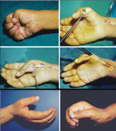
6. Preventing the joint extension by using the flexor digitorum sublimis tendon or by performing volar capsulorraphy: subli-mis tenodesis (Figure 10).
7. Advancing the interosseous muscles (13) 8. Ulnar motor neurectomy
9. Sling procedure (6)
10. Flexion osteotomy on the metacarpal
Surgical Treatment of “Thumb-in-Palm” Deformity:
“Thumb-in palm” deformity is one of the most common and complicated deformities of cerebral paralysis, and is caused by spasticity of the thumb adductor and flexor muscles. When the thumb is locked inside the palm, it cannot function. Also it hin-ders the grabbing function of the other fingers.
Parallel to the evolution of cerebral paralysis surgery, thumb op-erations have developed into dynamic soft tissue procedures, rather than static bone operations. Authors, including Goldner, Inglis and Keats, have succeeded in forming mobile thumbs by maintaining the muscular balance via tendon transfers and selec-tive joint arthrodeses (28).
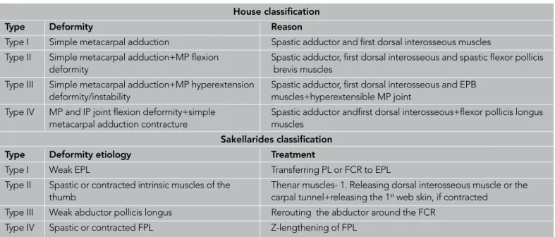
The objectives of the surgical treatment of thumb-in-palm defor-mity are to generate a strong lateral pinch on the mid-phalanx of the second middle finger -at fist form- and to maintain sufficient radial abduction during grabbing (29). The major challenge of the treatment is the multifactorial quality of the deformity. With the aim of making a systemic definition of the deformity and pro-posing a treatment plan, House (28) and Sakellarides (30) have formulated two different classification systems (Table 4).
The etiology of the thumb deformity is multifactorial. There are four main reasons for this form of deformity: 1) Spasticity of the
Figure 10. Augmentation of abductor and extensor tendons:
Preope-rative (upper row) and postopePreope-rative (lower row) views of the patient
House classification
Type Deformity Reason
Type I Simple metacarpal adduction Spastic adductor and first dorsal interosseous muscles
Type II Simple metacarpal adduction+MP flexion Spastic adductor, first dorsal interosseous and spastic flexor pollicis
deformity brevis muscles
Type III Simple metacarpal adduction+MP hyperextension Spastic adductor, first dorsal interosseous and EPB deformity/instability muscles+hyperextensible MP joint
Type IV MP and IP joint flexion deformity+simple Spastic adductor andfirst dorsal interosseous+flexor pollicis longus metacarpal adduction contracture muscles
Sakellarides classification Type Deformity etiology Treatment
Type I Weak EPL Transferring PL or FCR to EPL
Type II Spastic or contracted intrinsic muscles of the Thenar muscles- 1. Releasing dorsal interosseous muscle or the thumb carpal tunnel+releasing the 1st web skin, if contracted
Type III Weak abductor pollicis longus Rerouting the abductor around the FCR Type IV Spastic or contracted FPL Z-lengthening of FPL
Table 4. Surgical classifications of thumb-in-palm deformity
Figure 9. Correction of swan neck deformity with sublimis tenodesis
technique: Preoperative view of a 13-year-old patient with severe spas-ticity on the left hand (left-above)- Swan neck deformity occurred as a result of the “over correction” seen after lengthening and tendon trans-fer (right-above)- Immediate (left-below) and long-term (right-below) results after sublimis tenodesis surgery
adductor and flexor muscles, 2) Flaccid paralysis on the extensors and abductors, 3) Hypermobile metacarpophalangeal joint and
4) Contracture on the first web skin.
Assessing each of these points in the preoperative term would obviously facilitate the formulation of a surgical plan. The assess-ment involves physical examination, electromyography and diag-nostic nerve blocks. Usually, the increased tonus of the muscles and the thumb’s position at rest show which muscles are causing the deformity.
a. In the deformity related to the spasticity of the thenar mus-cles, there is flexion in the metacarpophalangeal joint and extension in the interphalangeal joint;
b. Deformity related to the spasticity of flexor pollicis longus muscle is concomitant especially with interphalangeal joint flexion and different degrees of flexions at the metacarpo-phalangeal joint;
c. The involvement of adductor pollicis muscle leads to flexion of the metacarpophalangeal joint and adduction of the first metacarpal bone towards the center of the palm;
d. The involvement of the opponens pollicis muscle or the first dorsal interosseous muscles leads to the adduction of the metacarpal bone in line with the palm; and thus the thumb gets trapped in the web space.
I. Treatment of adductor and flexor muscle spasticities:
Re-lease of adductor pollicis, first dorsal interosseous and flexor pollicis brevis muscles can be performed as muscle myotomy at the origin, insertion or between the two (10). Release at insertion is often avoided since it removes the entire functionality of the muscle. Release at origin, on the other hand, preserves the functionality to a certain extent via the adhesion of the muscle to the adjacent tissues. During muscle myotomy the muscle is kept in a tense po-sition, the tendinous part is cut, while the muscle fibers are protected.
Surgical techniques
a. Adductor / Flexor Release: An incision parallel to the thenar crease is made. The dissection is continued up to the the-nar muscles. Flexor pollicis brevis and abductor pollicis bre-vis are isolated at their origin on the transverse carpal liga-ment. They are released through the ligaliga-ment. Meanwhile, the proximal phalanx is brought to extension to facilitate the radial movement of the muscles. With proper splinting in the postoperative period, these muscles attach to their new loca-tion. If spastic, the adductor muscle is released during the same session. If the muscle is to be released from its origin, the flexor tendons of the second and third fingers and the neurovascular structures are retracted and stripped off over the third metacarpal bone. If the muscle is to be released from the insertion point, it is followed to the radial side and cut at the tendinous insertion point. In cases where the ad-ductor muscle is to be released without touching the flexor muscles, the adductor muscle is penetrated with an incision on the first web.
b. Releasing the dorsal interosseous muscle: An incision is made through the palpable ulnar border of the first metacarpal
bone. The radial nerve branches and the EPL tendon are re-tracted. The first dorsal interosseous muscle is a bipennate muscle. Its origin is in the first and second metacarpals. Its origin in the first metacarpal is released, while the segment in the second metacarpal is left untouched. This untouched segment helps the flexion and abduction of the second fin-ger. If the incision is taken to the MP joint distally, the abduc-tor can also be released through this incision.
c. FPL tendon lengthening: The FPL spasticity seen in Type IV deformity is treated with fractional lengthening or Z-length-ening on the FPL tendon proximal to the wrist, similar to the wrist flexors.
II. Supporting the abductor and extensor tendons: The
ten-don reinforcements and their objectives can be summa-rized as: APL –for the abduction of the first metacarpal, EPB –for the extension of the proximal phalanx and EPL – for the extension of the distal phalanx. The reinforcement can be performed in following forms: tendon plication, tendon transfer or fixing the tendon to a bone-perioste-um or to another tendon at a proper tension (tenodesis):
1) Tendon transfers: With the aim of maintaining the
thumb extension and taking the thumb out of the palm, transferring BR, PL, ECLR, ECRB, FCR, FCU and FDS tendons were recommended by several authors (10, 11). APL is the most important tendon effective on the carpometacarpal joint. Taking the APL tendon out of the first compartment and moving it volarly pro-vides a mechanical advantage for the abduction of the first metacarpal. In addition, suturing the PL tendon (end-to-side) to the volarly rerouted APL tendon may increase the abduction (28). If the extension vector is to be preserved, the same transfer can be performed by protecting the pulley of the first compartment. As an alternative, the APL is cut and its distal end is re-inforced with an appropriate motor, while the proxi-mal end is transferred to the EPB tendon (end-to-side) under tension. In this way, both metacarpal abduction and proximal phalanx extension are maintained. In cases where an appropriate motor muscle cannot be found for transfer, the APL is cut and its distal end is re-routed towards the volar side. Then it is fixed end-to-side to the FCR tendon. This may create a tenodesis effect and maintain metacarpal abduc-tion.
Figure 10 shows an example, where the above mentioned tech-niques are combined to reinforce the thumb abductor and exten-sor tendons.
2) Shortening APL and EPB tendons via plication 3) EPL Re-Routing: The EPL tendon, which passes around
the Lister’s tubercle, also has an adductor effect due to its path. Therefore, procedures radializing this route are expected to have a positive effect on abduction. Goldner et al. (26) removed the EPL tendon from its fibro-osseous channel, creating a new pulley over the radial styloid and moving the route of the tendon radi-ally. Later, Manske (31) took the EPL tendon through
the first dorsal retinacular compartment. This method eliminated the need for a new pulley. Modifying the method further, Rayan and Saccone (32) transected the EPL tendon proximal to the retinaculum, then anastomosed the distal EPL tendon slip taken through the first dorsal compartment, with the proximal slip. The authors reported that they maintained satisfac-tory abduction.
4) FPL Abductorplasty (Figure 11): The spastic FPL ten-don to be released is cut on the proximal phalanx. The distal segment is used for the stabilization of the IP joint in children. The proximal segment is pulled through a second incision on the wrist. A second sub-cutaneous tunnel is opened further radially and the tendon is radialized.
III. Hypermobile MP Joint: The stability of the
metacarpopha-langeal joint is crucial for the reinforcements on the EPL or EPB tendons. If the joint has a hyperextension exceeding 20º, reinforcement of these tendons will lead to hyperex-tension deformity of the MP joint. In order to prevent this, in such deformities where the MP joint is hyperextensible,
capsulodesis or arthrodesis of the MP joint is needed.
Cap-sulodesis can be attempted to avoid arthrodesis, especially in children younger than 13 (7). Filler et al. (33) have ob-tained long-term results by performing capsulodesis on 13 patients with cerebral palsy. According to their technique, the volar plate is cut and fixed to a further proximal position on the metacarpal, while the MP joint is kept at 30º flexion.
MP joint arthrodesis is recommended when capsulodesis is insufficient. During the arthrodesis, the growth plate on the epiphysis is preserved and, by performing joint cartilage resection only, growth is not disturbed in young children (approximately 4-5 year-old) (8).
IV. Releasing the first web skin: Any contracture on the skin
can be released by two- or four-flap Z-plasties.
CONCLUSION
Surgical management of the spastic upper limb in cerebral palsy requires meticulous evaluation and planning. It is important to know that upper extremity deformities are secondary manifesta-tions of the cerebral injury. Therefore, in addition to evaluation of upper extremity function, consideration should be given to the intelligence and motivation of the patient, and voluntary use of the upper extremity. Prior to surgery, the overall level of function needs to be considered. This is also important for the patients and their families, who should know that surgery is aimed at im-proving the upper extremity deformity, and not the primary disor-der. An appropriately indicated surgery can significantly contrib-ute to upper extremity function.
Conflict of interest: No conflict of interest was declared by the
authors.
REFERENCES
1. Bax M, Goldstein M, Rosenbaum P, Leviton A, Paneth N, Dan B, et al. Proposed definition and classification of cerebral palsy. Dev Med Child Neurol 2005; 47: 571-6. [CrossRef]
2. Dahlin LB, Komoto-Tufvesson Y, Salgeback S. Surgery of the spastic hand in cerebral palsy. Improvement in stereognosis and hand func-tion after surgery. J Hand Surg 1998; 23: 334-9. [CrossRef]
3. Manske PR. Cerebral palsy of the upper extremity. Hand Clin 1990; 6: 697-709.
4. Waters P, Van Heest A. Spastic hemiplegia of the upper extremity in children. Hand Clinics 1998; 14: 119-34.
5. Moberg IE. Reconstructive hand surgery in tetraplegia, stroke and cerebral palsy; some basic concepts in physiology and neurology. J Hand Surg 1976; 1: 29-34.
6. Zancolli EA, Zancolli ER Jr. Surgical management of the hemiplegic spastic hand in cerebral palsy. Surg Clin North America 1981; 61: 395-406.
7. Keats S. Surgical treatment of the hand in cerebral palsy. Correction of thumb-in-palm and other deformities. Report of nineteen cases. J Bone Joint Surg 1965; 47: 274-84.
8. Mital AM, Sakellarides HT. Surgery of the upper extremity in the retarded individual with spastic cerebral palsy. Orthop Clin North America 1981; 12: 127-41.
9. Skoff H, Woodbury DF. Management of the upper extremity in cere-bral palsy. J Bone Joint Surg 1985; 67: 500-3.
10. Goldner JL. Upper extremity tendon transfers in cerebral palsy. Or-thop Clin North Am 1974; 5: 389-414.
11. Beach WR, Strecker WB, Coe J, Manske PR, Schoenecker PL, Dailey L. Use of the Green transfer in treatment of patients with spastic cerebral palsy: 17- year experience. J Pediatr Orthop 1991; 11: 731-6. [CrossRef]
12. Manske PR. Cerebral palsy of the upper extremity. Hand Clin 1990; 6: 697-709.
13. Carlson MG. Cerebral palsy. In: Green DP, Hotchkiss RN, Pederson WC, Wolfe SW editors. Green’s Operative Hand Surgery 5th ed.
Pennsylvania: Churchill Livingstone; 2005. p.1197-234.
14. Strecker WB, Emanuel JP, Dailey L, Manske PR. Comparison of pro-nator tenotomy and propro-nator rerouting in children with spastic cere-bral palsy. J Hand Surg 1988; 13: 540-3. [CrossRef]
Figure 11. Correction of thumb-in-palm deformity with FPL
abductorp-lasty: Preoperative view and surgical technique b) Postoperative view of the patient shown in Figure 11a
15. Pletcher DFJ, Hoffer M, Koffman DM. Nontraumatic dislocation of the radial head in cerebral palsy. J Bone Joint Surg 1976; 58: 104-5. 16. Cheema TA, Firoozbakhsh K, De Carvalho AF, Mercer D.
Biomechan-ic Comparison of 3 Tendon Transfers for Supination of the Forearm. J Hand Surg 2006; 31: 1640-4. [CrossRef]
17. Sakellarides HT, Mital MA, Lenzi WD. Treatment of pronation con-tractures of the forearm in cerebral palsy by changing the insersion of pronator teres. J Bone Joint Surg Am 1981; 63: 645-52.
18. Gschwind C, Tonkin M. Surgery for cerebral palsy. Part I. Cassifica-tion and operative procedures for pronaCassifica-tion deformity. J Hand Surg Br 1992; 17: 391-5. [CrossRef]
19. Van Heest AE, Sathy M, Schutte L. Cadaveric modeling of the prona-tor teres rerouting tendon transfer. J Hand Surg Am 1999; 24: 614-8.
[CrossRef]
20. Özkan T, Tunçer S, Aydın A, Hoşbay Z, Gülgönen A. Brachioradia-lis re-routing for restoration of active supination and correction of forearm pronation deformity in cerebral palsy. J Hand Surg 2004; 29: 265-70. [CrossRef]
21. Ozkan T, Bicer A, Aydin HU, Tuncer S, Aydin A, Hosbay ZY. Brachia-lis muscle transfer to the forearm for the treatment of deformities in spastic cerebral palsy. J Hand Surg Eur Vol. 2012 Aug 22. [Epub ahead of print] [CrossRef]
22. Colton CL, Ransford AO, Lloyd-Roberts GC. Transposition of the tendon of the pronator teres in cerebral palsy. J Bone Joint Surg Br 1976; 58: 220-3.
23. Rayan GM, Young BT. Arthrodesis of the spastic wrist: J Hand Surg 1999; 24: 944-52. [CrossRef]
24. Inglis AE, Cooper W. Release of the flexor-pronator origin for flexion deformities of the hand and wrist and fingers in spastic paralysis J Bone Joint Surg Br 1966; 48: 847-57.
25. Goldner JL. Surgical reconstruction of the upper extremity in cere-bral palsy. Instr Course Lect 1987; 36: 207-35.
26. Hoffer HM, Lehman M, Mitani M. Long-term follow up on tendon transfers to extensors of the wrist and fingers in patients with cere-bral palsy. J Hand Surg Am 1986; 11: 836-40.
27. Swanson AB. Surgery of the hand in cerebral palsy and the swan-neck deformity. J Bone Joint Surg Am 1960; 42: 951-64.
28. House JH, Gwathmey FW, Fidler MO. A dynamic approach to the thumb-in-palm deformity in cerebral palsy. J Bone Joint Surg 1981; 63: 216-25.
29. Tonkin MA, Hatrick NC, Eckersley JRT, Couzens G. Surgery for cere-bral palsy. Part 3: Classification and operative procedures for thumb deformity. J Hand Surg 2001; 26: 465-70. [CrossRef]
30. Sakellarides HT, Mital AM, Matza RA, Dimakopoulos P. Classification and surgical treatment of the thumb-in-palm deformity in cerebral palsy and spastic paralysis. J Hand Surg 1995; 20: 428-31. [CrossRef]
31. Manske PR. Redirection of extensor pollicis longus in the treatment of spastic thumb-in-palm deformity J Hand Surg 1985; 10: 553-60. 32. Rayan GM, Saccone PG. Treatment of spastic thumb-in-palm
defor-mity: A modified extensor pollicis longus tendon rerouting. J Hand Surg 1996; 21: 834-9. [CrossRef]
33. Filler BC, Stark H, Boyes JH. Capsulodesis of the metacarpophalan-geal joint of the thumb in children with cerebral palsy. J Bone Joint Surg 1976; 58: 667-70.

