Photophysical and antimicrobial properties of new
double‑armed benzo‑15‑crown‑5 ligands and complexes
Serhat Koçoğlu1,2 · Hatice Ogutcu3 · Zeliha Hayvalı1
Received: 28 September 2018 / Accepted: 12 January 2019 / Published online: 24 January 2019 © Springer Nature B.V. 2019
Abstract
New double-armed crown ether ligands linked to pyridine derivatives have been synthesized and characterized. These macrocyclic ligands (1–5) have been synthe-sized by the reactions of 4′,5′-bis(bromomethyl)benzo-15-crown-5 with 3-hydroxy
pyridine derivatives. A series of Na+, K+ and Ag+ complexes (1a–5a, 1b–5b and
1c–5c) of the macrocyclic ligands have been prepared from sodium perchlorate, sodium picrate, potassium iodide, potassium picrate and silver nitrate salts,
respec-tively. The most suitable cation Na+ is bound to the 15-crown-5 cavity and 1:1
“fill-ing complexes” are formed (1a–5a) while the K+ cation interacts with the crown
ether cavity and forms sandwich-type complexes (1b–5b). The Ag+ complexes
(1c–5c) have been obtained with a pyridine moiety of the new crown ethers. New ligands undergo photophysical changes when bonding the cation. The influence of
metal cations such as Na+, Li+, K+, Fe3+, Cu2+, Ca2+, Ba2+ and Al3+ on the
spectro-scopic properties of the pyridine linked to the double-armed crown ether moiety was investigated in EtOH solution by means of absorption and emission spectrometry. The prepared compounds (1–5, 1a–5a, 1b–5b and 1c–5c) were evaluated for anti-bacterial and antifungal activities against pathogenic microorganisms. The results show that the antimicrobial activity of the synthesized compounds varying a degree of inhibitory effects on the growth of different tested pathogenic strains.
Keywords Crown ether · Alkali metal complexes · Silver(I) complexes · UV and fluorescence spectroscopy · Antimicrobial activity · Pathogenic microorganism
Electronic supplementary material The online version of this article (https ://doi.org/10.1007/s1116
4-019-03741 -3) contains supplementary material, which is available to authorized users. * Zeliha Hayvalı
1 Department of Chemistry, Faculty of Science, Ankara University, 06100 Ankara, Turkey 2 Science and Technology Application and Research Center, Bozok University, 66200 Yozgat,
Turkey
3 Department of Field Crops, Faculty of Agriculture, Kırşehir Ahi Evran University,
Introduction
Since the pioneer work of Pedersen on crown ethers, a significant number of
macro-cyclic crown ether compounds were synthesized [1–7]. Owing to their great
molecu-lar variety, these ligands are very quiet for selective complex formation. Especially,
crown ether rings are well known complexation groups in fluoroionophores [8–12].
They possess a cavity that allows them to reach high selectivity and sensitivity in
metal cation detection [8, 13–17]. Many metal cation sensors consist of a
fluoro-phore and a receptor unit, which are either separated by a spacer group or conju-gated to each other. While the photoinduced electron transfer (PET) sensors gen-erally have fluorophore–spacer–ionophore (guest binding site) moiety, the internal charge transfer sensors (ICT) consist of a receptor is part of the π-conjugated system
of the fluorophore [18]. Crown ether ligands exhibit ionophoric properties in
mem-branes, behaving very similarly to the biologically very important ionophore groups such as valinomycin, gramicidin, nonactin, which makes crown ether compounds especially useful in chemical and biological studies, and their pharmaceutical
poten-tials are very large [19–21]. Furthermore, these compounds, possessing additional
other functional groups, have emerged as one of the most influential architectural motifs for supramolecular compounds. The combination of pyridine derivatives with benzo-15-crown-5 group results in the design of the new ditopic compounds. The 15-crown-5 group can bind with alkali metal cations in the crown ether cavity, whereas the pyridine group binds transition and heavy metal ions with the N atom of the heterocyclic pyridine group. There are many studies in the literature of Ag(I) complexes bound to the N atom of the pyridine ligand with different binding modes
and coordination numbers [22–24]. Therefore, investigating the structures and
prop-erties of Ag(I)-pyridine coordination complexes would be a worthy and interesting work.
In this study, 15-crown-5 was chosen as an ionophore, and substituted pyridine
derivatives act as an additional chelating moiety (Scheme 1).
We have examined the effects of changing the type and position of the electron
donor or acceptor group (–CH3, Cl, Br, and NO2) on the pyridine ring (Scheme 2).
This means that the molecular structures of 2-nitropyridine derivatives are seldom
investigated according to 3- and 4-nitropyridine [25]. 3-Benzyloxy-2-nitropyri-dine compound is an important intermediate for the synthesis of asymmetric
cyanine dyes for the fluorescence detection of nucleic acids [25, 26].
2-Chloro-pyridine derivatives are key synthetic intermediates for many pharmaceutical and
commercially relevant products [27–29].
The 4′,5′-bis(bromomethyl)benzo-15-crown-5 and 3-hydroxy pyridine and derivatives were used as starting materials, and a series of new double-armed
crown ether compounds were prepared (1–5) (Scheme 2). The sodium (1a–5a),
potassium (1b–5b) and silver(I) complexes (1c–5c) were prepared by the
reac-tions of new ligands (1–5) with metal salts (Scheme 3). In this paper, we suggest
new ditopic ligands (1–5) in which pyridine groups are able to coordinate
transi-tion metal ion (Ag+), while the 15-crown-5 moiety binds with alkali metal ions
(Na+ and K+). All of the new crown ether compounds are described, and their IR,
mass, 1H- and 13C-NMR spectral data are reported.
The complex compositions of different metal cations (Na+, Li+, K+, Fe3+,
Cu2+, Ca2+, Ba2+ and Al3+) with the ligands (1–5) were determined by using
absorption and fluorescence study in EtOH. Because it was known that crown ether ligands could bind metal cations, we were motivated to test the existence
O O O O O Br Br N OH O O O O O O N O N N X OH O O O O O O N O N CH3 CH3 N OH C H 3 O O O O O O N O N O2N O2N (1) (2) (3-4) (5) X O O O O O O O N N X NO2 N OH (Cl, Br) KOH NaOH KOH NaOH
of metal ions by reviewing the changes of fluorescence intensity of new ligands (1–5). The variants of the emission spectra of this ditopic receptor detected upon different metal ion addition make the ligand convenient for multitasking sensors. Double-armed crown ethers are composed of two flexible side arm provides fur-ther coordination of a guest cation trapped in a crown efur-ther ring and side arms. So, multiple binding sites (crown ether group and side arms) are stronger cation binders in comparison to ordinary crown ethers, they offer great promise in the
area metal sensing and design of smart fluorescence-based sensor processes [30,
31].
New crown ether ligands and complexes were investigated for antimicrobial activity against pathogenic strains; Staphylococcus aureus, Listeria monocytogenes 4b, Escherichia coli, Salmonella typhi H, Staphylococcus epidermis, Micrococcus
luteus, Bacillus cereus and antifungal activity against Candida albicans.
Experimental
Reagents and equipments
The starting chemicals, tetraethylene glycol dichloride [32], benzo-15-crown-5
[1] and 4′,5′-bis(bromomethyl)benzo-15-crown-5 [33] were prepared according
to the cited literature. All solvents and pyridine derivatives (3-hydroxypyridine, 3-hydroxy-6-methylpyridine, 3-hydroxy-2-chloropyridine, 3-hydroxy-2-bromopyr-idine and 3-hydroxy-2-nitropyr3-hydroxy-2-bromopyr-idine) were purchased from Sigma-Aldrich Chemi-cal Company and used without any further purification. Alkali metal picrates were
prepared according to the literature method [34]. Perchlorate and picrate salts of
metal complexes with organic ligands are potentially explosive. All of the solvents
O O O O O O O N N R2 R2 Na+ R1 R1 X X- comp. ClO4- 1a, 2a pic- 3a-5a X -O O O O O OO N N R2 R2 R1 R1 O O O O O OO N N R2 R2 K+ R1 R1 X- comp. ClO4- 1b, 2b
pic- 3b-5b Comp.1a, 1b, 1c RH1 RH2
2a, 2b, 2c CH3 H 3a, 3b, 3c H Cl 4a, 4b, 4c H Br 5a, 5b, 5c H NO2 (1a-5a) (1b-5b) N+ O -O -O O O O O O O O N N R2 R2 Ag+ R1 R1 O O O O O O O N N R2 R2 Ag+ R1 R1 N+ O -O -O (1c, 2c) (3c-5c)
were used without further purification. Melting points were controlled on a
Gallen-kamp melting point platform. 1H NMR spectra were detected on a VARIAN
Mer-cury 400 MHz spectrometer. 1H NMR chemical shifts (δ) are given in ppm
down-field from Me4Si, determined by chloroform (δ = 7.26 ppm).13C NMR spectra were
detected on a VARIAN Mercury 400 MHz spectrometer. 13C NMR chemical shifts
(δ) are reported in ppm with the internal CDCl3 at δ 77.0 ppm as standard. IR
spec-tra were recorded on a Shimadzu Infinity FTIR spectrometer and were reported in
cm−1 units. Mass spectral analyses were performed on an Agilent Technologies
6224 TOF LC/MS spectrometer. UV–visible spectra were recorded on a HITACHI u2800 UV–Vis spectrophotometer. Fluorescence spectra were recorded on a Perkin Elmer LS 50 B Fluorescence Spectrometer.
Test microorganisms
The pathogenic bacterial cultures chosen were; Staphylococcus aureus ATCC25923,
Escherichia coli ATCC1280, Salmonella typhi H NCTC901.8394, Staphylococcus epidermis ATCC12228, Micrococcus luteus ATCC9341, Bacillus cereus RSKK-863, Listeria monocytogenes 4b ATCC19115 and yeast were used Candida albicans
Y-1200-NIH.
Detection of antimicrobial activity
The synthesized compounds (1–5, 1a–5a, 1b–5b and 1c–5c) were examined for their antimicrobial activity by the well diffusion method against five Gram-positive bacteria (S. aureus, S. epidermis, M. luteus, B. cereus, L. monocytogenes 4b) and two Gram-negative bacteria (S. typhi H, E. coli and yeast C. albicans). A variety of laboratory methods can be used to evaluate or screen the in vitro antimicrobial
activity of a pure compound [35–44]. The well diffusion method was applied for the
detection of antimicrobial activity.
All compounds were maintained dry at room temperature and dissolved (103 μM)
in DMSO. DMSO was found to have no antimicrobial activity against any of the tested organisms. 1% (v/v) of 24 h broth culture (pathogenic microorganisms)
con-taining 106 CFU/mL was placed in sterile petri dishes. Mueller–Hinton Agar (MHA)
(15 mL) kept at 45 °C was then poured into the petri dishes and allowed to solidity. Then wells of 6 mm diameter were punched carefully by using a sterile cork borer and were entirely filled with the synthesized compounds. The plates were incubated for 24 h at 37 °C. At the end of the incubation period, the mean value obtained for the two wells was used to calculate the zone of growth inhibition of each sample
[45–47]. Pathogenic bacterial cultures and yeast were tested for resistance to five
antibiotics produced by Oxoid Lt., Basingstoke, UK. These were: ampicillin (pre-vents the growth of Gram-negative bacteria), nystatin (binds to sterols in the fun-gal cellular membrane and alters the permeability allowing leakage of the cellular contents), kanamycin (used in molecular biology as an agent in isolating bacteria), sulphamethoxazole (a bacteriostatic antibacterial agent that interferes with folic acid
synthesis in susceptible bacteria), amoxicillin (aβ-lactam antibiotic used to treat
bac-terial infections caused by sensitive microorganisms) [48, 49].
Synthesis of the compounds
General procedure for the synthesis of new crown ether ligands (1–5)
The hydroxy-pyridine derivative (1.76 mmol) was dissolved in 5 mL DMF. Then, KOH (99 mg, 17.60 mmol) was added and the reaction mixture was stirred for 1 h. Subsequently, 4′,5′-bis(bromomethyl)benzo-15-crown-5 (400 mg, 0.88 mmol) in 5 mL DMF was added small portions to the solution. The reaction mixture was refluxed for 6 h, and the consumption of the starting material has been monitored using TLC (silica, eluent; THF). Then, the oily product was extracted from the
solu-tion CH2Cl2:water (1:1) and was recrystallized from n-hexane.
General procedure for the synthesis of sodium complexes (1a, 2a)
The corresponding crown ether (1 and 2) (1.44 mmol) and NaClO4 (176 mg,
1.44 mmol) were dissolved in EtOH (10 mL) and refluxed for 2 h. The crude com-plex was filtered and recrystallized from ethanol.
General procedure for the synthesis of sodium complexes (3a–5a)
The corresponding crown ether (3–5) (1.44 mmol) and sodium picrate (361 mg, 1.44 mmol) were dissolved in EtOH (10 mL) and refluxed for 2 h. The crude com-plex was filtered and recrystallized from ethanol.
General procedure for the synthesis of potassium complexes (1b, 2b)
The corresponding crown ether (1, 2) (1.44 mmol) and KI (120 mg, 0.72 mmol) were dissolved in EtOH (10 mL) and refluxed for 2 h. The crude complex was fil-tered and recrystallized from ethanol.
General procedure for the synthesis of potassium complexes (3a–5a)
The corresponding crown ether (3–5) (1.44 mmol) and potassium picrate (192 mg, 0.72 mmol) was dissolved in EtOH (10 mL) and refluxed for 2 h. The crude complex was filtered and recrystallized from ethanol.
General procedure for the synthesis of silver (I) complexes (1c–5c)
The corresponding crown ether (1–5) (1.00 mmol) and AgNO3 (170 mg, 1.00 mmol)
were dissolved in EtOH (10 mL) and mixed at room temperature for overnight. Then diethyl ether was added and the solution was mixed at the room temperature around
Results and discussion
Syntheses and structural characterisations
The new crown ether ligands (1–5) were successfully synthesized with the yield of 64%, 71%, 63%, 59% and 72%, respectively. Physical characterizations and experi-mental details of the ligands (1–5) and complexes (1a–5a, 1b–5b and 1c–5c) were given in Table S1.
The sodium complexes (1a–5a) were synthesized by using a solution of the
ligand in EtOH with NaClO4 (for 1a, 2a) or sodium picrate (for 3a–5a) salts. The
potassium complexes (1b–5b) were prepared by treating a solution of the ligand in EtOH with KI (for 1b, 2b) or potassium picrate (for 3b–5b) salts. The spectro-scopic results show that the stoichiometry of the sodium complexes (1a–5a) formed
between Na+:benzo-15-crown-5 was 1:1 (M:L). Because, Na+ ion is the best fit
cation for the 15-crown-5 macrocycle [50]. In the case of potassium complexes,
the sandwich complexes (1b–5b) were formed between K+ ion with the
benzo-15-crown-5 unit and the stoichiometry was 1:2 (M:L) [51]. The Ag(I) complexes
(1c–5c) were obtained in moderate yields by using one equivalent AgNO3 salt and
one equivalent new crown ether ligand in EtOH. The Ag(I) ion is of great interest in the synthesis of coordination chemistry. This is due to the flexibility of the coordina-tion sphere, the variety of coordinacoordina-tion numbers (2–4, rarely 5 or 6) and geometry
for the Ag(I) ion [52]. The Ag(I) binding properties of heteroditopic receptors 1–5
might be strongly dependent on the interactional behaviours of the substituent on the
pyridine ring position. Compound 2 containing -CH3 group in the pyridine ring at
position 5 while compounds 3, 4 and 5 containing Cl, Br and NO2 in pyridine ring,
respectively, at position 1 (Scheme 4) so Ag(I) complexes were obtained different
geometry. While Ag(I) can be obtained tetrahedrally coordinated in compounds 1 and 2, it forms a linear coordinated in compounds 3–5. The nitrate anion serves as a chelate ligand to the Ag(I) center for complexes 1c and 2c. In these complexes (1c and 2c), the silver center can be coordinated by two pyridine groups, while the nitrate ions play the role another ligand with two oxygen atoms. Similar bonding
modes have been observed previously [52]. The linear complexes 3c–5c were
pre-pared from two pyridine N atom with silver(I) ion. The mass spectra confirmed these assumptions. 10 11 O O 9 12 13 O O O 7 8 O 6 O 2 1 3 N 4 5 N R2 R2 R1 14 R1 Compound R 1 R2 (1) H H (2) CH3 H (3) H Cl (4) H Br (5) H NO2
All crown ether ligands (1–5) and complexes (1a–5a, 1b–5b and 1c–5c) were
stable in solution and the structures have been identified by FT-IR, 1H-NMR, 13
C-NMR and mass spectra. In particular, HRMS and C-NMR spectral results provided evidence for the formation and coordination of complexes.
FT‑IR spectra
The assignments along with the vibrational frequencies of new ligands (1-5) and complexes (1a–5a, 1b–5b and 1c–5c) are given in Table S2. IR spectra of the ligands
(1–5) show a broad band at 1281; 1202 and 1121; 1053 cm−1 for C–O–C aromatic
and C–O–C aliphatic crown ether stretching vibrations, respectively. In particular,
the peaks of the C–H aliphatic bond vibrations at 2945–2864 cm−1 and the peaks of
the aromatic C–C, C=C and C=N vibrations in the range of 1603–1344 cm−1 were
evaluated for the ligands. The bands at about 1603–1566 cm−1 and 1381–1344 cm−1
stems from the stretching vibrations of the skeleton C=N, which belongs to the pyridine group. These peaks have also supported the structures. In the IR spectrum 5, the asymmetric stretching νas(N=O) vibrations with strong intensity is located
at 1526 cm−1. The C–Cl stretching frequency is generally observed in the region
800–550 cm−1 [53, 54]. The IR band observed at 786 cm−1 has been assigned to
C–Cl stretching mode for compound 3. Three peaks were detected at approximately
990, 875 and 935 cm−1 in the spectra of compound 1–5, respectively. The first two
peaks were not detected in the spectra of the sodium and potassium complexes
con-firming conformational changes during a complexation event [55]. But three peaks
were observed in the spectra of the Ag(I) complexes (1c–5c). These results suggest that the complexation with Ag(I) cation can be formed by the aromatic side arms of the crown ether ring. The IR spectra of 1c and 2c show the characteristic vibrations
of N–O of coordinated nitrate groups at 1479 and 1492 cm−1, respectively.
NMR spectra
The structures of the crown ether ligands (1–5), sodium (1a–5a), potassium (1b–5b)
and silver(I) (1c–5c) complexes in solution were confirmed by 1H-NMR spectra (see
Supporting Information). The 1H-NMR spectral data are summarized in Table 1.
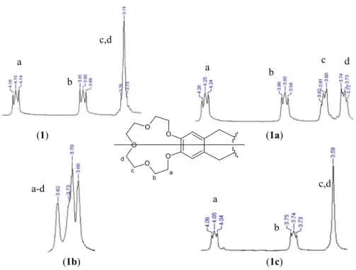
The 1H-NMR spectra and the integral ratio of the aliphatic and aromatic proton
peak for the ligands (1–5) and alkali metal complexes (1a–5a and 1b–5b) indicates
that the molecules are symmetric (Scheme 4). The signals for aliphatic methylene
protons of crown ether ligands (1–5) integrated to three multiple peak groups were
observed approximately at 3.73–4.18 ppm. The –CH2 protons for compounds (1–5)
were detected as a singlet at δ = 5.11, 5.09, 5.24, 5.25 and 5.30 ppm, respectively.
The –CH3 proton peak for compound 2 was observed at δ = 2.52 ppm. The signal
for H8 proton peaks was seen as a singlet at δ = 7.00, 6.99, 7.04, 7.07 and 7.04 ppm in 1–5, respectively. The multiple signals observed approximately at 7.09–8.36 ppm were attributed to the aromatic pyridine protons.
The 1H-NMR spectra of the sodium and potassium complexes (1a–5a and 1b–5b)
Table 1 1H-NMR spectr al dat a of lig ands ( 1– 5) ( δ, ppm) (in CDCl 3 ) (pr ot on numbers ar e giv en in Sc heme 4 ) s :sing le t, d double t, t tr iple t, dd double t of double ts, m multiple t Com pounds H1 H3 H4 H5 H6 H8 H10 H11 H12, H13 H14 (1 ) 8.36 (d; 2H) 7.20 (m; 4H) 8.23 (dd; 2H) 5.11 (s; 4H) 7.00 (s; 2H) 4.15 (t ; 4H) 3.90 (t ; 4H) 3.75 (t ; 8H) – 3J3-1 : 2.4 Hz 2J4-5 : 4.3 Hz 3J3-5 : 2.0 Hz (2 ) 8.30 (d; 2H) 7.20 (dd; 2H) 7.09 (d; 2H) – 5.09 (s; 4H) 6.99 (s; 2H) 4.15 (t ; 4H) 3.90 (t ; 4H) 3.75 (t ; 8H) 2.52 (s; 6H) 3J3-1 : 2.0 Hz 2J4-3 : 8.6 Hz 2J3-4 : 8.6 Hz 3J1-3 : 2.0 Hz (3 ) – 7.31 (d; 2H) 7.17 (dd; 2H) 7.98 (dd; 2H) 5.24 (s; 4H) 7.04 (s; 2H) 4.10 (m; 4H) 3.87 (m; 4H) 3.73 (m; 8H) – 2J4-3 : 8.0 Hz 2J5-4 : 4.7 Hz 2J4-5 : 4.7 Hz 2J3-4 : 8.0 Hz 3J3-5 : 1.2 Hz (4 ) – 7.28 (m; 2H) 7.20 (dd; 2H) 7.97 (dd; 2H) 5.25 (s; 4H) 7.07 (s; 2H) 4.11 (m; 4H) 3.87 (m; 4H) 3.74 (m; 8H) – 2J5-4 : 4.7 Hz 2J4-5 : 4.7 Hz 2J3-4 : 7.8 Hz 3J3-5 : 1.2 Hz (5 ) – 7.67 (d; 2H) 7.55 (dd; 2H) 8.10 (dd; 2H) 5.30 (s; 4H) 7.04 (s; 2H) 4.18 (t ; 4H) 3.91 (t ; 4H) 3.75 (t ; 8H) – 2J4-3 : 8.3 Hz 2J5-4 : 4.6 Hz 2J4-5 : 4.6 Hz 2J3-4 : 8.3 Hz 3J3-5 : 1.2 Hz
spectra of sodium and potassium complexes provide an explicit evidence for ligands
and their sodium and potassium complexes [55–57]. The small chemical shifts are
more pronounced for the methylene protons of the crown ether signals (Table 2). In
addition, three different multiple peaks for the ligands (1–5) were observed in the
peak region of the crown ether protons (OCH2–CH2O), while four multiple peaks
for the sodium complexes (1a–5a) and wide multiple peaks for the potassium
com-plexes (1b–5b) (Fig. 1).
The different appearance of four signals in 1H-NMR spectra of complexes
(1a–5a) may point out the extent of encapsulation of the sodium cation by the crown ether cavity. The extent of cation encapsulation in the cavity of the crown ether changes the conformation of the segments bearing crown ether protons. The cationic
diameter of the Na+ ion fits well with the size of the cavity of the 15-crown-5 moiety
and is expected to be attached inside the cavity, surrounded by the etheric oxygen.
However, the K+ cation is larger than the 15-crown-5 cavity and is preferred form
sandwich complex composition [58–60].
In 1H-NMR spectra of Ag(I) complexes (1c–5c), the shifts in the peaks of
aro-matic protons in the pyridine ring are more distinct (Table 3). For example, H3 and
H4 proton peaks were detected at 7.18 and 7.08 ppm as a multiplet for compound 2, same proton peaks were detected at 7.39 and 7.20 ppm as a quartet and doublet for
complex 2c. Similar changes have been observed for complexes 3c–5c. The –CH3
protons (H14) peak for complex 2c was observed at 2.40 ppm. This proton peak was observed at a chemical shift of 12 ppm lower than the free ligand. The aliphatic
–CH2 (H6) and aromatic H8 protons were observed as singlet peaks with small shifts
compared to the ligand. As a result, the binding of the Ag(I) ion to the pyridine N atom results in a more pronounced effect on the electronic structure of the pyridine protons, thereby significant shifts in pyridine proton signals while the crown ether
methylene proton signals remain unchanged (Fig. 1).
The 13C-NMR spectral data for ligands (1–5) were given in Table S3. The
spec-tra indicate that the molecules are symmetric (Scheme 4). Therefore, in the 13
C-NMR (decoupled) spectra of the ligands (1–5) four crown ether carbons (C10–C13)
were observed between 67.43 and 71.02 ppm. The –CH3 carbon (C14) peak in 2
was observed at 23.30 ppm. Aliphatic –CH2 carbons (C6) for compounds 1–5 were
detected in crown ether carbons peak region at 68.04, 68.24, 68.66, 68.70 and 69.14 ppm, respectively. In addition, other aromatic carbon peaks were detected in
the expected region and expected numbers. The 13C-NMR spectra of the sodium,
potassium and silver(I) complexes (1a–5a, 1b–5b and 1c–5c) are very similar to the corresponding ligand (1–5) spectra (Table S4).
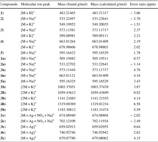
Ms spectra
We have reported that HRMS is a powerful tool in the determination of structures of new double armed benzo-15-crown-5 ligands (1–5), sodium (1a–5a), potas-sium (1b–5b) and silver(I) complexes (1c–5c). In keeping with this, the structures
of the ligands and complexes in CH3CN solution were examined by HRMS at
Table 2 1H-NMR spectr al dat a of sodium and po tassium com ple xes ( 1a –5a , 1b –5b ) ( δ, ppm) Com pounds H1 H3 H4 H5 H6 H8 H10 H11 H12 H13 H14 (1a ) a 8.36 (t ; 2H) 7.23 (m; 4H) 8.25 (m; 2H) 5.12 (s; 4H) 7.06 (s; 2H) 4.25 (t ; 4H) 3.95 (t ; 4H) 3.73 (t ; 4H) 3.81 (t ; 4H) – 3J3-1 : 1.8 Hz (2a ) a 8.21 (d; 2H) 7.14 (dd; 2H) 7.06 (d; 2H) – 5.07 (s; 4H) 7.04 (s; 2H) 4.23 (t ; 4H) 3.94 (t ; 4H) 3.72 (t ; 4H) 3.79 (t ; 4H) 2.52 (s; 12H) 3J3-1 : 2.8 Hz 2J4-3 : 8.5 Hz 2J3-4 : 8.5 Hz 3J1-3 : 2.8 Hz (3a ) a – 7.28–7.31 (dd; 2H) 7.17–7.20 (dd; 2H) 7.99–8.00 (dd; 2H) 5.21 (s; 4H) 7.06 (s; 2H) 4.14 (m; 4H) 3.96 (m; 4H) 3.77 (m; 4H) 3.81 (m; 4H) – 2J4-3 : 8.1 Hz 2J5-4 : 4.6 Hz 2J4-5 : 4.6 Hz 3J5-3 : 1.3 Hz 2J3-4 : 8.1 Hz 3J3-5 : 1.3 Hz (4a ) b – 7.62 (dd; 2H) 7.40 (dd; 2H) 7.95 (dd; 2H) 5.33 (s; 4H) 7.22 (s; 2H) 4.09 (t ; 4H) 3.77 (t ; 4H) 3.61 (m; 8H) – 2J4-3 : 8.1 Hz 2J5-4 : 4.5 Hz 2J4-5 : 4.5 Hz 3J5-3 : 1.5 Hz 2J3-4 : 8.1 Hz 3J3-5 : 1.5 Hz (5a ) a – 7.70 (d; 2H) 7.59 (dd; 2H) 8.13 (dd; 2H) 5.30 (s; 4H) 7.12 (s; 2H) 4.27 (m; 4H) 4.04 (m; 4H) 3.80 (t ; 4H) 3.83 (t ; 4H) – 2J4-3 : 8.5 Hz 2J5-4 : 4.4 Hz 2J4-5 : 4.4 Hz 2J3-4 : 8.5 Hz 3J3-5 :1.2 Hz (1b ) a 8.28 (d; 2H) 7.27 (m; 4H) 7.19 (dd; 4H) 8.18 (dd; 4H) 5.14 (s; 8H) 6.92 (s; 4H) 3.74 (m; 32H) – 3J3-1 : 3.1 Hz 2J3-4 : 8.4 Hz 2J4-5 : 4.5 Hz 2J5-4 : 4.5 Hz 3J3-5 : 1.4 Hz (2b ) b 8.16 (d; 2H) 7.31 (dd; 4H) 7.12 (m; 4H) – 5.13 (s; 8H) 7.11 (s; 4H) 4.04 (t ; 8H) 3.71 (t ; 8H) 3.57 (m; 8H) 3.59 (m; 8H) 2.35 (s; 12H) 3J3-1 : 3.1 Hz 2J4-3 : 8.6 Hz 2J3-4 : 8.6 Hz 3J1-3 : 3.1 Hz
Table 2 (continued) Com pounds H1 H3 H4 H5 H6 H8 H10 H11 H12 H13 H14 (3b ) a – 7.28 (m; 4H) 7.16 (dd; 4H) 7.97 (dd; 4H) 5.20 (s; 8H) 6.98 (s; 4H) 3.99 (m; 8H) 3.83 (m; 8H) 3.72 (m; 8H) 3.76 (m; 8H) – 2J5-4 : 4.7 Hz 2J4-5 : 4.7 Hz 2J3-4 : 8.2 Hz 3J3-5 : 1.2 Hz (4b ) b – 7.60 (dd; 4H) 7.39 (dd; 4H) 7.95 (dd; 4H) 5.32 (s; 8H) 7.22 (s; 4H) 4.08 (t ; 8H) 3.76 (t ; 8H) 3.59 (m; 8H) 3.61 (m; 8H) – 2J4-3 : 8.2 Hz 2J5-4 : 4.6 Hz 2J4-5 : 4.6 Hz 3J5-3 : 1.3 Hz 2J3-4 : 8.2 Hz 3J3-5 : 1.3 Hz (5b ) a – 7.81 (m; 4H) 7.58 (dd; 4H) 8.10 (d; 4H) 5.35 (s; 8H) 7.02 (s; 4H) 3.76 (m; 32H) – 2J5-4 : 4.2 Hz 2J4-5 : 4.2 Hz 2J3-4 : 8.4 Hz s sing le t, d double t, t tr iple t, dd double t of double ts, m multiple t a In CDCl 3 b In DMSO-d 6
peak in the pattern was selected and intensities of molecular ion peaks (found and
calculated) and error ratios (ppm) of the compounds are summarized in Table 4.
The isotope peaks pattern of the compounds (3, 4, 3a, 4a, 3b, 4b and 1c–5c)
are given as a detailed list in Table 5. High-resolution MS can replace elemental
analysis for chemical formula confirmation. Compound (1) spectrum show that
the molecular ion peak [M + H]+ at m/z 483.2146. In the mass spectra of other
ligands (2–5) the dominant peak at m/z 533.22467, 573.11581, 663.01264 and
595.16423, respectively corresponds to the ligand plus sodium [M + Na]+. In
addition, [M + K]+ peaks were observed in these spectra (It is very common to
see Na and K adducts in the HRMS-TOF spectra) (Fig. S1).
In the mass spectra of sodium complexes (1a–5a), the molecular ion peaks
corresponds to the ligand plus sodium ([M + Na]+). The sodium cation is bound
by the ion–dipole interaction to the crown ether cavity. The molecular ion
peak [2M + K]+ were detected at m/z 1003.37051, 1059.43612, 1141.21083,
1319.00369 and 1183.30812 for the other potassium complexes (1b–5b), respec-tively. These peaks are corresponding to the 1:2 (metal:ligand) complexes. In this work, the elements giving rise to significant M + 2 and M + 4 peaks are chlorine and bromine. A ratio of M to M + 2 and M + 4 of approximately 9:6:1 (for chlo-rine) and 1:2:1 (for bromine) indicates the presence of a two chlorine and bro-mine in compounds (3, 4, 3a, 4a, 3b and 4b) (Fig. S1).
(1) (1a) (1b) (1c) O O O O O a b c d c,d a b a b c d a-d c,d a b
Fig. 1 1H-NMR spectra of the crown ether protons [ligand (1); sodium complex (1a); potassium complex
Table 3 1H-NMR spectr al dat a of sil ver(I) com ple xes ( 1c –5c ) ( δ, ppm) s sing le t, d double t, t tr iple t, dd double t of double ts, m multiple t a In CDCl 3 b In DMSO-d 6 Com pounds H1 H3 H4 H5 H6 H8 H10 H11 H12 H13 H14 (1c ) b 8.31 (d; 2H) 7.45 (m; 2H) 7.31 (dd; 2H) 8.14 (dd; 2H) 5.18 (s; 4H) 7.14 (s; 2H) 4.05 (t ; 4H) 3.74 (t ; 4H) 3.59 (m; 4H) 3J3-1 : 2.7 Hz 2J3-4 : 8.6 Hz 2J4-5 : 4.7 Hz 2J5-4 : 4.7 Hz 3J3-5 : 1.2 Hz (2c ) b 8.18 (d; 2H) 7.41 (dd; 2H) 7.21 (d; 2H) – 5.14 (s; 4H) 7.11 (s; 2H) 4.04 (t ; 4H) 3.74 (t ; 4H) 3.80 (m; 4H) 2.40 (s; 6H) 3J3-1 : 3.1 Hz 2J4-3 : 8.6 Hz 2J3-4 : 8.6 Hz 3J1-3 : 3.1 Hz (3c ) a – 7.28–7.30 (m; 2H) 7.23 (m; 2H) 8.00 (dd; 2H) 5.24 (s;4H) 7.05 (s; 2H) 4.07 (m; 4H) 3.86 (m; 4H) 3.75 (m; 8H) 2J4-5 : 4.5 Hz 3J3-5 : 1.4 Hz (4c ) a – 7.31–7.34 (m; 2H) 7.18–7.21 (m; 2H) 7.99–8.00 (dd; 2H) 5.23 (s;4H) 7.01 (m; 2H) 4.08 (m; 4H) 3.86 (m; 4H) 3.74 (m; 8H) 2J4-5 : 4.7 Hz 3J3-5 : 1.2 Hz (5c ) a – 7.66 (d; 2H) 7.55 (m; 2H) 8.11 (d; 2H) 5.29 (s; 4H) 7.04 (s; 2H) 4.18 (t ; 4H) 3.92 (t ; 4H) 3.75 (m; 8H) 2J4-3 : 7.8 Hz 2J4-5 : 4.3 Hz
Table 4 Mass spectral data (in CH3CN)
Compounds Molecular ion peak Mass (found g/mol) Mass (calculated g/mol) Error ratio (ppm)
(1) [M + H]+ 483.21465 483.21317 − 3.06 (2) [M + Na]+ 533.22497 533.22641 − 2.70 [M + K]+ 549.19952 549.20035 − 1.51 (3) [M + Na]+ 573.11581 573.11717 2.37 [M + K]+ 589.08981 589.09111 2.21 (4) [M + Na]+ 663.01264 663.01409 2.19 [M + K]+ 678.98666 678.98803 2.02 (5) [M + Na]+ 595.16423 595.16529 1.78 (1a) [M + Na]+ 505.19482 505.19511 0.57 (2a) [M + Na]+ 533.22702 533.22641 − 1.14 (3a) [M + Na]+ 573.11444 573.11717 4.76 (4a) [M + Na]+ 663.01121 663.01409 4.34 (5a) [M + Na]+ 595.16325 595.16529 3.43 (1b) [2M + K]+ 1003.37051 1003.37439 3.87 (2b) [2M + K]+ 1059.43612 1059.43699 0.82 (3b) [2M + K]+ 1141.21083 1141.21555 4.14 (4b) [2M + K]+ 1319.00369 1319.01234 6.58 (5b) [2M + K]+ 1183.30812 1183.31474 5.59 (1c) [M + Ag + NO3 + Na]+ 674.08940 674.08804 − 2.02 (2c) [M + Ag + NO3 + Na]+ 702.12109 702.11934 − 2.49 (3c) [M + Ag]+ 659.02913 659.02955 0.64 (4c) [M + Ag]+ 746.92746 746.92942 2.62 (5c) [M + Ag]+ 679.07780 679.08062 4.15
Table 5 Molecular ion peaks (related to natural abundance) (g/mol)
M: Ligand
Compounds [M] [M + 2] [M + 4]
(3) 589.08981 [M + K] 591.08756 [M + 2 + K] –
(4) 676.98856 [M + K] 678.98666 [M + 2 + K] 680.98517 [M + 4 + K]
(3a) 573.11444 [M + Na] 575.11203 [M + 2 + Na] –
(4a) 661.01377 [M + Na] 663.01121 [M + 2 + Na] 665.01087 [M + 4 + Na]
(3b) 1141.21083 [2M + K] 1143.20895 [2M + 2 + K] – (4b) 1317.00495 [2M + K] 1319.00369 [2M + 2 + K] 1321.00123 [2M + 4 + K] (1c) 674.08940 [M + Na + Ag + NO3] 676.08951 [M + 2 + Na + Ag + NO3] – (2c) 702.12109 [M + Na + Ag + NO3] 704.12130 [M + 2 + Na + Ag + NO3] – (3c) 659.02913 [M + Ag] 661.02376 [M + 2 + Ag] – (4c) 746.92746 [M + Ag] 748.92633 [M + 2 + Ag] – (5c) 679.07780 [M + Ag] 681.07887 [M + 2 + Ag] –
In the mass spectra of silver(I) complexes (1c and 2c), the molecular ion peaks
correspond to the ligand plus silver nitrate and sodium ([M + Ag + NO3 + Na]+). A
ratio of M and M + 2 of approximately 1:1 indicates the presence of one silver(I)
ion in a complex (Tables 4, 5) (Fig. S2). The silver(I) ion is bound to the side arm of
the ligand (pyridine N atom) and nitrate anion (Scheme 3). The silver(I) complexes
(3c–5c), the molecular ion peaks corresponds to the ligand plus silver ([M + Ag]+).
In compounds 3c and 4c, unlike compounds 1c and 2c, the nitrate anion was not coordinated by the silver ion. The complexes 3c and 4c have the isotope peaks of the M and M + 2 from the chlorine, bromine and silver atoms (Fig. S2). The spec-trum of the silver complex 5c shows that the M and M + 2 peaks at 679.07780 and 681.07887, respectively. A ratio of the peaks is 1:1 as expected for silver(I) isotope peak patterns (Fig. S2).
Optical characteristics
Crown ether rings are well-known ionophores due to their metal binding ability. Crown ethers attached to a fluorophore group are named fluoroionophores and have a large structural variety that allows for high selectivity and sensitivity for metal ion detection. Synthesized ditopic ligands (1–5) centre hold various selectivities to metal ions, and having two binding units causes changes in the photophysical
properties of the complex receptor (Scheme 1). In this case, the substantial optical
changes upon complex formation can be suggested.
The UV–vis absorption and emission spectra were recorded at a concentration
of 5 × 10⁻⁵ M in EtOH. The room temperature UV–vis spectra of new ligands 1–5
exhibited one main absorption band at around λmax 292 nm (Fig S3). These peaks
correspond to the S0 → S1 transition of the benzo-15-crown-5 moieties [15]. All
compounds (1–5) exhibit absorption patterns that can be assigned characteristic of benzo-crown ethers. It has been seen that binding of different substituted pyridine units to the ligand (1–5) structure weakly affects the absorption wavelength band. The close values of intensities of the absorption bands of the ligands (1–4) and a small difference in the absorption maxima positions points at identical interactions of substituted pyridine units and benzo-15-crown-5 in the ground state. The UV–vis
behavior of compound 5 in the presence of the NO2 group in pyridine it prevents the
charge transfer from the donor crown ether to acceptor nitro pyridine moiety leading to a blue shift in the absorption band (Fig. S3).
The fluorescence emission spectra of ligands (1–5) in EtOH media were
inves-tigated and showed in Fig. 2. Compound 1 and 2 containing –H and –CH3
substi-tuted in the pyridine ring show the highest fluorescence intensity while compounds 3 and 4 containing –Cl and –Br substituted in pyridine ring have lowest fluorescence
intensity among the synthesized ligands (1–5) (Fig. 2). As is known, the spin–orbit
interaction of electronic states has a great influence on the optical properties of com-pounds. The presence of heavy atoms (Cl, Br) in the synthesized compounds (3 and 4) increases the inter-system crossing process, which causes the change in optical properties. This effect is known as the heavy atom effect. As a result, in the synthe-sized compounds (1–5), the fluorescence signal was increased for compounds 1 and
2 while decreasing for compounds 3 and 4 due to the intersystem crossing mecha-nism. The nitro group decreases the fluorescence signals due to their electron
with-drawing nature [15]. In general, nitro substituted rings have a lower LUMO energy
level due to the electron-withdrawing effect of the nitro group. Note that for the nitro substituted compound 5, very weak fluorescence in the emission spectra was observed compared to the other compounds (1–4), likely related to the significant decrease of the LUMO energy level and intramolecular charge transfer state process
was occurred [61, 62].
Our fluorescence investigations showed that introducing substituents (H, –CH3,
–Cl, –Br and –NO2) on the pyridine group may affect the fluorescence intensity
(Fig. 2).
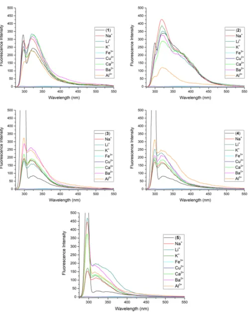
To investigate the metal selectivity of synthesized ligands (1–5), different metal
cations (Li+, Na+, K+, Fe3+, Cu2+, Ca2+, Ba2+ and Al3+) were added to these ligands
and fluorescence spectra were recorded (Fig. 3).
In metal selectivity studies of all synthesized ligands (1–5), different
fluores-cence intensities were recorded with each metal cations (Li+, Na+, K+, Fe3+, Cu2+,
Ca2+, Ba2+ and Al3+). As a common feature in all ligands (1–5) fluorescence was
quenched by the addition of Fe3 and Cu2+. Meanwhile, the weak fluorescence
emis-sion was detected for five ligands (1–5), with the upon addition of Li+, Na+ or K+
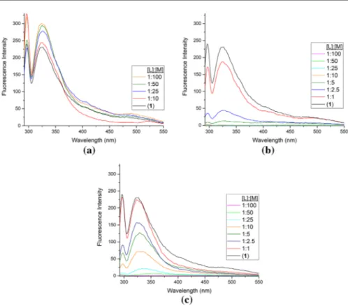
cations. To find the effect of joining together two ionophore units on metal cation complexation in solution, compound 1 has been chosen. The optical study of
com-pound 1 was performed with the addition of increasing amounts of Na+, Fe3+ and
Cu2+ ions (commonly selected ions) concentration in EtOH (Fig. 4).
The ligands (1–5) that are synthesized have two different binding sites (the crown
ether cavity and the pyridine N atoms) (Scheme 1). In this respect, it can be
con-sidered that the Na+ ion complex was formed from the crown ether cavity and the
Fig. 2 The fluorescence spectra of ligands (1–5); 25 °C; λexc: 292 nm; ligand concentration c: 5 × 10⁻⁵ in
transition metal ion (Fe3+ and Cu2+) complexes were formed from the pyridine N
atoms. In the titration study of compound 1 with three metal ions (Na+, Fe3+ and
Cu2+), only at about 325 nm peak intensity increase (for the addition of Na+ ion)
or decrease (for the addition of Fe3+ or Cu2+ ion) was observed (Fig. 4).
How-ever, a new peak was recorded at about 430 nm in the synthesized Ag(I) complex
(1c) (Fig. 5). This peak belongs to the complex. The lack of a new peak (at about
430 nm) in the titration studies of Fe3+ and Cu2+ ion suggests that the Fe3+ and Cu2+
Fig. 3 The effect of metal cations on the fluorescence spectra of ligands (1–5); metal salt anions: Na+
and K+: I−; others: NO
3−; 25 °C; λexc: 292 nm; ligand concentration c: 5 × 10⁻⁵ metal concentrations c:
Fig. 4 The fluorescence spectral changes of compound 1 during the addition of 1–100 equivalent of
a Na+, b Fe3+, c Cu2+. 25 °C; λ
exc: 292 nm; ligand concentration c: 5 × 10⁻⁵ metal concentration c:
5 × 10⁻³ M in EtOH
Fig. 5 The fluorescence spectrum of compound 1 and 1c 25 °C; λexc: 292 nm; complex concentration c:
ion complexes formed may have given inclusion complexes with the crown ether ring.
In compounds 1–5, pyridine group is deconjugated with benzo-15-crown-5 group and photoinduced electron transfer (PET) mechanism that may be occurred between
the benzo-15-crown-5 and pyridine fragments (Scheme 5). Intramolecular PET
mechanism is a well-known process through which the fluorescence of a fluorophore is quenched by electron transfer from the receptor (crown ether) to the fluorophore
group [63]. In the absence of metal cation (Fe3+ or Cu2+), the HOMO of the
recep-tor lies lower in energy than fluorophore and prevents an electron transfer from the donor to the acceptor, so fluorescence occurred. Binding of the sensor to its metal
cation (Fe3+ and Cu2+) lowers the receptor HOMO and LUMO energy level below
that of the fluorophore LUMO than that of the donor can transfer an electron to the
receptor’s LUMO, so fluorescence was quenched. By this reason for Fe3+ and Cu2+
complexation process was assigned oxidative-PET mechanism [63]. After
coordina-tion with alkali metal (Li+, Na+, K+) cation, the electron-rich element as oxygen
in crown group will be bound. Therefore, the PET action from the crown donor towards the side group is stopped and fluorescence is occurred for compounds 1–5. Antimicrobial activity
The synthesized compounds (1–5, 1a–5a, 1b–5b and 1c–5c) were screened for in vitro antibacterial and antifungal activity in DMSO as a test substance. The ligands (1–5) and complexes (1a–5a, 1b–5b and 1c–5c) were tested with the same concentrations in DMSO solution (0.1 µg/µL). Ligands, complexes and antibiotics indicated varying degrees of inhibitory effects on the growth of different tested both
gram negative and gram positive pathogenic bacteria and yeast (Figs. 6, 7).
Fig. 6 Antimicrobial activity of ligands (1–5) and complexes (1a–5a, 1b–5b and 1c–5c) and standard reagents (diameter of zone inhibition (mm). SXT25, sulphamethoxazole 25 µg; AMP10, ampicillin 10 µg; K30, kanamycin 30 µg; AMC30, amoxycillin 30 µg
Fig. 7 Antimicrobial activity of ligands (1–5) and complexes (1a–5a, 1b–5b and 1c–5c) and standard reagents (diameter of zone inhibition (mm). SXT25, sulphamethoxazole 25 µg; AMP10, ampicillin 10 µg; NYS100, nystatin 100 µg; K30, kanamycin 30 µg; AMC30, amoxycillin 30 µg
Based on Figs. 6 and 7 results, functional substitutions on the pyridine ring
selec-tively increase or decrease inhibition of ligand’s activity. The substituents (CH3,
NO2, Cl and Br) on the pyridine group seem to be a highly significant factor in
influ-encing the biological activity of the compound. For example, compound 2a showed the most activity against M. luteus (21 mm). However, halogen-substituted (Cl and Br) compounds (3 and 4) and their complexes (3a, 3b, 3c, 4a and 4b except 4c) have been no effect against M. luteus. In addition, L. monocytogenes, shows selec-tivity only for halogen-substituted ligands (3 and 4) and complexes (3a, 3b, 3c, 4a, 4b, 4c). This result demonstrates the importance of the substituent bound to the
structure for antimicrobial activity [64]. The compounds (3 and 4) that have
elec-tron-withdrawing groups Cl and Br were more active than those that have electron
donating group (CH3) in compound (2) [65]. However, NO2 substituted (electron
withdrawing group) compound 2 and complexes were interestingly inactive for L.
monocytogenes. Listeria monocytogenes, which is commonly found in nature in the
genus Listeria, is a highly pathogenic species for humans and animals.
All synthesized ligands (1–5) and complexes (1a–5a, 1b–5b and 1c–5c) showed best antibacterial activity against B. cereus. Compound 4 showed the most activ-ity against B. cereus (26 mm). All of potassium complexes (1b–5b) were not effec-tive against S. aureus. Compound 4c showed the most activity against S. epidermis (25 mm). S. epidermis infections are associated with intravascular devices (pros-thetic heart valves, etc.) and generally occur in pros(pros-thetic joints, catheters and large wounds. This pathogenic microorganism gains resistance against traditional antibi-otics every day. There is a need for more effective antibiantibi-otics to treat against this
disease [36].
Salmonella serovars cause very diverse clinical symptoms, from asymptomatic infection to serious typhoid-like syndromes in infants or certain highly susceptible
animals [35, 37]. Compounds 4, 4c, 5c (23 mm, 24 mm, 25 mm, respectively) and
especially 3c were the most potent growth inhibitors against S. typhi H with a zone value of 30 mm. Only compound 3b was not effective for S. typhi H. Ligands (1 and 2) and their complexes (1a, 2a, 1b, 2b, 1c and 2c) are completely inactive against E.
coli. Cl, Br and NO2 substituted (electron withdrawing groups on pyridine) ligands
and complexes (3, 3c, 4, 4b, 4c, 5, 5a, 5b and 5c) have moderate activity for E.
coli. It is well known that silver ions and silver-based compounds are highly toxic to
microorganisms showing strong biocidal effects on many species of bacteria
includ-ing E. coli [66–68]. However, it is reported that the concentration of the applied
compounds and the number of bacteria (CFU) may affect the antimicrobial
activ-ity [68]. In our study, this is thought to be the case (Fig. 7). The silver complexes
(1c–5c) have broad antimicrobial activity, which are generally considered the refer-ence standard for the comparison of the growth inhibitory effects of the test materi-als against Gram-positive bacteria B. cereus, S. epidermis and Gram-negative
bacte-ria E. coli (Figs. 6, 7).
Compound 2a showed the most activity against M. luteus (21 mm). Compound 4c showed the most activity against S. epidermis (25 mm) (Fig. 6). S.
epider-mis which infections are associated with intravascular devices (prosthetic heart
valves, etc.), but also generally occur in prosthetic joints, catheters and large wounds. These pathogenic microorganisms gain resistance against traditional
antibiotics every day. There is a need for more effective antibiotics for the
treat-ment of this disease [69]. Compound 4 showed the most activity against B. cereus
(26 mm).
Systemic fungal infections, including those by Candida albicans have emerged as important causes of morbidity and mortality in immune compromised patient
(Aids, cancer chemotherapy, organ or bond transplantation) [35]. All the
synthe-sized compounds demonstrated much activity against this yeast. Further, com-pounds 1b, 1c, 2c, 3, 3b, 3c, 4, 4a, 4b and 4c showed the most inhibition activity against C. albicans as compounds with zone values of 30–37 mm. Meanwhile, compound 4 (37 mm) showed the highest activity. In fact, all the synthesized ligands and complexes showed more activity against C. albicans than commercial
(standard) antifungal (positive control NYS100P) (Fig. 7).
Conclusions
In this study, a series of new double armed ligands (1–5) and sodium, potassium and silver(I) complexes (1a–5a, 1b–5b and 1c–5c) were synthesized and character-ized by using analytical and spectral techniques. Pyridine substituted crown ether compounds (1–5) were prepared by the reaction of 4′,5′-bis(bromomethyl)benzo-15-crown-5 with hydroxypyridine derivatives in basic media. The chemosensing behavior of 1–5 was carried out using fluorescence spectroscopy. The fluorescence experiments show that the fluorescence emission of 1–5 could be quenched by the
addition of Fe3+ and Cu2+ ion in the ligand. These fluorescence quenching
proper-ties of Fe3+ and Cu2+ ion present a potential for the design of fluorescence sensory
materials. The synthesized compounds (1–5, 1a–5a, 1b–5b and 1c–5c) have been found to exhibit antibacterial and antifungal activities at moderate to good levels both gram negative and gram positive bacteria. The antimicrobial activity of these compounds was also compared with commercial (standard) antibiotics. It was seen that the synthesized compounds were effective as the antibiotics and antifungal men-tioned. Furthermore, some of the synthesized compounds (S. typhi H; 3c, 4, 4c, 5c 30 mm, 23 mm, 24 mm, 25 mm respectively) have been found to be more effective than antibiotics and antifungal (C. albicans; all compounds, 23–37 mm). As a result, synthesized molecules may become potential candidates for the clinical trials.
Supporting information available
Tables; experimental details, IR and 13C NMR and also spectra; 1H-NMR, mass
and UV–vis spectra of compounds are provided as supplementary material.
Acknowledgements The authors gratefully acknowledge the financial assistance of the Scientific and Technical Research Council of Turkey (TUBITAK), Grant No. TBAG 210T122, and Ankara University Grant No. 17B0430004.
References
1. C.J. Pedersen, J. Am. Chem. Soc. 89, 7017 (1967) 2. N.S. Poonia, A.V. Bajaj, Chem. Rev. 79, 389 (1979)
3. F. Vögtle, E. Weber, in Crown Ethers and Analogs, ed. by S. Patai, Z. Rappoport (Wiley, Chichester, 1989), p. 207
4. Z. Hayvali, N. Gündüz, Z. Kiliç, E. Weber, J. Prakt. Chem. 341, 568 (1999) 5. C. Sousa, C. Freire, B. De Castro, Molecules 8, 894 (2003)
6. D. Liu, K. Tang, W. Liu, C. Su, X. Yan, M. Tan, Y. Tang, Dalton Trans. 39, 9763 (2010)
7. K. Sako, T. Kakehi, S. Nakano, H. Oku, X.F. Shen, T. Iwanaga, M. Yoshikawa, K. Sugahara, S. Toyota, H. Takemura, T. Shinmyozu, M. Shiotsuka, H. Tatemitsu, Tetrahedron Lett. 55, 749 (2014) 8. B. Valeur, I. Leray, Coord. Chem. Rev. 205, 3 (2000)
9. Q.Z. Yang, L.Z. Wu, H. Zhang, B. Chen, Z.X. Wu, L.P. Zhang, C.H. Tung, Inorg. Chem. 43, 5195 (2004)
10. E.N. Ushakov, M.V. Alfimov, S.P. Gromov, Macroheterocycles 3, 189 (2010) 11. H.S. Seo, S.H. Lee, J. Fluoresc. 21, 747 (2011)
12. L. Zhao, X. Chen, F. Guo, B. Gou, C. Yang, W. Xia, J. Lumin. 145, 486 (2014) 13. J.-P. Malval, R. Lapouyade, Helv. Chim. Acta 84, 2439 (2001)
14. S.K. Kim, M.Y. Bang, S.-H. Lee, K. Nakamura, S.-W. Cho, J. Yoon, J. Incl. Phenom. Macrocycl. Chem. 43, 71 (2002)
15. D. Şahin, H. Yılmaz, Z. Hayvalı, Res. Chem. Intermed. 42, 6337 (2016) 16. D. Şahin, Y. Süzen, Z. Hayvalı, Hetoroatom Chem. 25, 43 (2014) 17. H. Güler, Z. Hayvali, H. Dal, T. Hökelek, Polyhedron 31, 688 (2012) 18. K.K. Haldar, T. Sen, A. Patra, J. Phys. Chem. C 114, 4869 (2010) 19. Z. Hayvalı, H. Güler, H. Öğütcü, N. Sarı, Med. Chem. Res. 23, 3652 (2014) 20. G.W. Gokel, W.M. Leevy, E. Weber, Chem. Rev. 104, 2723 (2004) 21. M. Kralj, L. Tusek-Bozic, L. Frkanec, Chem. Med. Chem. 3, 1478 (2008)
22. P.L. Caradoc-Davies, L.R. Hanton, W. Henderson, J. Chem. Soc. Dalton Trans. 19, 2749 (2001) 23. Y. Kang, S.S. Lee, K.-M. Park, S.H. Lee, S.O. Kang, J. Ko, Inorg. Chem. 40, 7027 (2001) 24. C. Seward, J. Chan, D. Song, S. Wang, Inorg. Chem. 42, 1112 (2003)
25. W. Sun, Y. Cui, H. Liu, H. Zhao, W. Zhang, J. Mol. Struct. 1026, 133 (2012) 26. T. Nakamura, K. Takeuchi, JP Patent 2003-238832A, 2003
27. N. Kinarivala, P.C. Trippier, Tetrahedron Lett. 55, 5386 (2014)
28. T.F. Spande, H.M. Garraffo, M.W. Edwards, H.J.C. Yeh, L. Pannell, J.W. Daly, J. Am. Chem. Soc. 114, 3475 (1992)
29. J.K. Lynch, M.W. Holladay, K.B. Ryther, H. Bai, C.N. Hsiao, H.E. Morton, D.A. Dickman, W. Arnold, S.A. King, Tetrahedron-Asymmetr. 9, 2791 (1998)
30. J. Li, D. Yim, W.-D. Jang, J. Yoon, Chem. Soc. Rev. 46, 2437 (2017) 31. V.K. Gupta, S. Chandra, S. Agarwal, Indian J. Chem. 42, 813 (2003) 32. M.J. Calverley, J. Dale, Acta Chem. Scand. B. 36, 241 (1982)
33. B. Winkler, A.W.-H. Mau, L. Dai, Phys. Chem. Chem. Phys. 2, 291 (2000)
34. A. Bilgin, B. Ertem, P. Dinc Agın, Y. Gok, S. Karslıoglu, Polyhedron 25, 3165 (2006)
35. H. Öğütcü, N.K. Yetim, E.H. Özkan, O. Eren, G. Kaya, N. Sarı, A. Dişli, Pol. J. Chem. Technol. 19, 74 (2017)
36. C. Nithya, B. Gnanalakshmi, S.K. Pandian, Mar. Environ. Res. 71, 283 (2011) 37. U. Schillinger, F.K. Lucke, Appl. Environ. Microbiol. 55(8), 1901 (1989) 38. M. Balouiri, M. Sadiki, K.S. Ibnsouda, J. Pharm. Anal. 6, 79 (2016)
39. S. Magaldi, S. Mata-Essayag, C. Hartung de Capriles, C. Perez, M.T. Colella, C. Olaizola, Y. Ontiveros, Int. J. Infect. Dis. 8, 39 (2004)
40. C. Valgas, S.M. De Souza, E.F.A. Smânia, A. Smânia Jr., Braz. J. Microbiol. 38, 369 (2007) 41. Y. Xiang, X. Liu, C. Mao, X. Liu, Z. Cui, X. Yang, K.W.K. Yeung, Y. Zheng, S. Wu, Mater. Sci.
Eng. C 85, 214 (2018)
42. Z. Liu, Y. Zhu, X. Liu, K.W.K. Yeung, S. Wu, Colloids Surf. B 151, 165 (2017) 43. Y. Zhu, X. Liu, K.W.K. Yeung, P.K. Chu, S. Wu, Appl. Surf. Sci. 400, 14 (2017)
44. C. Mao, Y. Xiang, X. Liu, Z. Cui, X. Yang, K.W.K. Yeung, H. Pan, X. Wang, P.K. Chu, S. Wu, ACS Nano 11, 9010 (2017)
46. N. Sarı, N. Pişkin, H. Öğütcü, N. Kurnaz, Med. Chem. Res. 22, 580 (2013) 47. D. Nartop, N. Sarı, H. Öğütcü, Chin. J. Inorg. Chem. 30, 921 (2014) 48. A. Altundas, N. Sarı, N. Colak, H. Ögütcü, Med. Chem. Res. 19, 576 (2010) 49. D. Nartop, N. Sarı, A. Altundas, H. Ögütcü, J. Appl. Polym. Sci. 125, 1796 (2012)
50. M. Barboiu, A. Mefrfre, Y.-M. Legrand, E. Petit, L. Marin, M. Pinteala, A.V.D. Lee, Supramol. Chem. 26, 223 (2014)
51. N.S. Poonia, P. Bagdi, K.S. Sidhu, J. Incl. Phenom. 4, 43 (1986)
52. B. Antonioli, D.J. Bray, J.K. Clegg, K. Gloe, K. Gloe, O. Kataeva, L.F. Lindoy, J.C. McMurtrie, P.J. Steel, C.J. Sumby, M. Wenzel, Dalton Trans. 40, 4783 (2006)
53. G. Socrates, Infrared and Raman Characteristic Group Frequencies: Tables and Charts, 3rd edn. (Wiley, Chichester, 2001)
54. D. Lin-Vien, N.B. Colthup, W.G. Fateley, J.G. Graselli, The Handbook of Infrared and Raman Characteristic Frequencies of Organic Molecules (Academic Press, San Diego, 1991)
55. N. Ghildiyal, G.J. Nee Pant, M.S.M. Rawat, K. Singh, Spectrochim. Acta A 171, 507 (2017) 56. Y. Liu, J.R. Han, H.Y. Zhang, Supramol. Chem. 16, 247 (2004)
57. Z. Hayvalı, P. Köksal, J. Incl. Phenom. Macrocycl. Chem. 76, 369 (2013) 58. C.J. Pedersen, H.K. Frensdorff, Angew. Chem. Internat. Edit. 11, 16 (1972) 59. P.R. Mallison, M.R. Truter, J. Chem. Soc. Perkin 2, 1818 (1972)
60. V.W. Bhagwat, H. Manohar, N.S. Poonia, Inorg. Nucl. Chem. Lett. 17, 207 (1981) 61. R. Ziessel, L. Bonardi, P. Retailleau, G. Ulrich, J. Org. Chem. 71, 3093 (2006)
62. S. Imama-Reja, N. Kumar, R. Sachdeva, V. Bhalla, M. Kumar, RSC Adv. 3, 17770 (2013) 63. V. Bojinov, N. Georgiev, J. Chem. Technol. Metall. 46, 3 (2011)
64. F.R.F. Dias, J.S. Novais, T.A. do Nascimento Santos Devillart, W.A. da Silva, M.O. Ferreira, R.S. Loureiro, V.R. Campos, V.F. Ferreira, M.C.B.V. de Souza, H.C. Castro, A.C. Cunha, Eur. J. Med. Chem. 156, 1 (2018)
65. N.B. Reddy, G.V. Zyryanov, G.M. Reddy, A. Balakrishna, A. Padmaja, V. Padmavathi, C.S. Reddy, J.R. Garcia, G. Sravya, J. Heterocycl. Chem. https ://doi.org/10.1002/jhet.3435
66. Z. Xu, X. Wang, X. Liu, Z. Cui, X. Yang, K.W.K. Yeung, J.C. Chung, P.K. Chu, S. Wu, ACS Appl. Mater. Interfaces 9, 39657 (2017)
67. X. Xie, C. Mao, X. Liu, Y. Zhang, Z. Cui, X. Yang, K.W.K. Yeung, H. Pan, P.K. Chu, S. Wu, ACS Appl. Mater. Interfaces 9, 26417 (2017)
68. I. Sondi, B. Salopek-Sondi, J. Colloid Interface Sci. 275, 177 (2004)
69. A. Altundas, Y. Erdogan, H. Ögütcü, H.E. Kizil, G. Agar, Fresenius Environ. Bull. 25, 5411 (2016)
Publisher’s Note Springer Nature remains neutral with regard to jurisdictional claims in published