71
Case Study
Recurrence of Langerhans Cell Histiocytosis of Jaws: A Case Report with One Year Follow-up
Özlem Okumuş 1*, Vakur Olgaç 2, Semih Özbayrak 1
1Department of Dentomaxillofacial Radiology and Oral Medicine, Faculty of Dentistry, Altınbaş University, Istanbul, Turkey. 2Department of Cytology and Tumor Pathology, Institute of Oncology, Istanbul University, Istanbul, Turkey.
Submitted: February 15, 2019; Accepted: March 12, 2019
Abstract: Langerhans cell histiocytosis (LCH) is an unusual disorder characterized by high proliferation of Langerhans cells. A 27-year-old male patient was referred to the Oral Medicine Clinic, Faculty of Dentistry, Altınbaş University with a complaint of wound that does not heal for eight months. The clinical examination showed palatal mucosal ulceration extending to the bone at the upper right first molar region and ulceration at the lingual mucosa of the lower right first molar. The patient had been previously evaluated by a different dentist and was told that it was an aphthous lesion. A punch biopsy from the palatal mucosa and alveolar mucosa near the lesion revealed Langerhans cells and positive reaction to CD1 so the patient was diagnosed with LCH. The insignificant treatment trials may lead to diagnostic delay. Oral lesions may be the earliest symptom of LCH and in most cases; the oral cavity may be the only area involved. Awareness of lesions in the oral mucosa is important in achieving the accurate and early diagnosis and effective treatment plan.
Keywords: Langerhans cell histiocytosis; histiocytosis X; jaw; recurrence
Address of Correspondence: Özlem Okumuş - [email protected], ORCID:
orcid.org/0000-0002-5590-2357
Tel: +90(212)7094528, Department of Oral and Dentomaxillofacial Radiology, Faculty of Dentistry, Altınbaş University, Kartaltepe Mahallesi, İncirli Caddesi No: 11, 34147 Bakırköy, İstanbul, Turkey
1. Introduction
Langerhans cell histiocytosis (LCH) is formerly called histiocytosis X, is characterized by abnormal proliferation of Langerhans cells (histiocytes), with varying numbers of lymphocytes, eosinophils, plasma cells, neutrophils, and multi-nucleated giant cells (Pacino et al., 1999). LCH was previously grouped into three clinical forms according to the patient age and the distribution of lesion including eosinophilic granuloma, Hand-Schüller–Christian disease, and Letterer-Siwe disease (Chu, 2001). The new classification
72
of LCH is made based on the dissemination of the disease and is grouped into two groups: nonmalignant diseases such as unifocal and multifocal eosinophilic granuloma and malignant diseases, including Letterer-Siwe disease and histiocytic lymphoma. LCH commonly occurs in young adults and children (White and Pharoah, 2014).
The oral symptoms may be the first sign of LCH and the oral cavity may be the only affected area in some cases (Shirley and Thomton, 2000).The gingiva and hard palate are the most commonly affected sites in maxillomandibular involvement. The oral symptoms of LCH include ulcerative and bleeding gingiva, mobile teeth, and pain (Altay et al., 2017). The ulceration is typically extended to the underlying bone in the oral mucosal involvement. LCH is known to look like different diseases and its diagnosis may be particularly difficult owing to its broad clinical spectrum, ranging from a single lesion to a multisystemic involvement. There are few reports about the misdiagnosis of this disorder (Chen and Peron, 2000; Kılıç et al., 2011; Zhang et al., 2006).
This case report defines a case of Langer hans cell histiocytosis with oral cavity involvement and presents the importance of the awareness of the oral manifestations of LCH.
2. Description of the Case
A 27-year-old male patient was referred to Altınbaş University, Dentistry Faculty, Oral Medicine Clinic with a complaint of palatal ulceration with eight months of duration. There was no pain or bleeding in the ulcerated areas. The patient had been previously evaluated by a different dentist and was told that it was an aphthous lesion. A mouthwash and wound gel was prescribed but the lesion did not regress. The patient had no significant systemic disease but he had a gonorrhea treatment a year ago. The right first molar tooth of the mandible was also extracted due to the periodontal abscess.
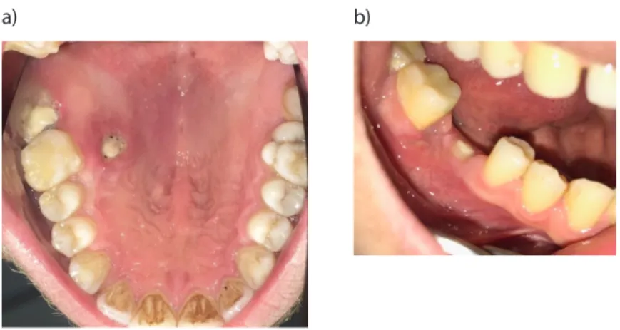
The clinical examination showed the necrotic area extending to the bone at the hard palate adjacent to the right first molar tooth (Figure 1a). The ulceration on the lingual mucosa and the necrotic area at the top of the alveolar crest was observed at the right first molar of the mandible (Figure 1b). Also the right and left submandibular lymphadenopathy were observed.
Radiologically, a periapical X-ray demonstrated vertical alveolar bone loss extending to the middle of mesial root of the second molar. The cone beam computed tomography (CBCT) was performed and it revealed the horizontal alveolar bone loss especially at the right premolar teeth region and the vertical alveolar bone loss at the mesial root of the right second molar tooth of the mandible. At the distobuccal root of the right first molar tooth of the maxilla, vertical alveolar bone destruction extending to the palatine bone was observed (Figure 2). Any other lesions in the remaining skull bones were not detected. The punch biopsy from the palatal mucosa and alveolar mucosa was performed under local anesthesia and the specimen in 10% formal saline was sent for histopathological examination at the Department of Cytology and Tumor Pathology, Institute of Oncology, Faculty of Medicine, Istanbul University. This study was performed according to the guidelines of the Declaration of Helsinki concerning ethical
73 principles for medical research involving human subjects and written informed consent was obtained
from the patient.
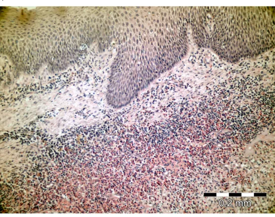
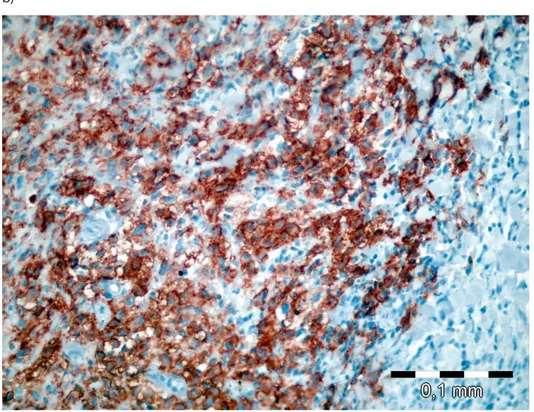
The histological and immunohistochemistry examination showed histiocytic cells, eosinophil polymorphs and positive reaction to CD1 (Figure 3a, 3b). According to these findings, the patient was diagnosed with LCH.
The patient was referred to the Department of Hematology, Faculty of Medicine, Istanbul University for further examination. The increased F-18 fluorodeoxyglucose (FDG) uptake was detected at the right first molar region both of maxilla and mandible and no other organ involvement was detected in the positron emission tomography (PET). The patient’s clinical and radiologic findings were evaluated; it was decided to start chemotherapy treatment. The patient underwent chemotherapy for 6 sessions with Vincristine sulfate 10 mg but the treatment was not completed since the patient didn’t show up for his sessions. The patient was admitted with a complaint of left femur pain after 6 months. The PET was performed and it revealed the hypermetabolic lytic lesion in the posterior ala of left iliac bone so radiotherapy was scheduled (Figure 4). The patient has applied 5 sessions of radiotherapy with the total radiation dose of 9 Gy iliac bone and 10 Gy tumor.
After the completion of radiotherapy treatment, in the follow up session in our department, the patient stated that the lesion in the mandible vanished after the first chemotherapy but the lesion at the palatal mucosa still not healed. It was seen as erythematous macular lesion with epithelized but incomplete keratinization and ill-defined margin (Figure 5). The patient is still under control 3-month intervals in department of hematology. Also he is asymptomatic for any other oral lessions with no further complaints of teeth mobility and pain.
Figure 1. a-b. The necrotic area extending to the bone on the right side of the hard palate (a). The ulceration on the lingual mucosa and the necrotic area at the top of the alveolar crest (b).
74
Figure 2. Axial view of CBCT, vertical bone destruction extending to the palatine bone at the distobuccal root of the right first molar tooth of the maxilla.
Figure 3. a-b. Langerhans cells and a large number of eosinophil polymorphs in the connective tissue (H&E X200) (a). Langerhans cells with strong positive staining by CD 1A primary antibody (CD1AX400) (b).
75 b)
76
Figure 5. Erythematous macular lesion with epithelized but incomplete keratinization at palatinal mucosa.
3. Discussion
LCH is diagnosed by evaluating clinical and radiological findings and confirmed with the histopathological and immunohistochemical examination. Among the histopathological findings, the infiltration of Langerhans cells, eosinophilic granulocytes, lymphocytes and giant cells are considered to be characteristic of the disease. The characteristic immunophenotype of LCH includes positive reaction to CD1a, S100 protein and langerin (CD207) (Eckardt and Schultze, 2003; Merglova et al., 2014).
When encountered in the oral cavity, the differential diagnosis of LCH poses a major challenge to the dentist because many clinical features of the disease resemble the more common conditions, including periodontal disease, malignancies and granulomatous or ulcerative lesions (Chu, 2001). Chen and Peron (Chen and Peron, 2000) reported an eosinophilic granuloma case that was misdiagnosed as a radicular cyst in a 28-year-old patient. In our case the patient has evaluated misdiagnosis as an aphthous lesion clinically and insignificant treatment trials were performed. The pathologic examination of the present case was useful for early diagnosis, treatment, and prevention of serious complications. A correct evaluation of medical and dental anamnesis, clinical examinations, radiographic examinations, histological and immunohistochemical analysis essential to the achieve the accurate diagnosis.
77 In the prognosis, the early detection of LCH plays an important role which is closely related to the
number of involved organs and the age of onset (Chu, 2001). This is especially important when the initial symptoms of LCH present in the oral cavity so it needs to be evaluated carefully. It is believed that the awareness of oral manifestations of LCH may help to clinicians greatly in reducing morbidity and mortality associated with this condition. Early diagnosis and effective treatment of LCH not only prevent the progression of the disease but also to prevent complications such as orthopedic impairment, skin scarring, hearing impairment, diabetes insipidus, and neuropsychological defects, liver cirrhosis, chronic pulmonary dysfunction, secondary malignancies (Bernstrand et al., 2007; Braier et al., 2002; Mittheisz et al., 2007; Nanduri et al., 2010).
The prognosis of LCH depends on the age at initial diagnosis as well as site and number of involved structures and organs. The treatment generally requires a multidisciplinary assessment. The rate of recurrence is 1,6-25 % (Koening, 2012). Abi-Akl et al.(Abi-Akl et al., 2017)reported a case which was previous history of LCH of the mandible, treated with radiotherapy and curettage and presented with recurrence in the temporal bone. In another case, Terada(Terada, 2013)reported a case which was previous history of LCH of the mandible, treated with surgical curettage and five years later presented with recurrence in the mandible and maxilla. In our case, the patient was admitted with a complaint of left femur pain after 6 months of chemotherapy and radiotherapy was applied. The patient is now asymptomatic and under control 3-month intervals in department of hematology.
The good integration of clinical, radiological examination, histological and immunohistochemical analysis may allow the clinician to reach the accurate diagnosis and form a multidisciplinary treatment plan. Oral lesions may be the earliest symptom of LCH and in most cases; the oral cavity may be the only area involved. Awareness of lesions in the oral mucosa is important in achieving the accurate and early diagnosis, effective treatment plan and prevention of complications.
Conflict of Interests
The authors declare that no conflict of interest
References
Abi-Akl, P., Haddad, G., Zaytoun, G. (2017). Temporal Bone Lesion: A Recurrence of Mandibular Langerhans Cell Histiocytosis. Ann Clin Case Rep, 2,1330.
Altay, M.A., Sindel, A., Özalp, Ö., Kocabalkan, B., Özbudak, İ.H., Erdem, R., et al. (2017). Langerhans Cell Histiocytosis: A Diagnostic Challenge in the Oral Cavity. Case Rep Pathol, 1691403.
Bernstrand, C., Cederlund, K., Henter, J.I. (2007). Pulmonary function testing and pulmonary Langerhans cell histiocytosis. Pediatr Blood & Cancer, 49, 323–328.
Braier, J., Ciocca, M., Latella, A., De Davila, M.G., Drajer, M., Imventarza, O. (2002). Cholestasis, sclerosing cholangitis, and liver transplantation in Langerhans cell Histiocytosis. Med Pediatr Oncol, 38,178–182.
78
Chen, N., Peron, J.M. (2000). The nonhealing of the buccal mucosa after tooth extraction. A propos a case of histiocytosis X. Rev Stomatol Chir Maxillofac, 101, 33–35 (abstract).
Eckardt, A., Schultze, A. (2003). Maxillofacial manifestations of Langerhans cell histiocytosis: A clinical and therapeutic analysis of 10 patients. Oral Oncol, 39, 687–694.
Kılıç, E., Er, N., Mavili, E., Alkan, A., Günhan, Ö. (2011). Oral mucosal involvement in Langerhans’ cell histiocytosis: Longterm follow-up of a rare case. Australian Dental Journal, 56, 433–436
Koenig, L.J. (2012). Diagnostic Imaging Oral And Maxillofacial, Amirsys, Canada.
Merglova, V., Hrusak, D., Boudova, L., Mukensnabl, P., Valentova, E., Hosticka, L. (2014). Langerhans cell histiocytosis in childhood – Review, symptoms in the oral cavity, differential diagnosis and report of two cases. Journal of Cranio-Maxillo- Facial Surgery, 42, 93–100.
Mittheisz, E., Seidl, R., Prayer, D., Waldenmair, M., Neophytou, B., Pötschger, U., Minkov, M., et al. (2007). Central nervous systemrelated permanent consequences in patients with Langerhans cell histiocytosis. Pediatr Blood & Cancer, 48, 50–56.
Nanduri, V., Tatevossian, R., Sirimanna, T. (2010). High incidence of hearing loss in long-term survivors of multisystem langerhans cell histiocytosis. Pediatr Blood & Cancer, 54,449–453.
Pacino, G.A., Serrat, A., Redondo, L.M., Verrier, A. (1999). Langerhans cell histiocytosis: clinical diagnostic features and current concepts. Med Oral, 4, 607-618.
Shirley, J.C., Thornton, J.B. (2000). Oral manifestations of Langerhans’ cell histiocytosis: review and report of case. ASDC J Dent Child, 67,293-296.
T., Chu. (2001). Langerhans cell histiocytosis. Australas J Dermatol, 42, 237–242.
Terada, T. (2013). Recurrent multifocal Langerhans cell histiocytosis of the mandible and maxilla in a 46-year-old man: a pathologic case report. Int J Clin Exp Pathol, 6, 939-942.
White, S., Pharoah, M.J. (2014). Oral Radiology Principles and Interpretation. 7. Edition, Elsevier, Canada. Zhang, K., Zeng, H., Chen, W.Q. (2006). Clinical features and diagnosis of Langerhans cell hyperplasia, Ai Zheng, 25, 88–91 (abstract).