Dose-dependent ultrastructural changes in rat cornea after oral methytphenidate administration
5
0
0
Tam metin
(2) Effects of methylphenidate in rat cornea ... Gozil et al. A. ttention-deficit hyperactivity disorder (ADHD) is the most prevalent adolescent psychiatric disorder affecting 3-7% of children, and characterized by a persistent pattern of alteration in one or more of the following behaviors: inattention, hyperactivity, and impulsivity.1 Methylphenidate, commonly known by brand name as Ritalin©, and amphetamine are the most frequently used treatments for ADHD, and like cocaine, they are the most commonly abused stimulant drugs.2-5 This psychostimulant binds and inhibits the dopamine transporter,6,7 like cocaine,8 and increases interstitial dopamine levels in rats. In neuronal and non-neuronal tissues, the membrane-bound receptors such as monoamine receptors, are mediating several responses to the endogenous catecholamines, which are epinephrine, norepinephrine, dopamine, and serotonin.9 Cavalotti et al10 showed D1-like and D2like dopamine receptors in sections of the rabbit cornea suggesting their possible role in the control of corneal functions while other researchers investigated their role on the retina.11 The use of the Ritalin© for the treatment of attention deficit/hyperactivity disorder has increased dramatically in recent years, since its first use in the 1950’s.12 Since then, only few studies have been made on the potential for serious side effects, such as mutagenicity and carcinogenicity, in animals or in humans.12 The great concern of the treatment of methylphenidate is that since in children the central nervous system (CNS) continues its maturation and growth well into the second decade of life, the risk therefore exists for adverse interactions between the developing CNS as well as cornea and long-term psychostimulant treatment.13-15 Our aim is to investigate dose-dependent ultrastructural changes in the rat cornea, to demonstrate possible toxicity of the long-term and high dose use of the methylphenidate. Methods. This study was conducted in Gazi University Faculty of Medicine, Department of Anatomy, Ankara, Turkey between March and May 2005. The experimental protocol was approved by the local Ethical Committee for animal studies. In the experimental protocol, 27 female Wistar albino rats with a weight of 110 g (±20), divided into 3 different dose groups (5mg/kg, 10 mg/kg, and 20 mg/kg) and their control groups, were used. Prepubertal (35 dayold) rats, as indicated in the literature, were treated orally with methylphenidate hydrochloride (MPH) dissolved in saline solution for 5 days per week over 3 months. We gave MPH orally since this is the route of administration used therapeutically for ADHD. The animals were synchronized to a light-dark cycle (lights on from 08:00 hours to 20:00 hours) beginning at least 2 weeks before the commencement of experiments.. These conditions were maintained for 12 weeks during March-May to avoid the possibility of seasonal rhythms affecting the findings. Tissue sampling. At the end of the third month, all the animals were anesthetized by ketamine hydrochloride (Ketalar, Parke-Davis, Istanbul, Turkey) 30 mg/kg intramuscularly. For muscle relaxation, 2% xylazine hydrochloride (Rompun, Bayer, Istanbul, Turkey) 6 mg/kg was used. Then, they were perfused with 1.25% glutaraldehyde, and 1% paraformaldehyde solutions. Following perfusion, the eye tissue was removed. Electron microscopic study. All tissues were fixed in 0.1M phosphate-buffer containing 2.5% glutaraldehyde for 2-3 hours, then they were post fixed in 1% osmium tetra oxide (OsO4), and dehydrated in a series of graded alcohols (50, 60, 70, 80, 90, 96, and 100% ethanol). After passing through propylene oxide, the specimens were embedded in Araldite CY 212, DDSA (2-dodecenyl succinic anhydride), BDMA (benzyl dimethyl amine), and dibutylphitalate. Semithin sections were cut and stained with toluidine blue, and examined with a BH2 Olympus light microscope. Ultra-thin sections were stained with uranyl-acetate and lead-citrate, and examined with a Carl Zeiss EM 900 transmission electron microscope (TEM). Results. Ultrastructural findings. In the control group, the endothelium cell volume and organelle composition appears in their normal structure. The ultrastructural evaluation of the corneal stroma shows the long spindle-shaped keratocytes with their oval nuclei and cytoplasm in their normal structure. Collagen fibers were forming bundles, which were layered circular and longitudinal over the corneal stroma. New synthesized collagen fibers were observed around the cell, and accepted as an indicator of active keratocyte existence. The corneal epithelium appears in its normal ultrastructural appearance with normal organelle composition and junctional complexes. In the low dose treated group (5mg/kg/day), the ultrastructural evaluation of the corneal endothelium cells show minimal hypertrophy with normal structure of the junctional complexes, and crystolysis of the mitochondrion was seen in these cells. The most remarkable changes observed in this group are the decrease of the keratocyte volume due to collagen fiber increase in the corneal stroma. An abundant amount of new synthesized collagen fibers was interpreted as an indicator of high activity of the cells. The nucleus shape was damaged with the degeneration of the cytoplasmic organelle structure. In addition, we observed crystolysis in some areas of the mitochondria and dilatation of the rough endoplasmic reticulum (rER) cisternae related to increased metabolic activity (Figure 1). The corneal epithelium shows www.smj.org.sa. Saudi Med J 2008; Vol. 29 (4). 499.
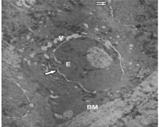
(3) Effects of methylphenidate in rat cornea ... Gozil et al. Figure 1 - Low dose treated group: M - crytolysis of the mitochondrion, K - keratocyte with decreased volume, CO - increased collagen fibers, ∗ - dilatation of the endoplasmic reticulum cisternae, (Uranyl acetate-lead citrate X3000).. Figure 3 - High dose treated group: M - mitochondrion, CO - collagen, V - vacuoles, K - keratocyte in the apoptotic process, → detachment between the cytomembranes of the nuclear membrane, (Uranyl acetate-lead citrate X3000). Figure 2 - Low dose treated group: E - epithelium, M - crystolysis of the mitochondrion, single arrow - detachment between the cytomembranes of the nuclear membrane, double arrow - disruption of the epithelial junctional complexes, V - vacuolization, BM - basement membrane, (Uranyl acetate - lead citrate X3000).. Figure 4 - High dose treated group: E - epithelium, BM - basement membrane, A - apoptotic body, (Uranyl acetate - lead citrate X3000).. evident degenerative changes such as vacuolization of the basal cells, disruption of the junctional complexes, and detachment between the cytomembranes of the nuclear membrane while surface cells and intermediary cells appear in their normal structure (Figure 2). In the curative dose treated group (10mg/kg/day), the corneal endothelium cells show a similar structure compared to the low dose treated group with highly active rER. Crystolysis of the mitochondrion was seen in some cells with hypertrophy of the cells. The junctional complexes show normal structure. The collagen fiber increase in. the corneal stroma was observed similar to the low dose treated group. Some keratocytes show activity as in the low dose group, however, others show increased activity. We also observed the active cells rER cisternae highly dilatated. The cell activity was more increased along these observations and crystolysis of the mitochondria was also prominent in this group. The corneal epithelium degenerative changes were increased compared to the low dose treated group. Especially in some areas, basal cell junctional complexes disruption was observed and these sites show large vacuolizations in the cells that. 500. Saudi Med J 2008; Vol. 29 (4). www.smj.org.sa.
(4) Effects of methylphenidate in rat cornea ... Gozil et al. were localized in the perinuclear site of the cytoplasm. Other degenerative changes, such as disruption of the junctional complexes, crystolysis of the mitochondria, and detachment between the cytomembranes of the nuclear membrane were also seen in this group similar to the low dose treated group, however, these changes were observed in all layers of the epithelium (data not shown). In the high dose treated group (20mg/kg/day), in the endothelial cell lines, we observed degenerated cells with disruption of their junctional complexes, and accepted this appearance as an indicator of the lysis and in some cells, crystolysis of the mitochondria were also seen with activity of the rER cisternae. The collagen fiber increase in the stroma was very prominent. A group of keratocytes was found to be swollen with detachment between the cytomembranes of the nuclear membrane and intracellular edema. Cellular organelle composition was not observed, and this finding observed in many cells was considered as a necrotic process. While a group of cells show changes related to apoptotic process, which is chromatin condensation in their nuclei, electron dense material accumulation, and crystolysis of the mitochondria in some area was also observed (Figure 3). The corneal epithelium, especially basal cell shows highly degenerative changes compared to other groups. A group of basal cell lines contained apoptotic bodies (Figure 4). We also observed in cells of all layers, chromatin condensation and ondulation in their nuclei. Crystolysis of the mitochondrion was also seen in this group. Discussion. Methylphenidate is routinely used for the treatment of ADHD.16 The pharmacodynamic of this psychomotor stimulant drug is primary activation of the noradrenergic and dopaminergic systems and has similar effects to cocaine and amphetamine.16 Pilon and Scheiffle17 reported a case of ulcerative keratitis associated with crack-cocaine abuse, and suggested that the practitioners should be aware of possible toxic effects of such agents on ocular tissues, such as corneal epithelial disruption, and stromal ulceration on cornea. Like cocaine in their case, Lu et al18 reported another case of Ritalin-associated cataract and glaucoma. The catecholamines dopamine, norepinephrine, and epinephrine are synthesized from the L-amino acid tyrosine, and are present in several nervous system areas, including the retina, uvea scleral tissue, and cornea.9,19,20 D1-like and D2-like receptors subfamilies of the dopaminergic receptors are members of the Gprotein-coupled receptor superfamily. While the D1like receptors can stimulate adenylyl cyclase, the D2like receptor subtypes inhibit adenylyl cyclase, and also stimulate mitogenesis and extracellular acidification.10 Several researchers demonstrated the presence of the. D1-like and D2-like receptors using autoradiographic techniques or freshly fixed human corneal tissue.9,10 Cavalotti et al,10 reported that the dopamine system is controlling the corneal function. Crosson et al,20 suggested that chloride secretion in the rabbit corneal epithelium can be modulated by preterminal dopamine receptors located on the sympathetic nerve fibers, therefore, dopamine stimulation appears to be a serial process mediated by the release of norepinephrine from sympathetic nerve terminals in the epithelium. In our study, we observed that all cells, prominently basal cells of the corneal epithelium show dose-dependent degenerative changes such as apoptotic bodies, chromatin condensation, and ondulation in their nuclei and crystolysis of the mitochondrion. In the stroma, the most evident finding was the increase of the collagen fiber. In addition to dose-dependent changes related to apoptotic process, which is chromatin condensation in their nuclei, electron dense material accumulation and pericellular edema in the cytoplasm were also seen. In the endothelial cell lines, disruption of the junctional complexes, vacuolization in the cell cytoplasm, and crystolysis of the mitochondria with rER cisternae activity were observed. As a result, Ritalin© is inducing an evident degeneration especially in epithelium cells, and this degeneration is occurring in the stroma and endothelial layer with increasing doses. Ultrastructural cell organelle composition degeneration with stromal fibrosis has a negative effect on cornea dehydration. In the light of these findings, we believe that the Ritalin© treatment doses need to be kept at a minimum to maintain healthy cornea ultrastructure and related physiology. References 1. Swanson JM, Sergeant JA, Taylor E, Sonuga-Barke EJ, Jensen PS, Cantwell DP. Attention-deficit hyperactivity disorder and hyperkinetic disorder. Lancet 1998; 351: 429-433. 2. Kollins SH, MacDonald EK, Rush CR. Assessing the abuse potential of methylphenidate in nonhuman and human subjects: a review. Pharmacol Biochem Behav 2001; 68: 611-627. 3. Swanson JM, Volkow ND. Serum and brain concentrations of methylphenidate: implications for use and abuse. Neurosci Biobehav Rev 2003; 27: 615-621. 4. Greenhill LL, Pliszka S, Dulcan MK, Bernet W, Arnold V, Beitchman J, et al. Practice parameter for the use of stimulant medications in the treatment of children, adolescents, and adults. J Am Acad Child Adolesc Psych 2002; 41(Suppl 2): S26-S49. 5. Thanos PK, Michaelides M, Benveniste H, Wang GJ, Volkow ND. Effects of chronic oral methylphenidate on cocaine self-administration and striatal dopamine D2 receptors in rodents. Pharmacol Biochem Behav 2007; 87: 426-433. 6. Schweri MM, Skolnick P, Rafferty MF, Rice KC, Janowsky AJ, Paul SM. [3H]Threo-(+/)-methylphenidate binding to 3,4dihydroxyphenylethylamine uptake sites in corpus striatum: correlation with the stimulant properties of ritalinic acid esters. J Neurochem 1985; 45: 1062-1070. www.smj.org.sa. Saudi Med J 2008; Vol. 29 (4). 501.
(5) Effects of methylphenidate in rat cornea ... Gozil et al 7. Gatley SJ, Pan D, Chen R, Chaturvedi G, Ding YS. Affinities of methylphenidate derivatives for dopamine, norepinephrine and serotonin transporters. Life Sci 1996; 58: 231-239. 8. Gerasimov MR, Franceschi M, Volkow ND, Gifford A, Gatley SJ, Marsteller D, et al. Comparison between intraperitoneal and oral methylphenidate administration: A microdialysis and locomotor activity study. J Pharmacol Exp Ther 2000; 295: 51-57. 9. Grueb M, Wallenfels-Thilo B, Denk O, Mielke J, Reinthal E, Rohrbach JM, et al. Monoamine receptors in human corneal epithelium and endothelium. Acta Ophthalmol Scand 2006; 84: 110-115. 10. Cavallotti C, Pescosolido N, Artico M, Feher J. Localization of dopamine receptors in the rabbit cornea. Cornea 1999; 18: 721-728. 11. Reis RAM, Ventura ALM, Kubrusly RCC, de Mello MCF, de Mello FG. Dopaminergic signaling in the developing retina. Brain Res Rev 2007; 54: 181-188. 12. El-Zein RA, Abdel-Rahman SZ, Hay MJ, Lopez MS, Bondy ML, Morris DL, et al. Cytogenetic effects in children treated with methylphenidate. Cancer Lett 2005; 230: 284-291. 13. Benes FM. Brain development, VII. Human brain growth spans decades. A J Psychiatry 1998; 155: 1489.. 14. Castner SA, Goldman-Rakic PS. Long-lasting psychotomimetic consequences of repeated low-dose amphetamine exposure in rhesus monkeys. Neuropsychopharmacology 1999; 20: 10-28. 15. Giedd JN, Blumenthal J, Jeffries NO, Castellanos FX, Liu H, Zijdenbos A, et al. Brain development during childhood and adolescence: a longitudinal MRI study. Nat Neurosci 1999; 2: 861-863. 16. LeBlanc-Duchin D, Taukulis HK. Behavioral reactivity to a noradrenergic challenge after chronic oral methylphenidate (ritalin) in rats. Pharmacol Biochem Behav 2004; 79: 641-649. 17. Pilon AF, Scheiffle J. Ulcerative keratitis associated with crackcocaine abuse. Cont Lens Anterior Eye 2006; 29: 263-267. 18. Lu CK, Kuang TM, Chou JC. Methylphenidate (Ritalin)associated cataract and glaucoma. J Chin Med Assoc 2006; 69: 589-590. 19. Cavallotti C, Pescosolido N, Pescosolido V, Iannetti G. Determination of D1 receptors in the human uveo scleral tissue by light microscope autoradiography. Int Ophthalmol 2001; 23: 171-179. 20. Crosson CE, Beuerman RW, Klyce SD. Dopamine modulation of active ion transport in rabbit corneal epithelium. Invest Ophthalmol Vis Sci 1984; 25: 1240-1245.. www.smj.org.sa Saudi Medical Journal Online features * Instructions to Authors * Uniform Requirements * STARD * Free access to the Journal’s Current issue * Future Contents * Advertising and Subscription Information All Subscribers have access to full text articles in HTML and PDF format. Abstracts and Editorials are available to all Online Guests free of charge.. 502. Saudi Med J 2008; Vol. 29 (4). www.smj.org.sa.
(6)
Şekil
Benzer Belgeler
Recurrent Coronary Thrombus in a Patient with Chronic Immune Thrombocytopenia with Treatment Using Eltrombopag. Garbe E, Andersohn F, Bronder E, Salama A, Klimpel A, Thomae M,
Objectives: To examine the effect of oral tetracycline and clarithromycin on the development of postoperative intra-abdominal adhesions in a rat uterine horn
Reduced ACTH(adrenocorticotrophic hormone), cortisol in blood have been found in human subjects and reduced CRF in hypothalamus, paraventricular nucleus has been shown
而 NO 的增加則是由於 iNOS 的正調節作用(up-regulation)所致。另外,預先以 tyrosine kinase 抑制劑(genistein 和 tyrphostin)、Ras-farnesyl transferase
Materials and Methods: One hundred patients between 0-17 years who were followed up due to scorpion stings or poisonings in Hatay Mustafa Kemal University, Faculty of
We present this case to emphasize that intrauterine vitamin D intoxication (maternal administration of vitamin D at 30 weeks of gestation, delivery at 34 weeks of
The results of this study revealed that the Board Commissioner Size, Proportion of Independent Commissioner, Managerial Capital Ownership, and Profitability did not significantly
Thus, this study aims to review the application of educational drones in various educational environments, different types and sizes of drones used in education, and
