Contents lists available atScienceDirect
Journal of Ethnopharmacology
journal homepage:www.elsevier.com/locate/jepSambulin A and B, non-glycosidic iridoids from Sambucus ebulus, exert
signi
ficant in vitro anti-inflammatory activity in LPS-induced RAW 264.7
macrophages via inhibition of MAPKs's phosphorylation
İrem Atay Balkan
a,1, Ayca Zeynep
İlter Akülke
b, Ye
şim Bağatur
b, Dilek Telci
b, Ahmet
Ceyhan Gören
c, Hasan K
ırmızıbekmez
a, Erdem Yesilada
a,⁎aYeditepe University, Faculty of Pharmacy, Department of Pharmacognosy and Phytotherapy, 34755 Ataşehir, İstanbul, Turkey bYeditepe University, Faculty of Engineering, Department of Genetics and Bioengineering, 34755 Ataşehir, İstanbul, Turkey cTUBITAK National Metrology Institute, Chemistry Group Laboratories, 41470 Gebze, Kocaeli, Turkey
A R T I C L E I N F O
Keywords: Sambucus ebulus Iridoids Sambulin A and B
Inducible nitric oxide synthase Mitogen activated protein kinases Cytokines
A B S T R A C T
Ethnopharmacological relevance: The leaves of Sambucus ebulus L. (Adoxaceae) are widely used in Turkish folk medicine particularly against inflammatory disorders. The fresh leaves after wilted over fire or the poultices prepared are directly applied externally to heal burns, edema, eczema, urticarial and abscess. Two iridoids were recently isolated (sambulin A, sambulin B) from the leaves of S. ebulus.
Aim of the study: This study aims to investigate the in vitro anti-inflammatory activities of these iridoids on LPS-induced RAW 264.7 macrophages.
Materials and methods: Raw 264.7 macrophages were treated with 12.5, 25 and 50 µg/ml Sambulin A and 6.25, 12.5 and 25 µg/ml Sambulin B and induced with 1 µg/ml lipopolysaccaharides (LPS). Effect of the compounds on nitric oxide (NO) production and cytokines (TNFα, IL-6) were determined by Griess and ELISA assays respectively. iNOS and the phosphorylation levels of MAPKs (ERK, JNK) were examined by Western Blot.
Results: Sambulin A and sambulin B inhibited 52.82% and 72.88% of NO production at 50 and 25 µg/ml concentrations respectively. The levels of iNOS were significantly decreased by both molecules, sambulin B at 25 µg/ml almost completely decreased iNOS levels (97.53%). Both molecules significantly inhibited TNFα productions. However, only sambulin B inhibited IL-6 production. Consequently, it was shown that sambulin B exerted its effect through the inhibition of ERK and JNK phosphorylations.
Conclusion: The prominent bioactivities exerted by two iridoids will contribute to explanation of the usage of S. ebulus in traditional medicine against rheumatoid diseases.
1. Introduction
Sambucus ebulus L. (Adoxaceae) or dwarf elder is a widespread annual herbaceous plant which is known as‘cüce mürver’ in Anatolia. Particularly the leaves of the plant are used against various types of inflammatory disorders in Turkish folk medicine (Sezik et al., 1992; Yesilada et al., 1999b). The fresh leaves are either externally applied on the affected body part after wilted over open fire or the poultice prepared by boiling in water to treat a wide range of dermatological problems including burns, infectious wounds, snake bites, edema, eczema, and urticaria, while its decoction is used to bath the affected
extremities to relieve rheumatic pain (Sezik et al., 1992; Yesilada et al., 1999b).
Previous experimental studies have indicated that S. ebulus leaves exert cytotoxic (Shokrazadeh et al., 2009), antiulcer (Yesilada et al., 2014), anti-Helicobacter pylori (Yesilada et al., 1999a), anti-microbial (Smee et al., 2011), anti-oxidative (Ebrahimzadeh et al., 2009) and wound-healing activities (Süntar et al., 2010). Anti-inflammatory and anti-nociceptive activities of the leaves have also been investigated in detail by using several in vivo experimental models such as: hot plate test, writhing test, tailflick test and carrageenan- or formalin-induced edema, adjuvant-induced chronic arthritis models (Yesilada, 1997;
http://dx.doi.org/10.1016/j.jep.2017.06.002
Received 13 March 2017; Received in revised form 24 May 2017; Accepted 2 June 2017
⁎Corresponding author.
1Current address:İstanbul Medipol University, School of Pharmacy, Department of Pharmacognosy, 34810, Beykoz, İstanbul, Turkey.
E-mail addresses:[email protected](İ.A. Balkan),[email protected](A.Z.İlter Akülke),[email protected](Y. Bağatur),[email protected](D. Telci), [email protected](A.C. Gören),[email protected](E. Yesilada).
Available online 10 June 2017
0378-8741/ © 2017 Elsevier B.V. All rights reserved.
Ahmadiani et al., 1998; Ebrahimzadeh et al., 2006). A wide range of phytochemicals have been isolated from the leaves of Sambucus species including lignans, iridoids, flavonoids, anthocyanins, and cyanogenins (Gross and Sticher, 1986; D’abrosca et al., 2001; Jordheim et al., 2007;Süntar et al., 2010;Atay et al., 2015).
Inflammation is an elaborated process that involves various path-ways. Signaling proteins which are synthesized during these pathways such as prostaglandins, leukotrienes, platelet-activating factors and cytokines such as IL-1α and TNFα are known to contribute to the progression of inflammation (Yesilada et al., 1997; Yesilada, 1997). Eventually antagonizing these inflammatory pathways would inhibit the productions of inflammatory mediators. Therefore these pathways are mainly targeted for the discovery of anti-inflammatory drug candidate. Key players in these pathways including enzymes such as inducible nitric oxide synthase (iNOS), cytokines such as interleukin-1 beta (IL-1β), tumor necrosis factor-alpha (TNFα) (Tincani et al., 2007) or mitogen activated protein kinases (MAPKs) are frequently employed as the main targets in anti-inflammatory research (Schmitz and Bacher, 2005;Yoon et al., 2010).
Macrophages regulate inflammation by using a wide range of pro-inflammatory mediators (Yoon et al., 2010; Choi et al., 2011; Lu et al., 2012). Lipopolysaccharides (LPS) which is the principal component of the outer membrane of gram-negative bacteria, is one of the most potent activators of macrophages (Schumann et al., 1990). Once macrophages treated with LPS are known to produce inflammatory mediators, such as nitric oxide (NO) and cytokines such as TNFα, IL-1 and IL-6 (Yoon et al., 2010; Choi et al., 2011; Lu et al., 2012). LPS activates MAPKs including extracellular signal-regulated kinases (ERK)-1 and -2, c-Jun N-terminal kinase (JNK), and p38. The activated MAPKs increase inflammatory disease states by regulating the biosynthesis of inflammatory mediators such as inducible nitric oxide synthase (iNOS) and TNFα (Geppert et al., 1994; Swantek et al., 1997; Chan and Riches, 2001). Raw 264.7 is a murine macrophage cell line and in vitro LPS-induced Raw 264.7 macrophage model is frequently used as an experimental model to determine the effect on inflammatory response (Yoon et al., 2010; Choi et al., 2011; Lu et al., 2012).
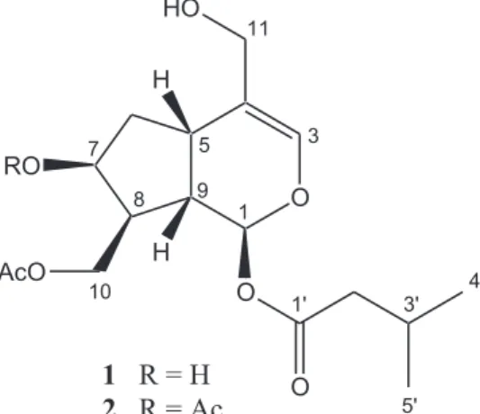
In our previous study, we reported the isolation and structure elucidation of two non-glycosidic iridoids from the methanol (MeOH) extract of S. ebulus leaves namely; sambulin A and sambulin B, the latter being a new compound (Atay et al., 2015). The chemical structures of these compounds were shown in Fig. 1. In the present study, the anti-inflammatory activity of sambulin A and sambulin B have been investigated further on several in vitro parameters using LPS induced Raw 264.7 macrophages. For this purpose, effects of the compounds on NO, TNFα, IL-6 levels were studied by Griess and ELISA methods. The variations in the iNOS protein expressions and phosphorylation levels of MAPKs (ERK1/2, JNK) in response to
Sambulin A and Sambulin B treatment were investigated by Western Blotting.
2. Materials and methods 2.1. Chemicals and reagents
Lipopolysachharides (LPS), dimethylsulfoxide (DMSO), HRP-con-jugated goat anti-mouse IgG and HRP-conHRP-con-jugated goat anti-rabbit IgG were purchased from Sigma-Aldrich (St. Louis, MO). Dulbecco's modified Eagle's medium (DMEM), Fetal bovine serum (FBS), peni-cillin-streptomycine was obtained from Invitrogen/Gibco (Grans Island, NY, USA). Antibodies for ERK1/2, ERK, phospho-JNK, JNK were purchased from Cell Signaling Technology (Beverly, MA, USA). ECL reagent was obtained from Amersham (Buckhinghamshire, UK). Enzyme linked immunosorbent assay (ELISA) kits for TNFα and IL-6 were from R & D Systems (MN, USA). iNOS and β-actin primary antibodies were purchased from Santa Cruz (CA, USA). Griess Reagent System was from Promega (CA, USA). L-NIL was obtained from Calbiochem, Sigma and Cayman respectively. WST-1 reagent was from Roche Applied Science (Mannheim, Germany).
2.2. Plant material
S. ebulus leaves were collected from Uludağ-Bursa (Turkey) in June 2009. The plant was identified by one of the authors (E. Yesilada). A voucher specimen is deposited at the Herbarium of Yeditepe University (YEF 09017). The plant material was dried under shade and powdered prior to extraction.
2.3. Extraction, isolation and structure elucidations of sambulin A and sambulin B
Sambulin A and sambulin B (Fig. 1) were isolated as described previously from the methanolic extract of S. ebulus leaves and their structures were elucidated by using 1-D and 2-D nuclear magnetic resonance spectroscopy (NMR), mass spectrometry (MS) techniques (Atay et al., 2015).
2.4. Cells and cell culture
The Raw 264.7 macrophages (ATCC TIB-71) were grown in DMEM supplemented with 10% FBS, 4 mML-glutamine, 100 IU/ml penicillin
and 100 µg/ml streptomycin at 37 °C in a humidified atmosphere containing 5% CO2.
2.5. WST-1 assay for cell viability
The non-toxic concentrations of the compounds were determined by using a WST-1 assay kit (Roche Applied Sciences) according to the manufacturer's instructions. WST-1, a tetrazolium-based salt dye, is reduced to a purple formazan salt by metabolically active cells, and it is directly quantified by spectrophotometric measurements. Raw 264.7 cells (22.500 cells/well) in 10% FBS-DMEM were seeded into 96-well plates and various concentrations of compounds were added to the wells with or without 1 µg/ml LPS, and incubated at 37 °C for 24 h. After the supernatant was removed, WST-1 was added directly to the cultures to afinal concentration of 5% (v/v), and cells were incubated at 37 °C for an additional 60 min. Absorbance was then read between 420 and 480 nm (λmax 450 nm) using a plate reader. All test
compounds were dissolved in DMSO and diluted with DMEM to the appropriate concentrations. Thefinal concentration of DMSO in the culture medium was not more than 0.1% (v/v).
2.6. Griess assay
Raw 264.7 cells (22.500 cells/well) in 10% FBS-DMEM were seeded into 96- well plates. The cells were co-incubated with the test compounds and 1μg/ml lipopolysaccharide (LPS). After that the plates were incubated at 37 °C for a total of 24 h and nitrite accumulated in the culture medium was measured by Griess Assay. Briefly, 50 μl of cell culture medium was mixed with 50μl of 1% (w/v) sulfanilamide in 5% (v/v) phosphoric acid and incubated for 10 min and then 50μl of 0.1% (w/v) naphtylethylenediamine-HCl (Promega) was added and then incubated at room temperature for another 10 min. The absorbance of thefinal solution was read at 550 nm using a microplate reader. The amount of nitrite in the test samples was calculated from the NaNO2
serial dilution standard curve.
2.7. Enzyme immunoassay for quantification of cytokines (TNFα, IL-6)
Raw 264.7 cells (22.500 cells/well) in 10% FBS-DMEM were seeded into 96- well plates. The cells were a pre-incubated with compounds for one-hour, and then stimulated with 1μg/ml LPS. The plates were incubated at 37 °C for a total of 24 h. Sandwich ELISA was used to determine the inhibitory effects of the isolated compounds on the production of cytokines (TNFα, IL-6) in LPS-induced Raw 264.7 cells. The supernatant was harvested and assayed according to the manufacturer's protocol for the relevant ELISA kit.
2.8. Western blot assay
Cells were washed once with ice-cold PBS and extracted by RIPA lysis buffer (1x TBS, 1% Nonidet P-40, 0.5% sodium deoxycholate, 0.1% SDS, 0.004% sodium azide-Santa Cruz) including 1 mM phenyl-methylsulfonyl fluoride, 1 mM Na ortho-vanadate and 1% protease inhibitor cocktail. The protein concentration was determined by Lowry assay after sonication. Forty micrograms of cellular protein were separated by 8% or 10% SDS-polyacrylamide gel electrophoresis. The proteins were electroblotted onto a nitrocellulose membrane and incubated with specific primary antibodies overnight with blocking solution (5% skimmed milk) at 4 °C. Blots were washed four times with Tween 20/Tris-buffered saline (TBST) and incubated with a 1:1000 dilution of secondary antibodies for 2 h at room temperature. Blots were again washed three times with TBST, and then visualized by ECL reagent (Amersham Life Science).
2.9. Statistical analysis
The data were recorded as mean ± standard error of the mean (SEM) of triplicate experiments. The data were analyzed by the PASW Statistics. The significance of the difference between means were determined by Mann Whitney- U test and p-values less than 0.05 were considered statistically significant.
3. Results
3.1. Effects of sambulin A and sambulin B on the viability of Raw 264.7 macrophages
Raw 264.7 macrophage cells were treated with 6.25, 12.5, 25 and 50 µg/ml sambulin A and 3.12, 6.25, 12.5, 25, 50 µg/ml sambulin B with or without LPS (1 µg/ml) for 24 h. As shown inFig. 2, sambulin B, when applied at concentrations higher than 25μg/ml, resulted in more than 40% decrease in the viability of Raw 264.7 macrophages, but sambulin A did not exert any toxic effects to cells up 50 μg/ml concentration. The chemical structure of Sambulin B only differs from sambulin A by the presence of an additional acetyl unit attached at the C-7 (OH) position. The difference between the cytotoxic effects of these
compounds might probably due to that additional acetyl moiety.
3.2. Effects of sambulin A and sambulin B on NO productions of Raw 264.7 macrophages
Raw 264.7 macrophage cells were co-incubated with sambulin A or sambulin B and LPS (1 µg/ml) for 24 h. As shown inFig. 3A, sambulin A inhibited the productions of NO by 2.88 ± 2.13%, 39.79 ± 4.55% and 52.82 ± 1.95% at 12.50, 25 and 50 µg/ml concentrations compared to the LPS-induced control group. Treatment of Raw 264.7 macrophages with sambulin B at 6.25, 12.50 and 25 µg/ml concentra-tions led to an inhibition up to 72.88 ± 1.71% (Fig. 3B). The inhibitory activity of sambulin B at 25 µg/ml concentration was close to the known NO inhibitor L-NIL (10μM) which inhibited 85.88 ± 1.63% of NO production. The inhibitory activity of sambulin B on NO synthesis was found to be higher than that of sambulin A, although its concentration was half to that of sambulin A.
3.3. Effects of sambulin A and sambulin B on iNOS protein levels of Raw 264.7 macrophages
In order to explain the inhibitory effects of sambulin A and sambulin B on NO production, the effects of these compounds on LPS-induced iNOS levels were investigated. As shown in Fig. 4, sambulin A treatment decreased the levels of iNOS enzyme up to 59.29 ± 6.70% at its highest concentration (50 µg/ml) when compared to the only LPS-treated control group suggesting that the inhibitory effect of Sambulin A on NO release might be due to the decrease in iNOS protein levels. On the other hand, sambulin B at 25 µg/ml concentration almost completely exhausted the iNOS protein levels in LPS-induced Raw 264.7 cells. The concentration dependent decrease in iNOS levels by sambulin B at 6.25, 12.5 and 25μg/ml concentrations, respectively, were found to be 43.49 ± 18.48%, 56.46 ± 9.65% and 97.53 ± 1.57% (Fig. 4B).
Fig. 2. Effects of sambulin A (SA) and sambulin B (SB) on the viability of Raw 264.7 macrophages. Cells were treated with the indicated concentrations of sambulin A and sambulin B with or without LPS (1 µg/ml) for 24 h. Cell viability was determined by WST-1 assay. Error bars represents the mean ± S.E.M. Values of *p≤ 0.05 **p ≤ 0.01 ***p≤ 0.001 vs. only media treated group were considered significant.
3.4. Effects of sambulin A and sambulin B on inflammatory cytokines The effects of compounds on LPS induced TNFα, and IL-6 produc-tions were investigated on LPS-induced Raw 264.7 macrophage cells using an ELISA assay. As mentioned above, Raw 264.7 macrophage cells were pre-treated with the compounds for one hour and then the cells were induced with LPS for a total of 24 h. Treatment with 1μg/ml LPS significantly increased TNFα levels in the culture medium (Fig. 5). Sambulin A exerted 23.53%, 28.96%, 36.21% inhibition at 12.5, 25 and 50μg/ml concentrations, while sambulin B provided a decrease of
16.75%, 21.32% and 34.20% in TNFα levels at 6.25, 12.5 and 25 μg/ml concentrations, respectively, when compared to the LPS-treated con-trol group (Fig. 5B). The inhibitory effects of the molecules on TNFα productions were found to be quite close to dexamethasone which provides 45.60% TNFα inhibition at 10 μM concentration.
On the contrary, sambulin A displayed a weak inhibitory activity on
Fig. 3. Effects of sambulin A (SA) and sambulin B (SB) on the productions of NO on LPS induced Raw 264.7 macrophages. Raw 264.7 cells were co-incubated with the indicated concentrations of sambulin A or and sambulin B and LPS (1 µg/ml) for 24 h. Media collected and NO levels were measured by Griess Assay respectively. Results were expressed as the percentage of only LPS treated control group. Error bars represents the mean ± S.E. M. *p≤ 0.05 **p ≤ 0.01 ***p ≤ 0.001compared to LPS treated group #p ≤ 0.05 ##p≤ 0.01 ###p ≤ 0.001 compared to non-induced group.
Fig. 4. Effects of sambulin A (SA) and sambulin B (SB) on iNOS protein levels on LPS induced Raw 264.7 macrophages. Raw 264.7 cells were co-incubated with indicated concentrations sambulin A or sambulin B and LPS (1 µg/ml) for 24 h. At the end of the 24 h cells were lysed and iNOS proteins were detected with Western Blot using specific antibodies A representative image of three independent experiments is shown. Relative band intensities were measured by Image J and signal was normalized toβ-actin. Results were expressed as the percentage of only LPS-treated control group. The data are means ± S.E.M. of three independent experiments. *p≤ 0.05 **p ≤ 0.01 ***p ≤ 0.001compared to only LPS treated group #p≤ 0.05 ##p ≤ 0.01 ###p ≤ 0.001 compared to non-induced group.
Fig. 5. Effects of sambulin A (SA) and sambulin B (SB) on the productions of TNFα and IL-6 on LPS induced Raw 264.7 macrophages.
IL-6 production which was around 11.33 ± 7.63% at 50 µg/ml concentration (Fig. 5C). However, IL-6 concentration was decreased by 59.12% when sambulin B applied to cells at 25μg/ml concentration. Dexamethasone at 5μM provided 77.24% inhibition.
3.5. Inhibitory effects of sambulin B on MAPK's phosphorylation Further investigations were carried out in order to explain the inhibitory effects of sambulin B on iNOS and cytokines, its effects on the MAPK phosphorylations were studied. For this purpose, Raw 264.7 macrophage cells werefirst treated with the indicated concentrations of sambulin B for two hrs and then stimulated with LPS (1 µg/ml) for 15 mins. Sambulin B inhibited ERK phosphorylation by 30.73 ± 13.19% and 73.29 ± 9.80% at 12.5 and 25 µg/ml concentration comparing to only LPS treated group (Fig. 6A). Similarly a concentra-tion-dependent decrease in phospho-JNK levels were observed in Raw 264.7 macrophages following sambulin B treatment (Fig. 6B). Sambulin B (2) inhibited JNK phosphorylation in a concentration-dependent manner. This molecule inhibited 36.17 ± 3.27%, 40.14 ± 8.19% of the phosphorylation of JNK at 12.5 and 25 µg/ml concentra-tions, respectively. Accordingly inhibition of JNK and ERK phosphor-ylations may be suggested as a possible mechanism for the anti-inflammatory activity of sambulin B.
Raw 264.7 cells were pre-incubated with the indicated concentra-tions of the molecules and stimulated with LPS (1 µg/ml) for a total of 24 h. Media collected and TNFα and IL-6 levels were measured by ELISA. Error bars represents the mean ± S.E.M. *p≤ 0.05 **p ≤ 0.01 ***p≤ 0.001compared to LPS treated group #p ≤ 0.05 ##p ≤ 0.01 ###p≤ 0.001 compared to non-induced group.
3.6. Discussion
Anti-inflammatory activity of S. ebulus leaves was previously investigated by several research groups and isolation of a number of active components were reported (Yesilada et al., 1997; Yesilada, 1997; Ahmadiani et al., 1998;Schwaiger et al., 2011). In these studies, MeOH and water extracts of aerial parts of S. ebulus were investigated for their in vivo anti-inflammatory and anti-arthritic activities when adminis-tered either orally or topically, and both extracts were shown to exert potent activity on carrageenan-induced edema and adjuvant-induced arthritis models. Chlorogenic acid was isolated as one of the active components of the MeOH extract (Yesilada et al., 1997). In another study, the MeOH extract and its hexane subextract from the leaves of S. ebulus were shown to possess moderate inhibitory activity on the
synthesis of cytokines IL-1α and IL-1β, while only chloroform sub-extract exerted weak inhibitory activity on TNFα production (Yesilada et al., 1997). Later ursolic acid was isolated from the diethyl ether subextract of ethanol extract of leaves, through bioassay-guided isola-tion procedures by monitoring the inhibiisola-tion of TNFα induced expres-sion of vascular cell adheexpres-sion molecule-1 (VCAM-1) on the surface of human umbilical vein endothelial cells (HUVECs) (Schwaiger et al., 2011). In another study, roots of the plant was also reported to possess significant anti-inflammatory activity on both acute and sub-acute in vivo models (Ahmadiani et al., 1998).
Iridoids are monoterpenic compounds bearing cyclopentanopyra-noid skeleton. This group of compounds have been shown to exert anti-hypertensive, hepatoprotective, choleretic, hypoglycaemic, hypolipi-demic, anti-spasmodic, anti-viral, immunomodulator and anti-in flam-matory activities in the previous studies (Ghisalberti, 1998). Iridoids such as monotropein, catalposide, scropolioside and harpagoside have been shown to inhibit the activation of NF-κB on LPS-induced macrophages. Moreover, these molecules provided significant reduc-tion in the levels of iNOS, COX-2 and various cytokines (An et al., 2002; Bas et al., 2007; Shin et al., 2013). S. ebulus have long been known to contain iridoids, in fact a number of new ‘‘valerian type’’ iridoids were recently isolated (Gross et al., 1986; Pieri et al., 2009; Tomassini et al., 2013). In our previous study, two‘‘valerian type’’ non-glycosidic iridoids were isolated from the methanol extract of S. ebulus leaves; sambulin A from the chloroform subextract and sambulin B from the hexane subextract of the leaves (Atay et al., 2015).
Sambulin A was found to be effective on LPS-induced NO produc-tion when applied to Raw 264.7 macrophages at 12.5, 25 and 50μg/ml concentrations. The NO production was inhibited up to 52.82% in accordance with the reduction in iNOS protein levels. It was also shown to be a potent inhibitor of TNFα and provided a reduction between 23% and 36%, depending on the concentration. On the other hand, it was only slightly active on IL-6 levels.
Sambulin B exerted a potent NO inhibition up to 73% when Raw 264.7 macrophage cells were incubated with 6.25, 12.5 and 25μg/ml. NO inhibitory effect of sambulin B was twice as much potent as sambulin A. Sambulin B differs from sambulin A by the presence of an additional acetyl unit attached at the C-7 (OH) position. Eventually it can be postulated that the presence of an additional acetyl group at C-7 position of the iridoid skeleton as in sambulin B increased the inhibitory activity against NO. Moreover, sambulin B provided up to 33% reduction in the TNFα concentration in the cell culture media. It was also found to be effective on IL-6 levels, at 25 μg/ml concentration, provided a 59.12% inhibition. Sambulin B at concentrations higher
Fig. 6. Effect of sambulin B (SB) on MAPK's (JNK and ERK) phosphorylation levels. (A) ERK phosphorylation (B) JNK phosphorylation. Raw 264.7 cells pre-incubated with the indicated concentrations of sambulin B and stimulated with LPS (1 µg/ml) for 15 min. At the end of the 24 h cells were lysed and proteins (p-ERK, p-JNK) were detected with Western Blot using specific antibodies A representative image of three independent experiments is shown. Relative band intensities were measured by Image J and signal was normalized to the corresponding total proteins (ERK, JNK). Results were expressed as the percentage of only LPS treated control group. The data are means ± S.E.M. of three independent experiments. * p≤ 0.05 **p ≤ 0.01 ***p ≤ 0.001compared to only LPS treated group #p ≤ 0.05 ##p ≤ 0.01 ###p ≤ 0.001 compared to non-induced group.
than 25μg/ml concentration resulted in more than 30% decrease in the viability of Raw 264.7 macrophages, while sambulin A was not toxic to cells up to 50μg/ml concentration suggesting that the acetyl group in sambulin B had also dissimilated the effect of molecule on cell viability. MAPKs are known to regulate the synthesis of many inflammatory mediators at the levels of translation and transcription (Johnson and Lapadat, 2002; Kaminska, 2005). This pathway was shown to regulate the production of LPS-induced iNOS and was found to be associated with COX-2 gene expression (Chen and Wang, 1999; Hwang et al., 1997; Kim et al., 2007). Moreover, MAPKs regulate the expressions of inflammatory mediators such as TNFα, IL-1β and IL-6 (Kaminska, 2005). In the present study, Sambulin B significantly inhibited ERK and JNK phosphorylations. For that reason, inhibition of JNK and ERK phosphorylations may be suggested as a possible mechanism for the in vitro anti-inflammatory activity of sambulin B.
The present study demonstrates the in vitro anti-inflammatory properties of two iridoid molecules, sambulin A and sambulin B, which were isolated from the leaves of S. ebulus for the first time. The demonstration of two active iridoids from S. ebulus leaves will contribute the explanation of its folkloric usage and the mechanism of its anti-inflammatory activity.
Conflict of interest
The authors declare no conflict of interest. Acknowledgements
This work was supported by The Scientific and Technological Research Council of Turkey [TUBITAK Project no: 110S197]. References
Ahmadiani, A., Fereidoni, M., Semnanian, S., Kamalinejad, M., Saremi, S., 1998. Antinociceptive and anti-inflammatory effects of Sambucus ebulus rhizome extract in rats. J. Ethnopharmacol. 61, 229–235.
An, S.J., Pae, H.O., Oh, G.S., Choi, B.M., Jeong, S., Jang, S.I., Oh, H., Kwon, T.O., Song, C.E., Chung, H.T., 2002. Inhibition of TNFα, IL-1β, and IL-6 productions and NF-κB activation in lipopolysaccharide-activated RAW 264.7 macrophages by catalposide, an iridoid glycoside isolated from Catalpa ovata G. Don (Bignoniaceae). Int. Immunopharmacol. 2, 1173–1181.
Atay,İ., Kırmızıbekmez, H., Gören, A.C., Yesilada, E., 2015. Secondary metabolites from Sambucus ebulus. Turk. J. Chem. 39, 34–41.
Bas, E., Recio, M.C., Máñez, S., Giner, R.M., Escandell, J.M., López-Ginés, C., Rios, J.L., 2007. New insight into the inhibition of the inflammatory response to experimental delayed-type hypersensitivity reactions in mice by scropolioside A. Eur. J. Pharmacol. 555, 199–210.
Chan, E.D., Riches, D.W.H., 2001. IFN-γ + LPS induction of iNOS is modulated by ERK, JNK/SAPK, and p38 MAPK in a mouse macrophage cell line. Am. J. Physiol-Cell. Physiol. 280, C441–C450.
Chen, C.C., Wang, J.K., 1999. p38 but Not p44/42 Mitogen activated protein kinase is required for nitric oxide synthase induction mediated by lipopolysaccharide in RAW 264.7 macrophages. Mol. Pharmacol. 55, 481–488.
Choi, R.J., Shin, E.M., Jung, H.A., Choi, J.S., Kim, Y.S., 2011. Inhibitory effects of kaurenoic acid from Aralia continentalis on LPS-induced inflammatory response in Raw 264.7 macrophages. Phytomedicine 18, 677–682.
D'abrosca, B., DellaGreca, M., Fiorentino, A., Monaco, P., Previtera, L., Simonet, A.M., Zarrelli, A., 2001. Potential allelochemicals from Sambucus nigra. Phytochemistry 58, 1073–1081.
Ebrahimzadeh, M.A., Mahmoudi, M., Salimi, E., 2006. Anti-inflammatory activity of Sambucus ebulus hexane extracts. Fitoterapia 77, 146–148.
Ebrahimzadeh, M.A., Nabavi, S.F., Nabavi, S.M., 2009. Antioxidant activities of methanol extract of Sambucus ebulus L.flower. Pak. J. Biol. Sci. 12, 447–450.
Geppert, T.D., Whitehurst, C.E., Thompson, P., Beutler, B., 1994. Lipopolysaccharide signals activation of tumor necrosis factor biosynthesis through the ras/raf-1/MEK/ MAPK pathway. Mol. Med. 1, 93–103.
Ghisalberti, E.L., 1998. Biological and pharmacological activity of naturally occurring
iridoids and secoiridoids. Phytomedicine 5, 147–163.
Gross, G.A., Sticher, O., 1986. Isosweroside, ein neues scoiridoidglucoside aus den Wurzeln des Zwergholunders Sambucus ebulus L. (Caprifoliaceae). Helv. Chim. Acta 69, 1113–1119.
Gross, G.A., Sticher, O., Anklin, C., 1986. Ein neues esteriridoidglucoside aus Sambucus ebulus L (Caprifoliacae). Helv. Chim. Acta 69, 156–162.
Hwang, D., Jang, B.C., Yu, G., Boudreau, M., 1997. Expression of mitogen-inducible cyclooxygenase induced by lipopolysaccharide. Biochem. Pharmacol. 54, 87–96. Johnson, G.L., Lapadat, R., 2002. Mitogen-activated protein kinase pathways mediated
by ERK, JNK, and p38 protein kinases. Science 298, 1911–1912.
Jordheim, M., Giske, N.H., Andersen, O.M., 2007. Anthocyanins in caprifoliaceae. Biochem. Syst. Ecol. 35, 153–159.
Kaminska, B., 2005. MAPK signalling pathways as molecular targets for anti-inflammatory therapy-from molecular mechanisms to therapeutic benefits. Biochim. Biophys. Acta 1754, 253–262.
Kim, H.G., Yoon, D.H., Lee, W.H., Han, S.K., Shrestha, B., Kim, C.H., Lim, M.H., Chang, W., Lim, S., Choi, S., Song, W.O., Sung, J.M., Hwang, K.C., Kim, T.W., 2007. Phellinus linteus inhibits inflammatory mediators by suppressing redox based NF-kappaB and MAPKs activation in lipopolysaccharide induced RAW 264.7 macrophage. J. Ethnopharmacol. 114, 307–315.
Lu, Y., Suh, S.J., Kwak, C.H., Kwon, K.M., Seo, C.S., Li, Y., Jin, Y., Li, X., Hwang, S.L., Kwon, O., Chang, Y.C., Park, Y.G., Park, S.S., Son, J.K., Kim, C.H., Chang, H.W., 2012. Saucerneol F, a new lignan, inhibits iNOS expression via MAPKs, NF-κB and AP-1 inactivation in LPS-induced Raw 264.7 cells. Int. Immunopharmacol. 12, 175–181.
Pieri, V., Schwaiger, S., Ellmerer, E.P., Stuppner, H., 2009. Iridoid glycosides from the leaves of Sambucus ebulus. J. Nat. Prod. 72, 1798–1803.
Schmitz, M.L., Bacher, S., 2005. Novel molecular targets in the search for anti-inflammatory agents. Phytochem. Rev. 4, 19–25.
Schumann, R.R., Leong, S.R., Flaggs, G.W., Gray, P.W., Wright, S.D., Mathison, J.C., Tobias, P.S., Ulevitch, R.J., 1990. Structure and function of lipopolysaccharide binding protein. Science 249, 1429–1431.
Schwaiger, S., Zeller, I., Polzelbauer, P., Frotschnig, S., Laufer, G., Messner, B., Pieri, V., Stuppner, H., Bernhard, D., 2011. Identification and pharmacological
characterization of the anti-inflammatory principal of the leaves of dwarf elder (Sambucus ebulus L.). J. Ethnopharmacol. 133, 704–709.
Sezik, E., Zor, M., Yesilada, E., 1992. Traditional medicine in Turkey. II. Folk medicine in Kastamonu. Int. J. Pharmacogn. 30, 233–239.
Shin, J.S., Yun, K.J., Chung, K.S., Seo, K.H., Park, H.J., Cho, Y.W., Baek, N.I., Jang, D., Lee, K.T., 2013. Monotropein isolated from the roots of Morinda officinalis ameliorates proinflammatory mediators in RAW 264.7 macrophages and dextran sulfate sodium (DSS)-induced colitis via NF-κB inactivation. Food Chem. Toxicol. 53, 263–271.
Shokrazadeh, M., Mirzayi, M., Saeedi, S., 2009. Cytotoxic effects of ethyl acetate extract of Sambucus ebulus compared with etoposide on normal and cancer cell lines. Pharmacogn. Mag. 5, 316–319.
Smee, D.F., Hurst, B.L., Wong, M.H., 2011. Effects of TheraMax on influenza virüs infections in cell culture and in mice. Antivir. Chem. Chemother. 21, 231–237. Süntar, I.P., Akkol, E.K., Yalcin, F.N., Koca, U., Keles, H., Yesilada, E., 2010. Wound
healing potential of Sambucus ebulus L. leaves and isolation of an active component, quercetin 3-O-glucoside. J. Ethnopharmacol. 129, 106–114.
Swantek, J.L., Cobb, M.H., Geppert, T.D., 1997. Jun N-terminal kinase/stress-activated protein kinase (JNK/SAPK) is required for lipopolysaccharide stimulation of tumor necrosis factor alpha (TNF-alpha) translation: glucocorticoids inhibit TNF-alpha translation by blocking JNK/SAPK. Mol. Cell. Biol. 17, 6274–6282.
Tincani, A., Andreoli, L., Bazzani, C., Bosiso, D., Sozzani, S., 2007. Inflammatory molecules: a target for treatment of systemic autoimmune diseases. Autoimmun. Rev. 7, 1–7.
Tomassini, L., Foddai, S., Ventrone, A., Nicoletti, M., 2013. Two new non-glycosidic iridoids from Sambucus ebulus. Nat. Prod. Res. 27, 2012–2015.
Yesilada, E., 1997. Evaluation of the anti-inflammatory activity of the Turkish medicinal plant Sambucus ebulus. Chem. Nat. Compd. 5, 539–540.
Yesilada, E., Gürbüz, I., Shibata, H., 1999a. Screening of Turkish anti-ulcerogenic folk remedies for anti-Helicobacter pylori activity. J. Ethnopharmacol. 66, 289–293. Yesilada, E., Gürbüz,İ., Toker, G., 2014. Anti-ulcerogenic activity and isolation of the
active principles from Sambucus ebulus L. herbs. J. Ethnopharmacol. 153, 478–483. Yesilada, E., Sezik, E., Honda, G., Takaishi, Y., Takeda, Y., Tanaka, T., 1999b. Traditional medicine in Turkey IX: folk medicine in north-west Anatolia. J. Ethnopharmacol. 64, 195–210.
Yesilada, E., Ustun, O., Sezik, E., Takaishi, Y., Ono, Y., Honda, G., 1997. Inhibitory effects of Turkish folk remedies on inflammatory cytokines: interleukin-1α, interleukin-1β and tumor necrosis factorα. J. Ethnopharmacol. 58, 59–73.
Yoon, W.J., Moon, J.Y., Song, G., Lee, Y.K., Han, M.S., Lee, J.S., Ihm, B.S., Lee, W.J., Lee, N.H., Hyun, C.G., 2010. Artemisia fukudo essential oil attenuates LPS-induced inflammation by suppressing NF-KB and MAPK activation in Raw 264.7 macrophages. Food Chem. Toxicol. 48, 1222–1229.