J Cardiovasc Thorac Res, 2019, 11(2), 164-166 doi: 10.15171/jcvtr.2019.28
http://jcvtr.tbzmed.ac.ir
Unexpected complications of vasospastic coronary artery disease and
its successful management
Gülay Gök1, Tufan Çinar2*
1Medipol University School of Medicine, Department of Cardiology, Istanbul, Turkey
2Health Science University, Sultan Abdülhamid Han Training and Research Hospital, Department of Cardiology, Istanbul, Turkey
Introduction
Vasospastic coronary artery disease (CAD) is thought to result from endothelin dysfunction, which impairs vasodilatation.1 Vasospastic CAD usually occurs during the percutaneous interventions and responds to conventional medical treatment. However, in rare conditions, it may involve multiple vessels, and it may be resistant to medical treatment, resulting in lethal complications, including acute myocardial infarction, ventricular arrhythmia, cardiopulmonary arrest, cardiogenic shock, and acute pulmonary edema.2,3 In this paper, a rare case of vasospastic CAD with multiple catastrophic complications and its successful management strategy is reported.
Case Report
A 44-year-old woman was admitted to the hospital, complaining of intermittent chest pain that began two weeks earlier. The chest pain’s strength gradually worsened the last few days. In her medical history, the patient had no history of hypertension, diabetes mellitus, smoking, dyslipidemia, or family history of cardiovascular disease. Two years ago, she had a history of coronary angiography, which revealed a significant occlusion of the mid-segment of the left anterior descending (LAD). At that time, a stent implantation was recommended; however, she refused it and was discharged from the
hospital with medical treatment. Upon admission this time, the patient’s physical examination was normal with a blood pressure of 125/80 mm/Hg and heart rate of 72 beats/min. Her electrocardiography (ECG) showed poor R-wave progression on the anterior leads. The laboratory analysis demonstrated that cardiac enzymes levels were above the normal limits. She was diagnosed with non-ST-segment elevation myocardial infarction, and medical treatment commenced. The patient’s treatment consisted of metoprolol (50 mg/d), isosorbide mononitrate ER (60 mg/d), atorvastatin (40 mg/d), enoxaparin (0.1 cc/kg), acetyl salic acid (300 mg/d), and clopidogrel (75 mg/d)]. Transthoracic echocardiography was performed providing a left ventricular ejection fraction of 30%-35%, global left ventricular hypokinesia, and mild mitral regurgitation. Therefore, we could not consider of calcium channel antagonists treatment. All medications were given at the maximum dosages the patient could tolerate. A coronary angiography via femoral approach was performed using a 6 French Judkins left 4 diagnostic catheter; it revealed a severe spasm of the distal LAD and obtuse marginal artery, which was not present in the previous catheterization (Figure 1). On the fifth injection to the left coronary artery, no-reflow occurred in the circumflex artery (CX) beyond its proximal part (Figure 2). At that time, the patient experienced severe chest pain and
*Corresponding Author: Tufan Çınar, Email: [email protected]
© 2019 The Author (s). This is an open access article distributed under the terms of the Creative Commons Attribution License (http://creativecommons. org/licenses/by/4.0), which permits unrestricted use, distribution, and reproduction in any medium, provided the original work is properly cited. Case Report Publishing Group TUOMS Article History: Received: 8 October 2018 Accepted: 10 May 2019 epublished: 18 May 2019 Keywords:
Vasospastic Coronary Artery Disease, Myocardial Infarction, Percutaneous Coronary Intervention, Complication Abstract
Vasospastic coronary artery disease (CAD) usually occurs during the percutaneous interventions and responds to conventional medical treatment. However, in rare conditions, it may be resistant to medical treatment, resulting in lethal complications, including acute myocardial infarction, ventricular arrhythmia, cardiopulmonary arrest, cardiogenic shock, and acute pulmonary edema. In this case report, a 44-year-old woman was admitted to the hospital with a diagnosis of non-ST-segment elevation myocardial infarction. During a diagnostic coronary angiography and in-hospital stays, multiple catastrophic complications due to vasospastic CAD occurred, and we were able to demonstrate a successful management strategy of these complications.
Article info
Please cite this article as: Gök G, Çınar T. Unexpected complications of vasospastic coronary artery disease and its successful management. J Cardiovasc Thorac Res 2019;11(2):164-166. doi: 10.15171/jcvtr.2019.28.
Complications of vasospastic coronary artery disease
J Cardiovasc Thorac Res, 2019, 11(2), 164-166 165 developed a complete atrioventricular block with a heart
rate of 35 beats/min. Her blood pressure fell to 73/50 mm/ Hg. Despite profound hypotension, intracoronary 500 μg of nitroglycerin and 1 mg of atropine were administered. However, an intracoronary injection of nitroglycerin was insufficient in relieving the spasm, so a percutaneous coronary intervention was planned. Initially, a temporary transvenous pacemaker was inserted in the right ventricle. Then, a 0.014-inch floppy was advanced through the CX. The mid-portion of the CX was dilated with a 1.5x9 mm balloon (Figure 3). The dilatation with the balloon eventually relieved the spasm and improved the blood pressure (Figure 4). After hemodynamic stabilization, she was transferred to the coronary care unit and was closely observed under the medical treatment. On the third day of hospital admission, she developed a pulseless ventricular fibrillation. She was defibrillated and needed endotracheal intubation. After 5 minutes of cardiopulmonary resuscitation, she was successfully returned to sinus rhythm. Her post-resuscitation ECG showed ST depression on the anterior leads (Figure 5). A continuous low dose of intravenous nitroglycerin was
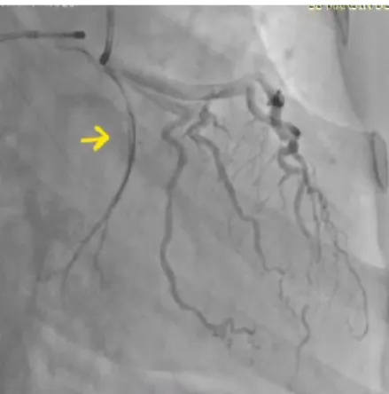
Figure 1. Coronary angiogram of left coronary artery in posterior-anterior cranial view showing a severe spasm of the distal left posterior-anterior
descending artery and obtuse marginal artery (arrow). Figure 3. The mid-portion of the circumflex artery was dilated using a 1.5x9 mm balloon.
Figure 4. After dilatation of the mid-portion of the circumflex artery with a balloon, the spasm was relieved (arrow).
Figure 2. Right anterior oblique caudal view showing an occurrence of no-reflow in the circumflex artery (arrows).
given, the ST segment depression on the anterior leads was resolved, and she was successfully extubated the second day. An implantable cardioverter defibrillator (ICD) was implanted due to ventricular fibrillation. Thereafter, she was discharged from the hospital with medical treatment, including, metoprolol (50 mg/d), isosorbide mononitrate ER (60 mg/d), atorvastatin (40 mg/d), aspirin (100 mg/d), and clopidogrel (75 mg/d). During a follow-up period of 30 months, the patient was without symptoms.
Discussion
The true incidence of vasospastic CAD has been unknown due to diagnostic challenges, but the incidence is higher in young populations, females, and Japanese people. The underlying pathophysiological mechanism of coronary artery spasm is not yet known. However, it is thought to be mainly because of endothelial dysfunction, which impairs vasodilatation. Cocaine abuse, smoking history, oxidative stress, inflammation, and genetic background may predispose one to vasospastic CAD.4 However, in this case report, the patient had no history of predisposing factors for vasospastic CAD.
Gök and Çınar
J Cardiovasc Thorac Res, 2019, 11(2), 164-166
166
made after observing significant stenosis in multiple and different locations compared to previous catheterization. Multisite and multi-vessel involvement without atherosclerotic lesions was a predictor of vasospastic CAD. During angiography, vasospastic CAD may be provoked by a catheter, guide wire insertion, balloon dilatation, and stent implantation or it may be a spontaneous phenomenon.5,6 In this case, the spontaneous induction of the CX spasm was predicted during diagnostic coronary angiography since the localization of the spasm was far away from the tip of the catheter. The localization of the spasm may be focal, multiple, or may include the entire coronary system.5 Patients with diffuse and multiple spasms are generally refractory to conventional medical therapy and have worse outcomes.7 Vasospastic CAD typically responds to coronary vasodilators; however, it can be refractory to medical therapy and may require balloon dilatation or stent implantation. In this case, the failure of intracoronary nitroglycerin treatment led us to opt for percutaneous coronary intervention.
The medical treatment of vasospastic CAD is calcium channel antagonists and nitrates because they reduce and resolve spasm attacks. The calcium channel antagonists suppress calcium ion (Ca2+) inflow into the vascular smooth muscle, and the nitrates metabolize to nitrite oxide, resulting in the relaxation of vascular smooth muscle and vasodilation. Because the patient had a low left ventricular ejection fraction, we could not give a calcium channel antagonists therapy. In the treatment of vasospastic CAD, there are still uncertainties about how to manage these patients and when to consider the use of ICD for primary prevention.4 When associated with multi-vessel involvement, vasospastic CAD has poor prognosis, as in this case.8 The previous studies strongly recommend ICD implantation in patients with vasospastic CAD for secondary prevention of sudden cardiac death, emphasizing the high recurrence risk of ventricular arrhythmias in those patients despite optimal medical treatment.9,10
Conclusion
Vasospastic CAD may have catastrophic results and it may be refractory to conventional medical therapy. These patients usually have poor prognosis and may require the consideration of ICD implantation.
Ethical approval
An informed consent was taken from the patient for publishing this case report.
Conflict of interest
All authors declare that they do not have conflict of interests.
Funding
The authors did not receive any specific funding for this work.
References
1. Lanza GA, Careri G, Crea F. Mechanisms of coronary artery spasm. Circulation. 2011;124(16):1774-82. doi: 10.1161/ CIRCULATIONAHA.111.037283.
2. Chuang YT, Ueng KC. Spontaneous and simultaneous multivessel coronary spasm causing multisite myocardial infarction, cardiogenic shock, atrioventricular block, and ventricular fibrillation. Circ J. 2009;73(10):1961-4. 3. Bromberg-Marin G, Mahmud E, Tsimikas S. Spontaneous
multivessel coronary vasospasm leading to cardiogenic shock. J Invasive Cardiol. 2007;19(4):E85-8.
4. Slavich M, Patel RS. Coronary artery spasm: Current knowledge and residual uncertainties. Int J Cardiol Heart
Vasc. 2016;10:47-53. doi: 10.1016/j.ijcha.2016.01.003.
5. Bell MR, Lapeyre AC, Bove AA. Angiographic demonstration of spontaneous diffuse three vessel coronary artery spasm. J Am Coll Cardiol. 1989;14(2):523-7. 6. 6, Wong A, Cheng A, Chan C, Lim YL. Cardiogenic Shock
Caused by Severe Coronary Artery Spasm Immediately after Coronary Stenting. Tex Heart Inst J. 2005;32(1):78-80.
7. Onaka H, Hirota Y, Shimada S, Kita Y, Sakai Y, Kawakami Y, et al. Clinical observation of spontaneous anginal attacks and multivessel spasm in variant angina pectoris with normal coronary arteries: evaluation by 24-hour 12-lead electrocardiography with computer analysis. J
Am Coll Cardiol. 1996; 27(1):38-44. doi:
10.1016/0735-1097(95)00423-8
8. Ahn JM, Lee KH, Yoo SY, Cho YR, Suh J, Shin ES, et al. Prognosis of Variant Angina Manifesting as Aborted Sudden Cardiac Death. J Am Coll Cardiol. 2016; 68(2):137-45.doi: 10.1016/j.jacc.2016.04.050.
9. Matsue Y, Suzuki M, Nishizaki M, Hojo R, Hashimoto Y, Sakurada H. Clinical implications of an implantable cardioverter-defibrillator in patients with vasospastic angina and lethal ventricular arrhythmia. J Am Coll Cardiol. 2012; 60(10):908-13. doi: 10.1016/j.jacc.2012.03.070.
10. Eschalier R, Souteyrand G, Jean F, Roux A, Combaret N, Saludas Y, et al. Should an implanted defibrillator be considered in patients with vasospastic angina?
Arch Cardiovasc Dis. 2014;107(1):42-7. doi: 10.1016/j.
acvd.2013.10.006.
Figure 5. An electrocardiogram obtained after post-resuscitation showing ST depression on the anterior leads.