Vol. 123, n. 3: 297-303, 2018
© 2018 Firenze University Press http://www.fupress.com/ijae
ITALIAN JOU R NAL OF ANATOMY AN D EM B RYOLOGY
DOI: 10.13128/IJAE-25423 * Corresponding author. E-mail: [email protected]
Research Article - Basic and Applied Anatomy
A dissection based study of the dimensions of the
thyroid cartilage in Anatolian population
Nujen Arya Sesen1, Alpen Ortug2,*, Gursel Ortug1
1 Bahcesehir University School of Medicine, Department of Anatomy, Istanbul, Turkey 2 Istanbul Medipol University School of Medicine, Department of Anatomy, Istanbul, Turkey
Abstract
Accurate knowledge of the dimension, shape, and morphology of larynx and its complex anat-omy is a prerequisite for performing surgery of the larynx. Population differences are important not only for planning surgical interventions but also for performing an operation. Previous mor-phometrical studies on the larynx had shown variable results obtained from different popula-tions, including Western and Eastern ones, yet information about larynx morphometry in Anato-lian population is limited. The purpose of this study is to make more statistical data available for Anatolian population and help clinicians for surgical approaches related to this area. Ten param-eters from anterior and lateral views were measured on 50 thyroid cartilages taken from autopsy specimens (28 male, 22 female). Specimens were removed and dissected under stereomicroscope after routine fixation procedure. Those with visible deformations were eliminated during dissec-tions. Statistical evaluations and comparisons were done for a comprehensive description. Sig-nificant sexual differences were observed. Mean values for all parameters were higher in males than in females. These results were compared with already existing anatomical data of other pop-ulations. This study on larynx specimens from different regions of Anatolia provides a general knowledge of thyroid cartilage dimensions. The obtained results can have great clinical influence on surgical approaches to the related area and give information for anthropomorphic studies. Key words
Morphometry, larynx, thyroid cartilage.
Introduction
Accurate knowledge of the dimension, shape, and morphology of the larynx and its complex anatomy is a prerequisite to perform surgical procedures for laryngeal operations (Kotian et al., 2014). Present anatomical and surgical textbooks cover the overall anatomy of the larynx and give important landmarks for operations but do not provide necessary morphometric information to perform a surgical procedure, especially considering the fact that population differences affect larynx dimensions to a great extent. Anatomical study of the larynx is needed not only to prevent com-plications but also to provide better surgical outcomes for the patients (Poornima and Dakshayini, 2017). Even though many studies concerning the dimensions of the human larynx and specifically thyroid cartilage are present in Western and Eastern populations, such as Europeans (Sprinzl et al., 1999; Zielinski, 2001; Kovac et al.,
is an official body of the Ministry of Justice, Turkey (Data proc. 2004/19 of January 19, 2004). Necessary permissions were taken and all dissections were performed in line with the Republic of Turkey laws. Fifty thyroid cartilages (28 males, 22 females) were used in total in this study after elimination of those with visible deformations during dissec-tions. The 28 males had ages ranging between 24 and 56 years with mean value 46 years and the 22 females were 32 years old on average, with a range 21-41 years.
Dissection method and measurements
Thyroid cartilages were removed from autopsy specimens with the rest of the larynx cartilages and then dissected under an S6 D stereomicroscope (Leica, Wetslar, Germany) after routine fixation procedure. Eight parameters from the anterior view and five parameters from lateral view were measured, as defined below in figures 1
Figure 1. Measurements performed on the anterior face of larynx. A1: Depth of superior thyroid notch from
ante-rior view. A2: Anteante-rior thyroid height. A3: Length of thyroid lamina. A4: Maximum height of lamina. A5: Distance between laryngeal prominence and base of superior horn. A6: Transverse distance between bases of inferior horn. A7: Anterior midvertical transverse distance. A8: Distance between the bases of superior and inferior horns.
and 2 and respective captions. A digital caliper (Altas 905, 150 mm), were used for measurements. Each parameter was measured double-blinded and in case of differ-ent values the average was recorded. Mean, minimum, maximum and standard devi-ation were calculated.
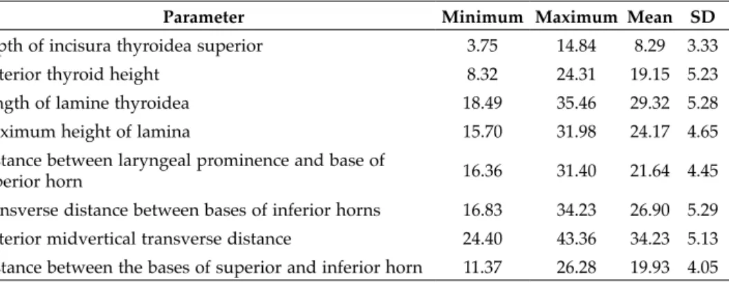
Results
In measurements from anterior view, the mean values of depth of superior thy-roid notch, length of thythy-roid lamina, maximum height of lamina, distance between laryngeal prominence and base of superior horn, transverse distance between bases of inferior horn, midvertical transverse distance, and distance between the bases of superior and inferior horns were greater for males than females; no significant dif-ference between males and females was found for anterior thyroid height. Tables 1 and 2 indicate the lowest, highest and mean values of these parameters for male and female specimens respectively.
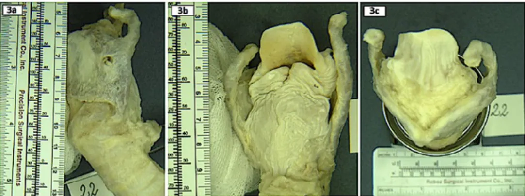
Also, at one of the thyroid cartilages, a remarkable variation of the left superior cornu was detected during dissections. This hook-like variation was only unilateral (Figure 3).
Table 3 indicates the lowest, highest and mean values of the measurements taken from the lateral view, i.e. depth of superior thyroid notch, thyroid height, length of
Figure 2. Measurements performed on the left (a) and right (b) side of larynx. L1: Depth of superior thyroid
notch from lateral view. L2 : Anterior thyroid height from lateral view. L3 : Length of thyroid lamina from lateral view. L4 : Maximum height of lamina from lateral view. L5 : Horizontal distance between the laryngeal prominence and posterior edge of the lamina.
thyroid lamina, maximum height of lamina, horizontal distance between the laryn-geal prominence and posterior edge of the lamina. These measurements were not dif-ferentiated by sex.
Discussion
In the present study, the depth of incisura thyroidea superior (A1) was found 8.29 ± 3.33 mm for male and 5.54 ± 0.31 mm for female. This parameter was observed as 9.7 ± 3.36 mm in Western Indian population disregarding sexual difference (Joshi et al., 2015), 10.47 ± 1.39 mm for males and 7.92 ± 1.09 mm for females in Thais popu-lation (Viravud and Palakornkul, 2014), 9.0 ± 1.9 mm for males and 6.4 ± 1.4 mm in European population (Sprinzl et al., 1999), 11.20 ± 2.75 mm for males and 9.70 ± 2.73 mm for females in Indian population (Jain and Dhall, 2008).
The anterior thyroid height (A2) was measured here as 19.15 ± 5.23 mm in male specimens and 21.57 ± 4.81 mm in females. Interestingly, this value was the only one that was higher in females than in males. Joshi et al. (2015) observed that in Western
Anterior midvertical transverse distance 24.40 43.36 34.23 5.13 Distance between the bases of superior and inferior horn 11.37 26.28 19.93 4.05
Table 2. Measurements from anterior view in females (N = 22; data in mm; SD: standard deviation).
Parameter Minimum Maximum Mean SD
Depth of incisura thyroidea superior 5.29 5.89 5.54 0.31
Anterior thyroid height 16.33 25.78 21.57 4.81
Length of lamina thyroidea 5.24 32.59 27.77 5.24
Maximum height of lamina 13.01 27.47 20.55 7.25
Distance between laryngeal prominence and base of
superior horn 18.11 23.78 20.14 3.16
Transverse distance between bases of inferior horns 19.94 28.61 25.45 4.80 Anterior midvertical transverse distance 29.77 36.65 34.08 3.75 Distance between the bases of superior and inferior horn 14.75 20.20 18.03 2.89
Indian population the average height of thyroid from anterior view was 16.71 ± 2.5 mm. Jain and Dhall (2008) found this value to be 16.40 ± 2.66 for males and 13.40 ± 3.20 for females in Indian population. In Thais, measures were 18.08 ± 1.42 mm for males and 16.20 ± 2.91 mm for females in the study of Viravud and Palakornkul (2014). This parameter was observed as 23.8 ± 3.9 mm for males and 15.0 ± 2.1 mm for females by Sprinzl et al. (1999).
The length of lamina thyroidea (A3) for Anatolian population was measured as 29.32 ± 5.28 mm in males and 27.77 ± 5.24 mm in females in the current study. These values are similar to those observed in other populations: 28.9 ± 2.6 mm for males and 21.9 ± 2.2 mm for females in European population (Sprinzl et al., 1999), 26.85 ± 1.99 mm for males and 22.70 ± 1.82 mm for females in Thais (Viravud and Palakornkul, 2014), 27.50 ± 2.96 mm and 22.70 ± 3.71 mm respectively for males and females in Indian population (Jain and Dhall, 2008).
The maximum height of lamina (A4) was found 24.17 ± 4.65 mm in males and 20.55 ± 7.25 mm in females in this study. In the study of Joshi et al. (2015) in Western Indian population, this parameter was measured on the right side and the left side separately and values were 26.73 ± 4.27 mm and 26.80 ± 3.05 mm respectively. Sprinzl
Figure 3. Unilateral hook-like variation of superior cornu of thyroid cartilage was observed on the left side.
a) Lateral view showing left cornu, b) Posterior view showing asymmetry, c) Superior view showing asym-metry
Table 3. Measurements from lateral view, in both sexes together (N = 50; data in mm; SD: standard
devia-tion).
Parameter Minimum Maximum Mean SD
Depth of superior thyroid notch from lateral view 6.67 10.78 9.17 1.52 Anterior thyroid height from lateral view 15.31 32.52 19.55 6.43 Length of lamina thyroidea from lateral view 23.42 42.87 29.75 6.82 Maximum height of lamina from lateral view 19.69 39.15 29.0 7.52 Horizontal distance between the laryngeal prominence
The midvertical transverse distance between bases of superior and inferior horn from anterior view (A7), the distance between the bases of inferior horn and superior horn (A8) was measured 19.93 ± 4.05 mm in males and 18.03 ± 2.89 mm in females..
As for the distances measured from lateral view, only L5, which is from the laryngeal prominence to posterior edge of lamina, was found stated in the literature as ‘breadth of lamina’. This measurement also has great variance both between genders and races. In our study, mean value was found 34.50 ± 4.94 mm regardless of sex. However, accord-ing to a previous study the smallest value for this measurement was found to be 29.60 ± 8.00 mm for males and 26.20 ± 4.90 mm for females (Indian) and the largest 41.90 ± 3.38 mm for males and 31.20 ± 2.73 mm for females (German; Jain and Dhall, 2008).
Unilateral variation of superior cornu of thyroid cartilage was only one among all dissected specimens. Some of the superior cornu variations are related to many symptoms such as dysphagia, odynophagia, throat pain or foreign body sensation (Mortensen et al., 2009). This abnormality is also called superior thyroid cornu syn-drome. It is stated that laryngologists should be aware of this variation (Wojtowicz et al., 2015).
Statistical evaluations and comparisons were done for a comprehensive descrip-tion. Significant sexual differences were observed. In accordance with previous stud-ies, mean values for all parameters were higher in males compared to females except for the anterior thyroid height for which the opposite holds true. These results were compared with already existing anatomical data of other populations. Comparisons have shown an overall accordance with the values measured in other populations, especially close values were detected in the study of Sprinzl et al., 1999 in European population. This result is comprehensible because Anatolian population is considered as White-Caucasian anthropologically smaller in body size than Western European populations and larger than Asian populations.
Conclusion
This study on larynx specimens from different regions of Anatolia provides infor-mation about thyroid cartilage dimensions. Apart from the significant sex differences, an exceptional variation of the left superior horn was observed in one of the thyroid cartilages. Measurements were compared with already existing anatomical data of other populations. Use of different measuring points, ethnological differences and artifacts due to formalin fixation may lead to different results in larynx morphometry studies. As mentioned by Kovac et al. (2010) population-related factors can explain
the minor differences between the results of this study and those of other researchers, which increase the importance to provide a detailed description of the dimensions of thyroid cartilage in each population. The present results can have great clinical influ-ence in surgical approach to the related area. Also, the results might give information for anthropomorphic studies.
Conflict of interest
This research did not receive any specific grant from funding agencies in the pub-lic, commercial, or not-for-profit sectors.
Acknowledgement
The larynx reproduced in figure 3 was also used - photographed from a different perspective - to show the presence of double foramen thyroideum in a human larynx.
References
Jain M., Dhall U. (2008) Morphometry of the thyroid and cricoid cartilages in adults. J. Anat. Soc. India 57: 119-112.
Joshi M.M., Joshi S.S., Joshi S.D. (2015) Morphometric study of thyroid cartilages in Western India. Int. J. Anat. Res. 3: 1028-1033.
Jotz G.P., Stefani, M.A., da Costa Filho, O.P., Malysz, T., Soster, P. R., Leao, H. Z. (2014) A morphometric study of the larynx. J. Voice 28: 668-672.
Kaur R., Singla, R.K., Laxmi, V. (2014) The morphology and morphometry of adult human thyroid cartilage - a cadaveric study in North Indian population. CIBTech. J. Surg. 3: 18-26.
Kotian, R.S., Nayak, V., Souza, A.S.D., Souza, A.D. (2014) Morphology of human lar-ynx: an anatomical study. Çukurova Med. J. 39: 779-783.
Kovac, T., Popovic, B., Marjanovic, K., Wertheimer, V., Kovacevic, M., Nikolic, V., Jo-Osvatic, A., Radic, R. (2010) Morphometric characteristics of thyroid cartilage in people of Eastern Croatia. Coll. Antropol. 34: 1069-1073.
Mortensen M., Ivey C. M., Iida M., Woo P. (2009) Superior thyroid cornu syndrome: an unusual cause of cervical dysphagia. Ann. Otol. Rhinol. Laryngol. 118: 833-838. Poornima G.C., Dakshayini K.R. (2017) A study of morphometry of adult human
lar-ynx and its importance in clinical applications. Int. J. Anat. Res. 5: 3713-3717. Sprinzl GM., Eckel HE., Sittel C., Pototschnig C., Koebke J. (1999) Moprhometric
measurements of the cartilaginous larynx: an anatomic correlate of laryngeal sur-gery. Head Neck 21: 743-750.
Viravud Y., Palakornkul V. (2014) A metrical study of the laryngeal skeleton in adult Thais. Siriraj Med. 66 (Suppl): S63-S66.
Wojtowicz P., Szafarowski T., Kukwa W., Migacz E., Krzeski A. (2015) Extended supe-rior cornu of thyroid cartilage causing dysphagia and throat pain. J. Med. Case, 6: 134-136.
Zielinski R. (2001) Morphometrical study on senile larynx. Folia Morphol. 60: 73-78.
View publication stats View publication stats


![["Ömer Seyfettin" adlı televizyon programı hakkında İstanbul TV Müdürü Vural Tekeli ile Taha Toros arasındaki yazışmalar]](data:image/gif;base64,R0lGODlhAQABAIAAAP///wAAACH5BAEAAAAALAAAAAABAAEAAAICRAEAOw==)