Türk Kardiyol Dern Arş - Arch Turk Soc Cardiol 2012;40(7):612-614 doi: 10.5543/tkda.2012.70750
Shone’s complex with dextrocardia and situs inversus totalis:
a case report
Dekstrokardi ve situs inversus totalisle birlikte görülen Shone kompleksi:
Olgu sunumu
Department of Cardiology, Ufuk University Faculty of Medicine, Ankara;
#Department of Cardiology, Türkiye Yüksek Ihtisas Education and Research Hospital, Ankara
Berkay Ekici, M.D., Aycan Fahri Erkan, M.D., Yeliz Sökmen, M.D.,# Omaç Tüfekçioğlu, M.D.#
Summary– Parachute mitral valve complex is an unusual congenital anomaly that has been described by Shone et al. It is characterized by a parachute deformity of the mitral valve associated with additional forms of left heart anoma-lies, such as aortic valvular stenosis and coarctation of the aorta. A 21-year-old female who was referred to our depart-ment because of progressive dyspnea on effort and at rest and minimal cyanosis is presented in this case report. On cardiac auscultation, the patient had a grade III/VI pansys-tolic murmur best heard at the lower left sternal border. The chest X-ray demonstrated dextrocardia and mild cardiomeg-aly. Echocardiographic evaluation revealed Shone’s com-plex, including parachute mitral valve anomaly.
Özet– Paraşüt mitral kapak kompleksi, Shone ve arkadaş-ları tarafından tanımlanan nadir bir doğumsal anomalidir. Mitral kapak paraşüt şeklinde biçim bozukluğuna uğramış olup, anomali aort kapak darlığı ve aort koarktasyonu gibi sol kalbe ait bazı doğumsal bozukluklarla birlikte bulunur. Bu olgu sunumunda, egzersiz ve istirahatte ilerleyici nefes darlığı gelişen ve minimal siyanozu olan 21 yaşında kadın hasta bildirildi. Kalbin oskültasyonunda, en iyi sternumun sol alt kenarında duyulan III/VI. derecede pansistolik üfü-rüm saptandı. Göğüs radyografisinde dekstrokardi ve hafif kardiyomegali görüldü. Ekokardiyografik değerlendirme, paraşüt mitral kapak anomalisini içeren Shone kompleksini gösterdi.
612
he finding of a parachute mitral valve (PMV) in an adult patient is extremely rare. Most patients are diagnosed in childhood with the pathologic com-plex known as Shone’s anomaly, which includes a PMV and aortic coarctation (68% of patients), atrial septal defect (54% of patients), ventricular septal de-fect (46% of patients), aortic valve stenosis (32% of patients), subaortic stenosis (20% of patients), and left ventricular hypoplasia (19% of patients).[1] Fewer
than 50 cases of Shone’s complex have been reported in the literature, and it has previously only been ob-served in children.[2]
CASE REPORT
A 21-year-old female patient was referred to our hospital for evaluation of dyspnea, a systolic
mur-mur, and minimal cyanosis. She experienced progres-sive dyspnea on effort and
at rest. Cyanosis had been diagnosed when she was 2 years old but her family did not seek medical at-tention for socio-economic reasons. Cyanosis was not apparent at rest. She had no history of smoking or significant pulmonary disease history before this diagnosis. On cardiac auscultation, the patient had a grade III/VI pansystolic murmur best heard at the lower left sternal border, with radiation to the right lower sternal border. The electrocardiogram revealed sinus rhythm, right ventricular hypertrophy, and sinus tachycardia with marked right axis deviation. A chest X-ray showed dextrocardia and mild cardiomegaly with increased pulmonary vascularity. Ventilation/
T
Received:February 20, 2012 Accepted: April 03, 2012
Correspondence: Dr. Berkay Ekici. Ufuk Üniversitesi Mevlana Bulvarı (Konya Yolu) No: 86-88, 06520 Balgat, Ankara, Turkey. Tel: +90 - 312 - 481 90 73 e-mail: [email protected]
© 2012 Turkish Society of Cardiology
Abbreviation:
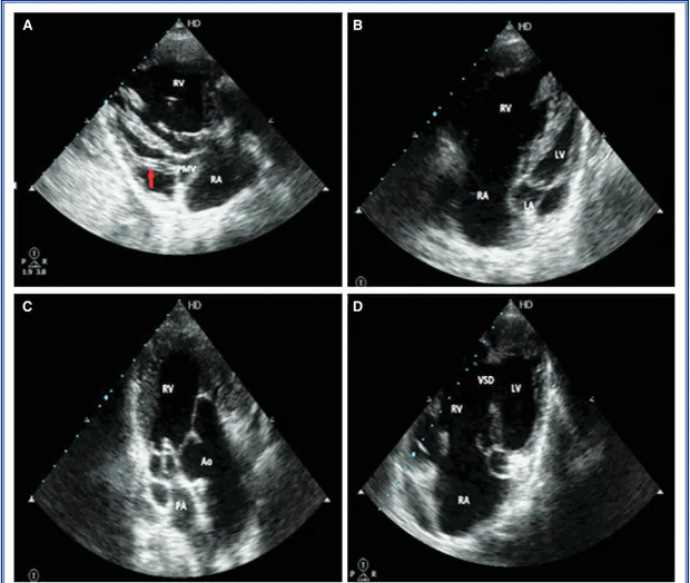
perfusion imaging and spiral computed tomography excluded pulmonary embolism. Transthoracic echo-cardiography revealed dextrocardia. Moreover, the morphological right atrium was on the left side of the heart, and the morphological left atrium was on the right side of the heart. Concordance atrioventricular connection, PMV, supramitral membrane, insertion of all the mitral valve’s chordae tendineae into a single papillary muscle and ventricular septal defect were observed. Additionally, moderate tricuspid valve in-sufficiency, rudimentary left ventricle, double outlet right ventricle, and mild pulmonary stenosis were de-tected. The aorta was in front of the pulmonary trunk (Fig. 1). Cardiac catheterization was offered but the patient refused this procedure. Abdominal ultrasono-graphic examination revealed that abdominal viscera were all located in the contralateral side of their
nor-mal location (situs inversus totalis). Enalapril was ad-ministered in order to decelerate negative remodeling. The patient comes for periodic follow-up visits twice a year. At the follow-up evaluation, the patient’s New York Heart Association class remains stable and no significant disease progression was observed.
DISCUSSION
In 1963, Shone et al.[3] originally described the
devel-opmental complex that included PMV, supravalvular ring of the left atrium, subaortic stenosis, and coarcta-tion of the aorta and reported that the degree of mitral valve involvement appeared to be the main factor de-termining outcome in these patients. This hypothesis has been confirmed in two reports of the long-term surgical outcome in patients with multiple left heart obstructions and mitral valve involvement.[4,5]
Shone’s complex with dextrocardia and situs inversus totalis 613
Figure 1. (A) Concordance atrioventricular connection, supramitral membrane, insertion of all the mitral valve’s chordae tendineae into a single papillary muscle (arrow) (B) rudimentary left ventricle, (C) double outlet right ventricle, mild pulmonary stenosis, (D) and ventricular septal defect were observed. PMV: Para-chute mitral valve; LA: Left atrium; RA: Right atrium; LV: Left ventricle; RV: Right ventricle; VSD: Ventricular septal defect; Ao: Aorta; PA: Pulmonary artery.
A
C
B
This condition is characterized by insertion of all the chordae tendineae into a single papillary mus-cle group. The chordae are generally shortened and thickened, and the anatomy of the papillary muscles is highly variable. The anterolateral, or rarely both papillary muscles, may be completely absent; or two identifiable but partially fused papillary muscles may be present.[6] Commonly associated conditions
in-clude isolated or combined supramitral ring, aortic subvalvular or valvular stenosis, coarctation of the aorta, or the complete ‘Shone complex’ of anoma-lies. Right ventricular outflow tract obstruction has also been described. Median longevity approaches 10 years of age, correlating best with left ventricular size and poorly with mitral valve orifice size.[7]
Patients with a PMV who reach adulthood usually present with dyspnea. PMV patients have a wide spec-trum of hemodynamic anomalies of transmitral flow.
[8-10] However, PMV may be incidentally diagnosed
during echocardiography with normal hemodynamics across the mitral valve.[11] Medical or surgical
treat-ment is not indicated in such patients. Patients with hemodynamically significant stenosis or regurgitation need to undergo mitral valve replacement or repair.[12]
Only hemodynamically significant and/or symptom-atic associated congenital cardiac lesions should be surgically corrected.
The most interesting aspect of this case is that the patient reached adulthood without receiving any medical therapy and was diagnosed in the third de-cade of her life. Clinicians must be alert for the other components of Shone’s complex when they encounter a mitral valve anatomy consistent with PMV.
Acknowledgments
There is no conflict-of-interest in this study. REFERENCES
1. Schaverien MV, Freedom RM, McCrindle BW. Independent factors associated with outcomes of parachute mitral valve in
84 patients. Circulation 2004;109:2309-13.
2. Prunier F, Furber AP, Laporte J, Geslin P. Discovery of a para-chute mitral valve complex (Shone’s anomaly) in an adult. Echocardiography 2001;18:179-82.
3. Shone JD, Sellers RD, Anderson RC, Adams P Jr, Lillehei CW, Edwards JE. The developmental complex of “parachute mitral valve,” supravalvular ring of left atrium, subaortic ste-nosis, and coarctation of aorta. Am J Cardiol 1963;11:714-25.
4. Bolling SF, Iannettoni MD, Dick M 2nd, Rosenthal A, Bove EL. Shone’s anomaly: operative results and late outcome. Ann Thorac Surg 1990;49:887-93.
5. Brauner RA, Laks H, Drinkwater DC Jr, Scholl F, McCaffery S. Multiple left heart obstructions (Shone’s anomaly) with mitral valve involvement: long-term surgical outcome. Ann Thorac Surg 1997;64:721-9.
6. Oosthoek PW, Wenink AC, Wisse LJ, Gittenberger-de Groot AC. Development of the papillary muscles of the mitral valve: morphogenetic background of parachute-like asymmetric mi-tral valves and other mimi-tral valve anomalies. J Thorac Cardio-vasc Surg 1998;116:36-46.
7. Ruckman RN, Van Praagh R. Anatomic types of congeni-tal mitral stenosis: report of 49 autopsy cases with consid-eration of diagnosis and surgical implications. Am J Cardiol 1978;42:592-601.
8. Glancy DL, Chang MY, Dorney ER, Roberts WC. Parachute mitral valve. Further observations and associated lesions. Am J Cardiol 1971;27:309-13.
9. Shapira OM, Connelly GP, Shemin RJ. Ischemic papillary muscle dysfunction in an adult with a parachute mitral valve. J Cardiovasc Surg (Torino) 1995;36:163-5.
10. Fitzsimons B, Koch CG. Parachute mitral valve. Anesth Analg 2005;101:1613-4.
11. Patsouras D, Korantzopoulos P, Kountouris E, Siogas K. Iso-lated parachute mitral valve as an incidental finding in an as-ymptomatic hypertensive adult. Clin Res Cardiol 2007;96:38-41.
12. Coles JG, Williams WG, Watanabe T, Duncan KF, Sherret H, Dasmahapatra HK, et al. Surgical experience with reparative techniques in patients with congenital mitral valvular anoma-lies. Circulation 1987;76:117-22.
Türk Kardiyol Dern Arş
614
Key words: Mitral valve; parachute mitral valve; Shone’s complex.
Anahtar sözcükler: Mitral kapak; paraşüt mitral kapak; Shone kompleksi.