1 Department of Radiology, Medical School, Dicle University, Diyarbakir, Turkey 2 Department of Radiology, Medical School, Yuzuncuyil University, Van, Turkey 3 Department of Radiology, Medical School, Baskent University, Adana, Turkey 4 Department of Radiology, Medical School, Akdeniz University, Antalya, Turkey
Yazışma Adresi /Correspondence: Cihad Hamidi,
Dicle University, Medical School, Department of Radiology, Yenişehir 21280 Diyarbakır Email: [email protected] Geliş Tarihi / Received: 01.04.2013, Kabul Tarihi / Accepted: 11.07.2013
Copyright © Dicle Tıp Dergisi 2013, Her hakkı saklıdır / All rights reserved
ORIGINAL ARTICLE / ÖZGÜN ARAŞTIRMA
The diagnostic efficacy of low-dose cervical and thoracic CT in multiple trauma
patients
Multipl travmada servikal ve torakal düşük doz spiral BT’nin tanısal etkinliği
Cemil Göya1, Alpaslan Yavuz2, Cihad Hamidi1, Çağatay Andiç3, Mehmet Guli Çetinçakmak1,Memik Teke1, Can Özkaynak4 ÖZET
Amaç: Amacımız çoklu travma hastalarının tanısında,
düşük doz bilgisayarlı tomografinin (BT) değerini araştır-maktı.
Yöntemler: Acil servise çoklu travma nedeniyle başvuran
74 hasta (30 kadın, 44 erkek) çalışmaya dahil edildi. Di-rekt röntgen ve düşük doz BT ile servikal bölge ve toraks bölge yaralanmaları değerlendirildi. Hastalar hastaneden taburcu edilene kadar, gidişatları, cerrahi bulguları, ek olarak radyolojik bulguları kaydedildi.
Bulgular: Radyografinin duyarlılığı ve özgüllüğü
sırasıy-la torakal patolojilerde, %50, %95; servikal travmasırasıy-larda ise %0-40 ve %95-100 idi. BT’nin duyarlılığı ve özgüllüğü sırasıyla 100 ve %80-100; torakal travmalarda %95-100 ve %96-%95-100 olarak bulundu.
Sonuç: Servikotorakal travmaların değerlendirilmesinde
düşük doz BT, daha duyarlıdır. Düşük doz BT ile hasta takibinin de değiştiği saptandı. Servikotorasik bölge trav-malarının değerlendirilmesi ve takibinde, başlangıç olarak düşük doz BT, radyografik yönteme tercih edilebilir.
Anahtar kelimeler: Düşük doz BT, röntgen, çoklu
trav-malar
ABSTRACT
Objective: The aim of this study was to evaluate the
ini-tial diagnostic efficacy of low-dose computed tomography (CT) in multi-trauma patients.
Methods: 74 patients (44 male, 30 female; average age:
36.9 years) accepted to the emergency unit with the rea-son of multiple trauma which is included in this study. Cer-vical and thoracic injury sites were initially evaluated with portable X-Ray and low-dose CT. The Patients’ progress, surgical findings and additional radiologic examination re-sults were recorded until the patients were discharged.
Results: The sensitivity and specificity of X-Ray graphies
were 50% and 95% for thoracic traumatic pathologies; 0-40% and 95-100% for cervical injuries respectively. CT examination sensitivity and specificity values were 95-100% and 80-95-100% for thoracic injuries; 95-95-100% and 96-100% for cervical injuries respectively.
Conclusion: Low-dose CT examination is more sensitive
than X-Ray graphs for the evaluation of cervicothoracic traumatic injuries. We recommend to prefer low-dose CT as an initial radiologic examination for managing cervico-thoracic trauma cases because of its higher diagnostic capability than X-Ray graphy.
Key words: Low-dose CT, X-Ray, multiple traumas
INTRODUCTION
Trauma is the primary cause of death in healthy young adults between 1 and 44 years of age [1,2]. The leading factor in reducing mortality and in-creasing the quality of life in the period of following the trauma in these patients is a rapid and precise
diagnosis. There are already numerous methods in use throughout the diagnostic process beginning with the clinical examination, continuing with the radiological examination and laboratory tests, and reaching towards invasive diagnostic methods when necessary. However, there is no algorithm to date
on which a consensus has been reached by every organization [3,4]. The most commonly known guideline on this subject is the Advanced Trauma Life Support (ATLS) Student Manual published by the American College of Surgeons [5].
Trauma patients have various features, which make a timely and precise diagnosis difficult. The clinical examination methods which lead to accu-rate diagnoses in conscious patients are rendered invalid in unconscious patients [6] Also, the fact that more than one system has been subjected to the trauma increases the number of the diagnostic methods required and the time spent for the diag-nosis. Problems that are more prominent may mask less obvious problems such as intra-abdominal pa-thologies, which are not recognizable at first sight but may turn out life threatening. All these reasons necessitate a more objective diagnostic method that may be used as a standard.
CT is already in use in emergency trauma pa-tients. There are already a number of studies about its application with or without contrast agents for the diagnosis and the prediction of the posttraumat-ic process in cervposttraumat-ical spinal trauma [7-9], thoracposttraumat-ic trauma [10-14], and abdominal trauma [15-18].
Low-intensity helical CT has first been used as a scanning method for the early diagnosis of lung cancers [19,20]. Having the advantages of CT like low radiation levels and short scanning time, this method has been found to be superior to postero-an-terior (PA) lung radiography as a scanning method [20].
In this study, the current approach to the neck and thorax comprising the clinical examination and radiographic methods described in the ATLS Stu-dent Manual [5] is compared with the low-dose CT scans used in the primary evaluation of emergency trauma patients.
METHODS
Patients who were presented to the emergency room due to multiple trauma injuries were clinically eval-uated and the obtained data were recorded. The re-corded clinical data of the patients included the age, sex, systolic and diastolic blood pressure, heart rate, respiratory rate and the Glasgow coma scale.
A total of 114 patients who were presented to the emergency department due to multi-trauma were
included in the evaluation. Among these patients, 40 were excluded due to inadequate CT or radio-graphic image quality or shortcomings in the clini-cal follow-up, leaving a total of 74 study patients. A total of 74 patients (44 males and 30 females) were enrolled in the study. The ages of the patients varied between 12 and 78 (mean age, 36.9 years).
For the initial radiographic assessment, lateral cervical and AP chest radiographs were obtained in the supine position using a portable X-ray device. In cases where the patient’s condition necessitated, posterior and oblique cervical, and antero-posterior thoracolumbar radiographs were later add-ed to the standard procadd-edure. However, the results of these radiographs are not included in the study, since our aim was to compare the CT images with the radiographs, which are considered standard for the patient under emergency conditions.
The parameters used in the Shimadzu portable X-ray device were 6.3 mAs and 64-72 kV for the cervical radiographic assessments and 10-20 mAs and 70-80 kV for the AP radiographs.
The CT images were taken with the Toshiba Xpress and Xvision (Toshiba Medical, Tokyo, Ja-pan) devices in all the patients included in the study.
The cervical low dose helical CT was assessed with a primary helical volume of 110 mm (120 kV, 50 mA), followed by a secondary helical volume of 60 mm (120 kV, 100mA). The mA value in Xpress was the lowest value authorized by the device. The first helical volume comprised the upper and mid-cervical region while the second cervical heli-cal volume was planned to include the area of the shoulder joint and the claviculae. Possible artefacts were tried to be prevented with higher mA values. For the cervical CT, a rate of 7 mm/rotation and 5 mm collimation were chosen and the cross-sections were reconstructed with 3 mm distance.
For the thoracic low-dose helical CT, the pa-rameters were selected as 120 kV and 50 mA. The thorax was scanned on a single helical volume at a rate of 20 mm/rotation and 10 mm collimation; and the cross sections were reconstructed with 8 mm distance. Thus, the pitch values were selected as 1.4 for cervical CT and 2 for thoracic CT. No oral or intravenous agents were used.
The time elapsed between the request of the ra-diographic examination and the start of the X-ray
procedure was 95 seconds. The total duration of the lateral cervical and AP chest radiographs was 60 seconds. The time until the patient arrived to the CT was 120 seconds and the duration of the CT scan was approximately 60 seconds including all 3 heli-cal volumes.
The X-rays and CT images were independently evaluated by two radiologists in case of differing interpretations, consensus was sought between the radiologists.
The clinical, CT and radiographic data of the patients were entered to the Statistical Package for Social Science (SPSS) (Version 10.0) software. In the neck area, patients were assessed under the sub-headings cervical injury, cervical vertebral fracture, cervical soft tissue injury and cervical vertebral dislocation. Under the thoracic trauma heading, as-sessments were made under the costal fracture, cla-vicular fracture, scapular fracture, sternal fracture, thoracic vertebral compression fracture, transverse process fracture, vertebral instability, pneumotho-rax, hemothopneumotho-rax, mediastinal injury (pneumome-diastinum, hemome(pneumome-diastinum, esophageal injury), pulmonary contusion and subcutaneous emphy-sema subheadings. Imaging was carried out using low dose helical CT and radiography, and the data obtained from these two methods were compared with the help of McNemar’s test.
RESULTS
According to the final clinical outcomes, the patho-logical findings detected in the cervical area were cervical vertebral fractures in 5 patients, soft tissue injuries of the neck in 8 patients, cervical disloca-tion in 5 patients and other injuries classified under the heading of cervical injury in 12 patients. The majority of these pathological findings could not be diagnosed using radiography (only 8% could be di-agnosed), while almost all of them were detected through CT (100%). Some pathological imaging findings were given in Figure 1-4. The findings are summarized in Table 1.
When evaluated according to the final clinical outcomes, the cervical radiological examinations had a 0-40% sensitivity and 95-100% specificity. The findings are summarized in Table 2.
When thoracic pathologies were assessed in general, the sensitivity of radiography was found
as 50% while its specificity was 95%. When these values are assessed in detail, the sensitivity values among the thoracic pathologies were found as 50% in costal fractures, 4% in hemothorax, 21% in pneu-mothorax, 38% in pulmonary contusions, and 17% in subcutaneous emphysema; while the sensitivity and specificity in scapular and sternal fractures and mediastinal injuries were inadequate. The speci-ficity of CT in these pathologies was 98%, 100%, 96%, 100%, 92% and 100%, respectively. The other details of the findings including the CT values are presented in Table 2.
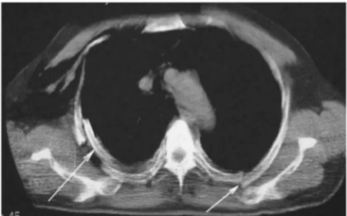
Table 1. Computerized tomography (CT) and X-Ray
radi-ography pathologic findings
CT Radio-X-Ray graphy Final Clini-cal Out-comes Cervical injury 12 1 12
Cervical vertebra fracture 5 2 5
Cervical soft tissue injury 5 0 5
Cervical dislocation 5 1 5 Thoracic trauma 33 17 34 Rib fracture 21 7 22 Claviculae fracture 9 5 9 Scapula fracture 5 0 5 Sternum fracture 2 0 2 Vertebra trauma 17 0 17 Burst fracture 5 0 5
Transvers process fracture 11 0 11
Vertebral instability 1 0 1 Pneumothorax 14 3 14 Hemothorax 23 1 23 Mediastinalinjury 4 0 4 Pulmonary contusion 21 8 21 Subcutaneous emphysema 11 4 11
We investigated the difference between radiog-raphy and CT in terms of the final clinical outcome according to the McNemar’s test and summarized the results we obtained in Table 2.
The p-values were calculated as p = 0.625 for cervical vertebral fractures, p = 0.219 for cervical dislocations, and as p = 0.006 for cervical injuries. In the comparison of the remaining cervical soft tissue injuries, no appropriate p-value could be ob-tained since no pathological findings were detected through radiography (Table 2).
In terms of the thoracic injuries, a value of p = 0.063 was observed in clavicular fractures, while this value was p = 0.125 in mediastinal injuries. Thus, both values were found to be statistically in-significant. On the other hand, a value of p = 0.001
Table 2. Sensitivity,
specificity and P val-ues of Computerized tomography (CT) and X-Ray Radiog-raphy to final clinical outcome
Pathologic Entity SensitivityX-Ray Radiography CT P Value
% Specificity% Sensitivity% Specificity%
Cervical injury 8 95 100 97 0.006
Cervical vertebra fracture 40 99 100 100 0.625
Cervical soft tissue injury 0 100 100 96
----Cervical dislocation 20 99 100 99 0.219 Thoracic trauma 50 95 97 80 0.001 Rib fracture 32 98 95 100 0.001 Clavicula fracture 50 100 100 98 0.063 Scapula fracture 0 100 100 100 ----Sternum fracture 0 100 100 100 ----Vertebra trauma 0 100 100 96 ----Burst fracture 0 100 100 100
----Transvers process fracture 0 100 100 97
----Vertebral instability 0 99 100 100 ----Pneumothorax 21 100 100 97 0.001 Hemothorax 4 96 100 90 0.001 Mediastinal injury 0 99 100 97 0.125 Pulmonary contusion 38 92 100 83 0.001 Subcutaneous emphysema 17 100 100 97 0.004
was calculated in costal fractures, pneumothorax, hemothorax and pulmonary contusions; while this value was p = 0.004 in subcutaneous emphysema. These p-values pointed to a statistical significance.
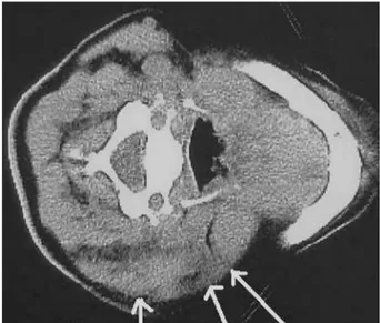
Figure 1. Left hemithorax sited minimal pneumothorax
(arrowhead), contusion (white arrow) and pleural effusion (black arrow) were detected by low-dose CT
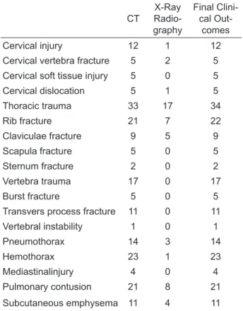
Figure 2. Soft tissue injury and edema at left side of the
Figure 3. Rib fracture detected by low-dose CT (black
arrow)
Figure 4. Fracture and subluxation at C2 vertebra (black
arrow)
DISCUSSION
In the assessment of the trauma patients, conven-tional CT is a great development in comparison to radiography. However, helical CT and Multi-Detec-tor Computed Tomography (MDCT) provide even greater advantages than conventional CT in patients with severe trauma. These advantages include the shorter duration of the scan and reconstruction, op-timal vascular enhancement, and less misregistra-tion- and motion artifacts. Also, the reformat and 3-dimensional reconstruction images are of a higher quality [21]. On the other hand, the low-dose CT is superior to the standard CT in terms of both the dose and the duration.
In spite of all the radiological developments, routine radiography is still the primary method used in multiple trauma patients in emergency rooms [22]. The approach described in the ATLS Student Manual published by the American College of Sur-geons5 also features lateral cervical radiograph, AP chest radiograph and AP pelvic radiograph.
However, some authors report that in the evalu-ation of the cervicothoracic region, radiography leads to a diagnostic result in only 50% to 70% of the cases. Some others report a failure rate of 23% to 57% in the diagnosis of cervical vertebral frac-tures [23]. Although no significant difference be-tween radiography and CT was observed in the cer-vical vertebral fractures, CT was still numerically superior in terms of the dislocations and soft tissue injuries in our study.
The diagnostic sensitivity of X-Ray exams for spinal trauma cases (especially for cervical trauma) were between 39% and 94% (with variable specific-ity values) in previously published studies in the lit-erature [24-30]. Pia et al. reported that; plain X-Ray graphy as an initial radiologic examination for clini-cally significant cervical injuries caused delays for an accurate diagnose in 5-23% of cases [31]. MDCT is the most effective method to evaluate the cervical bone injuries of blunt trauma patients today [21,31-37]. In our study; there was no statistical significant difference between the sensitivity and specificity of plain films and CT for determination of cervi-cal vertebra fracture, dislocation and cervicervi-cal soft tissue injuries (P values) but CT has the numerical superiority.
Among the spinal cord injuries, 85% occur at the time of the trauma, while 5-10% occurs im-mediately after the trauma [23]. Therefore, the ex-aminations must be carried out without moving the patient. However, this is practically impossible in radiography. On the other hand, the entire scan of the cervicothoracic region may be completed in a single position in the CT.
Also, although it is a known fact that although the cervicothoracic region is one of the most impor-tant areas in terms of the evaluation of the trauma, radiography may have shortcomings in that area due to the dose and superposition. Low-dose helical CT prevents superpositions and minimizes shoulder ar-tifacts.
CT is known to be more successful in the char-acterization of possible intrathoracic pathologies (e.g. injuries of the aorta, heart, pericardium, me-diastinum, diaphragm and lungs).38In our study, CT led to a statistically significantly difference in all the intrathoracic finding groups except for one. All of these are important in terms of the treatment (pneumothorax, hemothorax, pulmonary
contu-sions). The fact that no statistical difference was observed in the group of mediastinal injuries was a result of the limited number of the patients; and all the four patients were correctly diagnosed through CT, while this number was zero with radiography.
Conventional radiography is generally accept-ed the primary diagnostic process for chest trauma patients today 39-40 but other studies reported that CT is more effective as a beginning scanning method for management of emergency chest trauma cases [41-45]. In polytrauma patients, it is difficult to get direct radiographs and the results are non-diagnostic. The method has limitations originating from the superposition of the structures forming the thoracic wall, the dose and reasons related to the trauma patient (the position of the patient and the restricted mobility, etc.). The best examples of this are the vertebral, sternal and scapular fractures. Any pathological findings in these groups are reason to modify the treatment modality. In a retrospective study, it has been observed that 22% of the thoracic vertebral fractures were overlooked.38In our study, CT led to a statistically significant difference com-pared to radiography in costal fractures. Although there was no statistically significant difference in the other groups (Table 2), a clear difference is vis-ible in the number of the patients (Table 1). While CT could reveal all the cases in scapular, sternal and vertebral fractures, radiography could not show any of the conditions.
In this study, there was no significant difference between the time that elapsed until the radiography and the CT. Keeping in regard the diagnostic op-portunities provided by the CT, this point may rep-resent a superior aspect of the method.
Regarding from a financial point of view, radi-ography is doubtlessly cheaper than CT. However, the diagnostic superiority of CT may influence the treatment positively and may indirectly reduce the length of the hospital stay by preventing complica-tions. In addition, polytrauma patients are usually in an altered mental state and therefore undergo com-puted brain tomographies. Similarly, thoracic insta-bility or pelvic fractures are also indications for CT. Performing the cervicothoracic examination within the same CT session may be more time-efficient.
The limitation of this study was; our depart-ment has helical CT, which was used in this report
thus we could not have the chance to take technical advantages of most widely-used MDCT machines. Low-dose protocols can be easily applied to MDCT and similar studies can be work out with different parameters. In certain centers, CT scans performed on various systems in severe trauma patients are preferred to an examination starting with radiogra-phy. However, the number of the studies consider-ing cost-effectiveness is still inadequate [38].
In conclusion, in the traumas of the cervicotho-racic region, low-dose helical CT may be preferred to radiography due to the diagnostic superiorities as a starting method.
REFERENCES
1. Ruiz E. Initial Approach to the trauma patient In: Tintinalli JE, Ruiz E, Krome RL, eds. Emergency Medicine: A Com-prehensive Study Guide. 4th ed. New York, NY: McGraw Hill; 1127-1131:1996.
2. Centers for Disease Control and Prevention, National Center for Injury Prevention and Control. Web-based Injury Sta-tistics Query and Reporting System (WISQARS) [online] (2007) [cited 2011 Mar 4]. Available from URL: http:// www.cdc.gov/injury/wisqars.
3. Harris JH Jr. What is the minimum number of plain ra-diographs necessary to evaluate the cervical spine in pa-tients who have had trauma? Questions and answers. AJR 1994;163:217-218.
4. West OC, Anbari MM, Pilgram TK, Wilson AJ. Acute cer-vical spine trauma: Diagnostic performance of single-view versus three-single-view radiographic screening. Radiology 1997;204:819-823.
5. Alexander RH, Proctor HJ. Advanced Trauma Life Support (ATLS™) Course for Physicians. Chicago, Ill: American College of Surgeons, 1993.
6. Velmahos GC, Theodorou D, Tatevossian R, et al. Radio-graphic cervical evaluation in the alert asymptomatic blunt trauma victim: much ado about nothing? J Trauma 1996;40:768-774.
7. Pech P, Kilgore DP, Pojunas KW, Haughton VM. Cervical spine fractures: CT detection. Radiology 1985;157:117-120.
8. Blacksin MF, Lee HJ. Frequency and significance of frac-tures of the upper cervical spine detected by CT in patients with severe neck trauma. AJR 1995; 165:1201-1204. 9. Berne JD, Velmahos GC, El-Tawil Qalid, et al. Value of
complete cervical helical computed tomographic scanning in identifying cervical spine injury in the unevaluable blunt trauma patient with multiple injuries: a prospective study. The Journal of Trauma: Injury, Infection and Critical Care 1999; 47:896-903.
10. Gavant ML, Menke PG, Fabian T, et al. Blunt traumatic aortic rupture: detection with helical CT of the chest. Radi-ology 1995;197:125-133.
11. Worthy SA, Kang EY, Hartman TE, et al. Diaphragmatic rupture: CT findings in 11 patients. Radiology 1995; 194:885-88.
12. Unger JM, Schuchmann GG, Grossman JE, Pellett JR. Tears of the trachea and bronchi caused by blunt trauma: radiologic findings. AJR 1989;153:1175-1180.
13. Wagner RB, Crawford WO, Schimpf PP. Classifica-tion of the parenchymal injuries of the lung. Radiology 1988;167:77-82.
14. Kuhlman JE, Pozniak MA, Collins J, Knisely BL. Ra-diographic and CT findings of blunt chest trauma: aor-tic injuries and looking beyond them. Radiographics 1998;18:1085-1106.
15. Shuman WP. CT of blunt abdominal trauma in adults. Radi-ology 1997;205:297-306.
16. Becker CD, Gal I, Baer HU, Vock P. Blunt hepatic trauma in adults: correlation of CT injury. Grading with outcome. Radiology 1996;201:215-220.
17. Benya EC, Bulas DI, Eichelberger MR, Sivit CJ. Splenic injury from blunt abdominal trauma in children: follow-up evaluation with CT. Radiology 1995;195:685-688. 18. Novelline RA, Rhea JT, Bell Timothy. Helical CT of
ab-dominal trauma. Radiol Clin North Am 1999;37:591-612. 19. Kaneko M, Eguchi K, Ohmatsu H, et al. Peripheral lung
cancer: screening and detection with low-dose spiral CT versus radiography. Radiology 1996;201:798-802.
20. Henschke CI, McCauley DI, Yankelevitz DF, et al. Early Lung Cancer Action Project: overall design and findings from baseline screening. Lancet 1999;354:99-105.
21. Le Blang SD, MD, Nunez DB. Radiol Clin North Am 1999;37:515-532.
22. Fritz M. et al: Imagıng of chest trauma: radiological pat-terns of injury and diagnostic algorithms. Eur J Radiol 2003;48:61-70.
23. H. Imhof, M Fuchsjager. Traumatic injuries: Imaging of spi-nal injuries: Eur Radiol 2002;12:1262-1272.
24. Stiell IG, Wells GA, Vandemheen KL, et al. The Canadian C-spine rule for radiography in alert and stable trauma pa-tients. JAMA 2001;286:1841-1848.
25. Frankel HL, Rozycki GS, Ochsner MG, et al. Indications of obtaining surveillance thoracic and lumbar spine radio-graphs. J Trauma 1994;37:673-676.
26. Widder S, Doig C, Burrowes P, et al. Prospective evaluation of computed tomographic scanning for spinal clearance of obtunded trauma patients: preliminary results. J Trauma 2004;56:1179-1184.
27. Blackmore CC, Ramsey SD, Mann FA, et al. Cervical spine screening with CT in trauma patients: a cost effectiveness analysis. Radiology 1999;212:117-125.
28. Brandt MM, Wahl WL, Yeom K, et al. Computed tomo-graphic scanning reduces costs and time of complete spine evaluation. J Trauma 2004;56:1022-1028.
29. Tins BJ, Cessar-Pullicino VN. Imaging of acute cer-vical spine injuries: review and outlook. Clin Radiol 2004;59:865-880.
30. Blackmore CC, Mann FA, Wilson AJ. Helical CT in the pri-mary trauma evaluation of the cervical spine: an evidence based approach. Skeletal Radiol 2000;29:632-639. 31. Sundgren PC, Philipp M, Pavel VP, Maly P. Spinal Trauma.
Neuroimag Clin N Am 2007;17: 73-85.
32. Diaz JJ Jr, Gillman C, Morris JA Jr, et al. Are five view plain films of the cervical spine unreliable? A prospective evalu-ation in blunt trauma in patients with altered mental status. J Trauma 2003;55:658-663.
33. Griffen MM, Frykberg ER, Kerwin AJ, et al. Radiographic clearance of blunt cervical spine injury: plain radiograph or computed tomography scan? J Trauma 2003;55:222-226. 34. Holmes JF, Mirvis SE, Panacek EA, et al. For theNEXUS
Group. Variability in computed tomography and magnetic resonance imaging in patients with cervical spine injuries. J Trauma 2002;53:524-529.
35. Kligman M, Vasili C, Roffman M. The role ofcomputed to-mography in cervical spine injury due to diving. Arch Or-thop Trauma Surg 2001;121:139-1341.
36. Schenarts PJ, Diaz J, Kaiser C, et al. Prospective compari-son of admission computed tomographic scan and plain films of the upper cervical spine in trauma patients with altered mental status. J Trauma 2001;51:663-668.
37. Berne JD, Velmahos GC, El Tawil Q, et al. Value of com-plete cervical helical computed tomographic scanning in identifying cervical spine injury in the unavailable blunt trauma patient with multiple injuries: a prospective study. J Trauma 1999;47:896-902.
38. Luis A. Rivas, et al: Multislice CT in thorasic trauma:Radiol Clin Am 2003;41:599-616.
39. Wicky S, Wintermark M, Schnyder P, et al. Imaging of blunt chest trauma. Eur Radiol 2000;10:1524-1538.
40. Mirvis SE. Imaging of acute thoracic injury: the advent of MDCT screening. Seminars inUltrasound, CT & MR 2005;26:305-331.
41. Mirvis SE. Imaging of acute thoracic injury: the advent of MDCT screening. Seminars inUltrasound, CT & MR. 2005;26:305-331
42. Sroka NL, Combs J, Mood R, Henderson V. Scout an-teroposterior and lateral CT scans as ascreening test for thoracolumbar spine injury in blunt trauma. Am Surg 2007;73:780-743.
43. Magu S, Yadav A, Agarwal S. Computed Tomography in Blunt Chest Trauma. Indian J Chest Dis Allied Sci 2009;51:75-81.
44. Mirvis S. Diagnostic imaging of acute thoracic injury. Sem-inars in Ultrasound, CT & MR. 2004;25:156-179. 45. Omert L, Yeaney W, Protetch J. Efficacy of thoracic
com-puterized tomography in blunt chest trauma. Am Surg 2001;67:660-664.