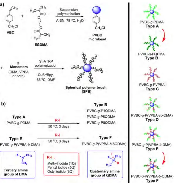
Synthesis and Antibacterial Activities of Boronic Acid-Based Recyclable
Spherical Polymer Brushes
Abstract: Crosslinked poly(4-vinylbenzyl chloride) (PVBC) microbead was prepared by suspension polymerization. Various spherical polymer brushes (SPBs) were pro-duced by grafting polymeric chains on their surfaces via surface initiated-atom trans-fer radical polymerization (SI-ATRP) using 4-vinylphenyl boronic acid (VPBA), 2-(dimethylamino)ethyl methacrylate (DMA), and quaternized DMA (QDMA). PVBC-g-PDMA, PVBC-g-PQDMA, PVBC-g-PVPBA, b-DMA), PVBC-g-P(VPBA-co-DMA) and PVBC-g-P(VPBA-b-QDMA) SPBs were characterized using nuclear mag-netic resonance spectroscopy, attenuated total reflection Fourier transform infrared spectroscopy, thermogravimetric analysis, and scanning electron microscopy. Anti-bacterial activities of the synthesized SPBs were investigated against Escherichia
coli and Staphylococcus aureus in nutrient and nutrient free media. Although PVBC-g-P(VPBA-b-DMA) SPB provided high antibacterial activity in the nutrient containing media due to its antibacterial, anti-biofilm and anti-QS properties, PVBC-g-P8QDMA SPB was found to be more effective in nutrient free media. Considering repeatable antibacterial activity, the PVBC-g-P(VPBA-b-8QDMA) SPB has advantageous over PVBC-g-P(VPBA-b-DMA) and PVBC-g-P8QDMA SPBs for long-term applications such as wastewater treatment in fluidized bad system. Keywords: antibacterial microbead, suspension polymerization, SI-ATRP, spherical polymer brushes, 4-vinylbenzyl chloride, 4-vinylphenyl
boronic acid, 2-(dimethylamino)ethyl methacrylate.
1. Introduction
Antibacterial solid surfaces are being used in hospital, dental equipments,1 water purification,2,3 food preservation,4 textiles,5
wood,6 paper,7, 8 shoes,9 and many other places to reduce the risks
of microbial infections. Killing (active) and prevention (passive) are main methods to inhibit bacterial population with antibac-terial surfaces.10 There is a direct interaction between the
anti-bacterial surface and bacteria in killing mechanism. On the other hand, prevention involves steric and/or electrostatic repulsion, low surface energy and anti-quorum sensing (anti-QS) proper-ties of surface that prevent the increase in bacteria population. Among the solid surfaces and drug delivery systems,11 polymer
beads (PBs) owns a special place in antibacterial applications. They have surface modification capabilities as they can have dif-ferent functionalities using surface initiated-atom transfer rad-ical polymerization (SI-ATRP) and surface-initiated reversible addition-fragmentation chain-transfer (SI-RAFT) polymeriza-tion, hydrolysis and condensation reactions and grafting tech-niques to obtain the antibacterial beads.3,5-7,12-15 Most popular
preferred techniques for surface polymerization are controlled living radical polymerizations such as ATRP. As an example, SI-ATRP has particular advantages. It requires less stringent experi-mental conditions and has great tolerance for various functional monomers. It also provides a strong bond between the substrate and polymer chains on the surface, well-controlled chain length and compositions with a great number of architectures. Its suc-cessful applications for the preparation of well-defined polymer brushes on different substrates, such as silicon, silica, gold, poly-mers have been well reported by various research groups.16,17
Micro-PBs are easily recoverable and reusable. Suspension polymerization has been used to produce antibacterial PBs of sized between 10 and 1000 μm size having various functional groups on their surfaces such as amine, carboxyl, hydroxyl, gly-cidyl and halogens.18-25
Polymeric form of 2-(dimethylamino)ethyl methacrylate (DMA) and its quaternized forms (QDMA) are well-known for their high antibacterial activities.2,26-28 There are several reports and reviews
published on the antibacterial effectiveness of the stated polymer and similar polymeric structures.15,29,30 Boron has great
physi-cal and chemiphysi-cal properties which offer an opportunity to dis-cover new pioneering drug disdis-covery areas. Boron therapeutics reveal different modes of inhibition against different biological targets.31 Antibacterial SPBs of nearly 250 µm diameter have been
produced by the quaternization of PDMA (PQDMA) brushes on
Hüseyin Cicek*,1
Gökhan Kocak2
Özgür Ceylan3
Vural Bütün4
1Department of Chemistry, Muğla Sitki Koçman University, Muğla, 48000, Turkey 2Department of Chemistry, Adiyaman University, Adiyaman, 02040, Turkey 3Food Quality Control and Analysis Program, Ula Ali Kocman Vocational School,
Muğla Sitki Koçman University, Muğla, Ula, 48147, Turkey
4Department of Chemistry, Eskisehir Osmangazi University, Eskisehir, 26480, Turkey
Received May 1, 2018 / Revised October 3, 2018 / Accepted November 19, 2018
Acknowledgments: The authors would like to express their thanks to the Scientific and Technological Research Council of Turkey (TUBITAK, grand no. 213M181) for financial support.
their surfaces using SI-ATRP tecniques.2 The polymer produced
from the 4-vinylphenyl boronic acid (VPBA), a boron containing monomer, has been used in various boronic acid affinity stud-ies.23,32-37 They are also prone to show antibiofilm properties as
they boronic acids show anti-QS activities.38,39 In the literature,
there are a great number of reports on the antibacterial activity of boron based compounds, but studies on the antibacterial activity of boron-containing polymers are very limited. In our previous study, we have reported antimicrobial and anti-quo-rum sensing activities of dyes containing new diazaborine and phenylboronic acid based copolymer.40,41 As far as we know
from literature, this study is a pioneering study that demon-strates the antibacterial effect of boron-based recycled poly-meric microbeads.
This study was aimed to obtain antibacterial spherical poly-mer brushes (SPBs) carrying both killing and preventive prop-erties. PVBC microbeads carrying DMA and VPBA copolymeric brushes were synthesized by SI-ATRP. DMA residues on blocks were quaternized to provide antibacterial activities to the particles. The synthesized PBs can be recycled as antibacterial inhibitors.
2. Experimental
2.1. Materials
4-Vinylbenzyl chloride (VBC, 90.0%, Milwaukee, WI, USA), VPBA (Aldrich), ethylene glycol dimethacrylate (EGDMA, Aldrich, 98.0%), 2-(dimethylamino)ethyl methacrylate (DMA, Aldrich, 98.0%), 2,2'-azobis(2-methylpropionitrile) (AIBN, 98.0%, SIAL, St. Louis, MO, USA), 2,2'-bipyridyl (Bpy, Alfa Aesar, 99.0%), CuBr (Aldrich, 99.9%), methyl iodide (SIAL, 99.0%), pentyl iodide (Aldrich, 98.0%), octyl iodide (Aldrich, 98.0%,), dimethylformamide (DMF, Merck, 99.8%), chloroform (CHCl3, SIAL, 99.0%), dichloromethane (CH2Cl2,
SIAL, 99.8%), tetrahydrofuran (THF, SIAL, 99.9%), ethanol (EtOH, SIAL, 99.8%), anhydrous diethyl ether (SIAL, 99.5%), poly(vinyl alcohol) (PVA, g mol-1, average M
w=85,000-124,000, 87.0-89.0%
hydrolyzed, Aldrich), methanol (MeOH, 99.0%, Merck, Darmstadt, Germany), and ethylenediaminetetraacetic acid (EDTA, SIAL, 98.5%) were bought from the stated commercial sources. All monomers were passed through a silica gel or basic alumina column prior to polymerization to remove the inhibitor.
2.2. Instruments and measurements
Scanning electron microscope (SEM) images of samples was cap-tured by Jeol JSM-7600F (Tokyo, Japan) to determine the anti-biofilm properties of bacteria, yeast and dyes that contained copolymers. Samples were Au/Pd coated to provide conductiv-ity. Approx. 100 particles were counted on SEM photographs to determine the number average diameter (Dn) with Eq. (1).
(1) where Ni is the number of particles of diameter Di (µm).
Thermogravimetric analysis (TGA) of all microbeads with increasing temperature were obtained by TGA 400 (Perkin Elmer, Waltham, MA, USA) with 10.0oC min-1 heating rate under N
2 (g)
atmosphere. Attenuate total reflection-Fourier transform infra-red (ATR-FTIR) measurements were carried out using Perkin Elmer Spectrum 100 spectrometer (Perkin Elmer, Waltham, MA, USA). The pictures of microbeads and spherical polymer brushes (SPBs) were determined with E100 optical microscope (Nikon, Tokyo, Japan). Boron-11 NMR experiments were conducted using a JEOL ECZ500R (JEOL Co., Tokyo, Japan) NMR spectrometer. 2.3. Preparation of PVBC microbeads and their derivat-ization with polymeric brushes
The crosslinked PVBC microbeads were prepared by suspen-sion polymerization (Figure 1(a)).42 VBC (8.0 mL, 51.1 mmol),
EGDMA (2.4 mL, 12.5 mmol), and AIBN (35.0 mg, 0.2 mmol) were dissolved in toluene (12.0 mL). The reaction mixture was purged with nitrogen (15 min) to remove oxygen and dispersed in an aqueous medium, prepared by dissolution of PVA (0.4 g) in water (130.0 mL). Polymerization was carried out in a mag-netically stirred (1000 rpm) 2-neck round bottom flask equipped with a reflux condenser and sealed with rubber septum (250.0 mL) at 78 °C for 24 h. The prepared PVBC microbeads were washed exhaustively (water 2 h, EtOH and THF) to remove the unwanted moieties and dried in vacuo at room temperature for two days. Six different SPBs were prepared using SI-ATRP technique2,3,6,26,28,42,43
by changing the sequence of the monomers added to the polym-erization environment (See Table S1 for reagents used).
To obtain PDMA on PVBC microbeads by SI-ATRP, PVBC microbeads (2.0 g) were first added into a magnetically stirring (400 rpm) reaction mixture in a glass flask (50.0 mL) containing DMA (8.0 mL, 46.5 mmol), Bpy (0.37 g, 0.23 mmol), CuBr (0.16 g, 0.11 mmol) and DMF (5.0 mL). Nitrogen was flushed for 20 min to remove the dissolved oxygen. The flask was sealed, polymer-ization was carried out at 65 °C for 48 h. After the reaction was terminated, the SPBs were washed several times with a 10% EDTA solution for 24 h to remove copper. Finally, all SPBs were subjected to exhaustive washing with THF, 0.01 M HCl, water and MeOH. PVBC-g-PDMA (Type A) SPB was obtained after drying in vacuo at room temperature for 48 h. PVBC-g-PVPBA (Type C) SPB was obtained similarly, except VPBA was used instead of DMA monomer (See Table S1 for reagents used).
A typical modified procedure for the synthesis of PVBC-g-P(VPBA-b-DMA) SPB (Type E) is as follows: PVBC microbeads (2.0 g) were added into a reaction mixture containing VPBA (0.2 g, 1.35 mmol), Bpy (0.37 g, 0.23 mmol), CuBr (0.16 g, 0.11 mmol) and DMF (5.0 mL). The reaction mixture was purged with nitrogen for 20 min to remove the dissolved oxygen. The flask was sealed and polymerization was carried out at 65 °C for 28 h while the mixture stirred at 400 rpm. DMA (8.0 mL, 46.5 mmol) was added to the reaction medium under nitrogen bridge, allowed to pro-ceed for further 24 h. PVBC-g-P(VPBA-b-DMA) SPB (Type E) was obtained after washing (same as the one used to wash homopoly-mer spherical brushes). SPB carrying random brushes (Type D) have been synthesized by reacting the two monomers simulta-neously.
The Type A and Type E SPBs containing PDMA were quater-nized with methyl iodide, pentyl iodide and octyl iodide (Figure 1(b)). It was desired to check the change in the antibacterial Dn=∑ DNi i/ N∑ i
activity of the beads having alkyls of various lengths. Quaterni-zation of the polymer brushes containing DMA (QDMA) was carried out by stirring these SPBs (2.0 g) with excess of alkyl iodides (5.0 mL) at 50 °C for 72 h under a nitrogen atmosphere in a 50.0 mL glass flask.7,27,44 After exhaustively washed with THF
and MeOH, the synthesized quaternized SPBs were dried in vacuo at room temperature for 72 h (See Table S2 for codes of quaternized spherical brushes).
2.4. Antibacterial activity
Antibacterial activity of the microbeads was performed according to the literature,2 in phosphate buffer saline (PBS); with and
without nutrient.
Two different antibacterial tests were performed with nutri-ent-containing PBS. First, the aim was to determine the amount of SPBs needed to completely inhibit 8.0×10⁵ colony forming
unit per milliliter (CFU/mL) initial bacteria population after 24 h incubation. For this, Escherichia coli ATCC 25922 (American type culture collection, Mananas, VA, USA) and Staphylococcus
aureus ATCC 25923 (American type culture collection, Mananas, VA, USA) were separately cultivated in 50.0 mL of a 3.1% yeast-dextrose broth. Dry SPBs were added into this inocula to get the final SPBs concentrations ranging from 8.0-32.0 mg/mL for E. coli and 1.0-4.0 mg/mL for S. aureus. Incubation temperature was kept constant at 30oC and 35oC for E. coli and S. aureus,
respec-tively. The SBP concentration, at which all bacteria in the medium were inhibited, was determined as the minimum inhibition concentration (MIC) of SBP.
Another target was to follow antibacterial reduction with time in nutrient containing media. E. coli or S. aureus was cultivated in 50.0 mL containing 3.1% yeast-dextrose broth with initial bacterial concentration of 4.0×10⁷ CFU/mL. 30 mg of SPBs were
added to 20.0 mL of the stated medium to obtain 1.5 mg/mL of final SPB concentration. The mixtures were incubated at 200 rpm and 37 °C. Absorbance of the medium was kinetically recorded on a UV-visible spectrophotometer at 600 nm to follow bacte-ria multiplication.
In antibacterial tests with nutrient-free PBS, E. coli was culti-vated in 50.0 mL of a 3.1% yeast-dextrose broth (containing
Figure 1. Synthetic pathway of microbeads and SPBs. a)
9
CH2 I+
,,.,_ CH2CI VBC 0+
Monomers (OMA, VPBA or both) S1-ATRP polymerization CuBr/Bpy, 65 °C, DMF Suspension polymerization8
CHiCI PVBC microbeadSpherical polymer brush
(SPB)
b)
Type A PVBC-g-PDMA Type E PVBC-g-P(VPBA-b-DMA) Tertiary amine group of DMA R-1 50 °C, 3 days R-1 50 °C, 3 days R-1 : Methyl iodide (10.) Perityl iodide (SQ} Octyl iodide (80) Type B. PVBC-g-P1 QDMA PVBC-g-P5QDMA PVBC-g-P8QDMA Type F PVBC-g-P(VPBA-b-8QDMA).
-x
+f H3 'N ' 'R H3C Quaternary amine group of CDMA PVBC-g-PDMA Type A~~
~.. ,t)
PVBC-g-PQDMA Type B*
PVBC-g-PVPBA TypeC IIPVBC-g-P(VPBA-co-DMA) Type D PVBC-g-P(VPBA-b-DMA) Type E
*
)
PVBC-g-P(VPBA-b-QDMA) Type F10.0 g/L peptone, 8.0 g/L beef extract, 5.0 g/L sodium chloride, 5.0 g/L glucose, and 3.0 g/L yeast extract of pH 6.8) at 37 °C. The E. coli cells were centrifuged at 5,000 rpm for 10 min. The pre-cipitated cells were washed twice with a sterile PBS and resus-pended in nutrient-free PBS to obtain an initial cell concentration of 1×10⁶ CFU/mL. 30 mg of SPBs were added into 20.0 mL of
the stated PBS suspension and incubated at 200 rpm and 37 °C. The viable E. coli cells were counted by using surface spread-plate method. In this method, first 1.0 mL of bacteria culture was taken at predetermined time intervals to prepare serial dilutions with PBS. 0.1 mL of the diluted sample from each serial dilution was then spread onto solid growth agar plate. The num-bers of viable colonies were counted manually; real number was calculated as 1×10⁶ CFU/mL by considering the dilution factor.
The same method used for E. coli was applied for S. aureus anti-bacterial activity determination. Two times tested microbeads were used in antibacterial experiments. The particles were sonicated three times (before the tests) in EtOH/THF (1:1, v/v) and allowed to stand for 10 min under UV. SPBs were imaged with SEM after the surfaces were coated with gold before and after purification.
3. Results and discussion
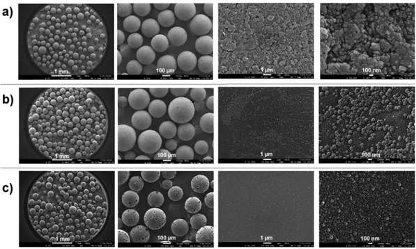
3.1. Characterization of PVBC microbeads and SPBs Using Eq. (1), SEM photographs provided the average size of PVBC microbeads (Figure 2(a)) as 241.9±51.8 µm. Figure 2 shows the modified surface of PVBC microbeads. The widest pore on PVBC microbeads surface (Figure 2(a)) is approximately 50 nm. The pores get closed and aggregates formed when the PVPBA brushes were added onto it (Figure 2(b)). The complete disap-pearance of the pores on the surface of the PVPBA brushes by the addition of the PDMA blocks can be considered a sign of successful surface modification. Pits were formed on the surface
of PVBC-g-P(VPBA-b-DMA) SPB (Figure 2(c)) due to the impact of the magnetic stirrer on the particle surface after long stirring. In spite of these pits, microbeads could be used in antibacterial tests because they did not lose their integrity.
The ATR-FTIR spectra of the PVBC microbeads, PVBC-g-PVPBA, PVBC-g-PDMA, and PVBC-g-P(VPBA-b-DMA) SPBs are presented in Figure 3. The absorption bands were observed at 668 (stretch C-Cl), 1728 (stretch C=O, ester), 1614 (stretch C=C, phenyl ring), and 2820-2925 cm-1 (stretch C-H). Stretching B-O band around
1350 cm-1 related to -B(OH)
2 supports the successful grafting of
VPBA on the PVBC.45,46 There is no significant difference between
other peaks (Figure 3(a) and (b)) as the structures of VBC and VPBA monomers are similar, as well as due to very low proportion of VPBA in the copolymer structure. Amine groups on PDMA provided a weak peak around 1600 cm-1 (stretch C-N) after
modifi-cation (Figure 3(c)). The sharpness of this peak decreased as the amount of PVPBA increased as block polymer before PDMA block on the PVBC microbeads (Figure 3(d)).
The presence of PVPBA segments at the particle surface can
Figure 2. SEM images of microbeads at different magnifications (from left to right 25×, 100×, 10000×, and 50000×); (a) PVBC microbead, (b) PVBC-g-PVPBA, and (c) PVBC-g-P(VPBA-b-DMA) SPBs.
Figure 3. ATR-FTIR spectra of; (a) PVBC microbeads, (b) PVBC-g-PVPBA, (c) PVBC-g-PDMA, and (d) PVBC-g-P(VPBA-b-DMA) SPBs.
3500 2000
2500 ·1)
be observed on naked eye by the reaction of phenyl boronic acid with 1,2-diols (Figure 4(f)). For this purpose, quercetin (as a model 1,2-diol) was interacted with PVBC-g-PVPBA SPB at pH 8.5.47
Digital photographs of the particles, before and after interaction with quercetin, are presented in Figure 4. The gray color of the PVBC beads (Figure 4(a)) did not change after the interaction with quercetin (Figure 4(c), left tube), but PVBC-g-PVPBA beads (Figure 4(b)) turned into yellow when interacted with querce-tin (Figure 4(d) and (e)). This yellow color indicates that quercequerce-tin has been captured by the boron atoms present on the surface of PVBC-g-PVPBA.
ATR-FTIR spectra of PVBC-g-PDMA and its quaternized forms with different alkyl lengths are given in Figure S1(a)-(c). Quat-ernization increased the intensity of C-H stretch peak around 2820-2925 cm-1 depends on the length of alkyl chain. FTIR
spec-tra of PVBC with random or block copolymeric brushes of PVPBA, PDMA and P8QDMA are presented in Figure S2. Although there is no significant difference between block and random polymeric forms of VPBA and DMA (Figure S2(b) and (c)), the effect of alkyl length is clearly visible (Figure S2(a)) in the form of band inten-sity of CH2 around 2820-2925 cm-1. The peak appeared at
3200-3600 cm-1 in case of SPBsis -OH stretching vibration of H 2O
(Figure S2(b) and (c)).
The broad signal in the range 0-40 ppm observed in 11B
solid-state NMR spectrum of PVBC-g-P(VPBA-co-DMA) SPB (Figure S3) shows the presence of boronic acid in the structure of SPB. In the 11B NMR spectrum of PVBC-g-P(VPBA-b-DMA) SPB, the PVPBA
block could not be observed in the structure because it remained in the interior.
The TGA curves of PVBC microbeads and its modified forms are presented in Figure 5. PVPBA arms on PVBC-g-PVPBA SPB decreased the thermal degradation temperature of PVBC microbe-ads.48-50 PVBC-g-PDMA SPB showed lower maximum rate of the
degradation temperature (the first step at Tmax~150oC) with three
steps. TGA results we obtained with the thermal decomposition
behavior of the PDMA homopolymer (the first step at ~167oC,
the second step at ~306 oC and the third step at ~403 oC) is quite
similar with the data given in the literature.7,51 On the other hand,
the PVPBA homopolymer is degraded in one step and the decompo-sition temperature (Tmax) is determined as 451oC in the previous
study.52 As reported in the literature, PVPBA is more thermally
resistant than PDMA.7,51,52 The thermal degradation of
PDMA-containing SPBs showed large mass decreases at ~300 and ~450oC, while the thermal decomposition temperature of PVBC
and PVBC-g-PVPBA microspheres was determined at higher temperatures (Tmax 430-450oC).
3.2. Antibacterial activity
Antibacterial activities of the modified PVBC microbeads in
nutri-Figure 4. Optical microscope photograph of PVBC microbeads; (a) before treatment with quercetin, (b) PVBC-g-PVPBA SPB before treatment with quercetin, (c) PVBC beads after treatment with quercetin, (d) PVBC-g-PVPBA SPB after treatment with quercetin, (e) PVBC-g-PVPBA SPB after treatment with quercetin (bar:1 mm =25 µm), and (f) complex formation of PVPBA-based SPBs and quercetin (1,2-diol) in aqueous solution.
Figure 5. TGA curves of PVBC, g-PVPBA, g-PDMA, and PVBC-g-P(VPBA-b-DMA) SPBs. VPBA Unit pH>8.5
-·
...··
100 90 80 70..
60 .c ti>~
50 ~ 0 40 30 20 10 Boronic acid-quercetin complex OHs-o
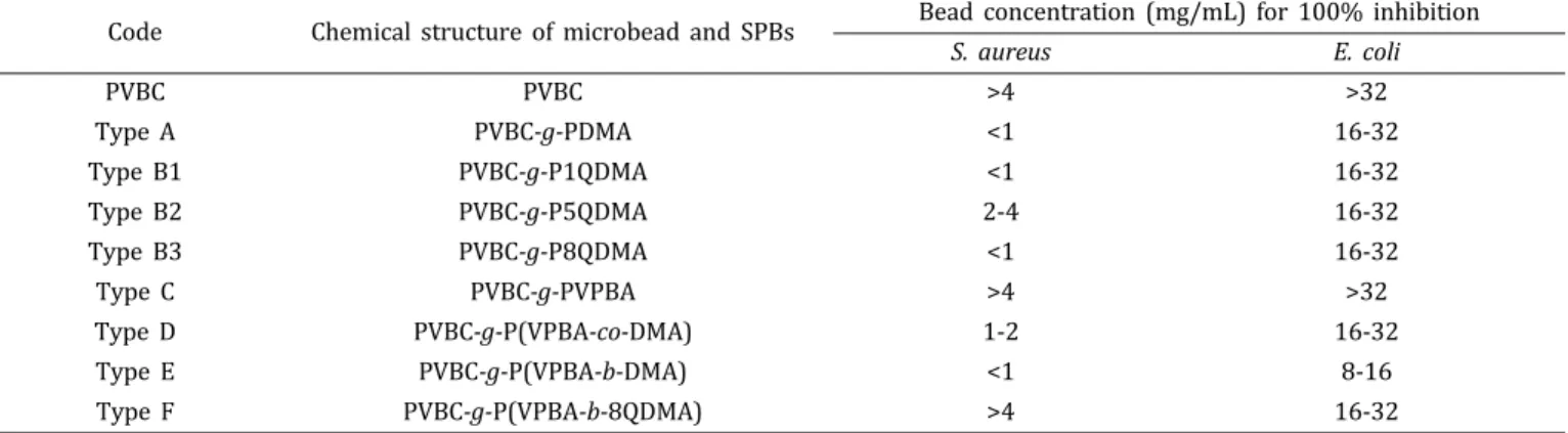
d OH PVBC-g-PVPBA SPB PVBC-g-P(VPBA-b-DMA) SPB 100 200 300 400 500 600 Temperature (0C)ent-containing PBS are presented in Table 1. Smaller particle concentration for 100% inhibition in nutrient-containing PBS means higher antibacterial activity. Particle concentration larger than 4.0 and 32.0 mg/mL for S. aureus and E. coli, respectively, were not tested since they are insignificant as reported earlier.2
Table 1 reflects that PVBC microbeads and PVBC-g-PVPBA SPB did not show considerable antibacterial activity as was expected. On the other hand, PVBC-g-PDMA, PVBC-g-P1QDMA, and PVBC-g-P8QDMA required lower SPB concentration for inhibit 100% of bacteria, which means they have higher anti-bacterial activities. Literature shows that when PDMA is quat-ernized with longer alkyl chains (8-12 carbons), it reflects higher antibacterial activity.2,26-29,53Although increase in alkyl length in
the quaternization of PVBC-g-PDMA SPB caused approximately similar antibacterial activity against both bacteria, PVBC-g-P5QDMA SPB presented less antibacterial activity against S. aureus with higher SPB concentration (4.0 mg/mL).
A suitable ratio of hydrophobicity/polarity and solubility are needed to obtain the highest antibacterial activity against S. aureus. Since polarity and solubility in aqueous solvents decreases with increasing alkyl group, negative effects may be more than posi-tive if the hydrophobicity of PVBC-g-P5QDMA SPB increased. Penetration of polymeric arms into the cell wall of the S. aureus with the suitable balance between those effects is needed to gain higher antibacterial activity. While the changes in alkyl group length of PVBC-g-P5QDMA SPB did not change inhibition per-formance of E. coli, this changes found to be effective on inhibi-tion S. aureus.
Antibacterial activities of PVBC microbeads carrying DMA and VPBA copolymeric brushes were varied in random and block configurations of the monomers in the brushes. PVBC-g-P(VPBA-b-DMA) SPB provided substantially high antibacterial activity against both bacteria as compared to PVBC and PVBC-g-PDMA SPB (Table 1). Random distribution of VPBA and DMA residues in PVBC-g-P(VPBA-co-DMA) SPB reduced antibacte-rial activity as compared to PVBC-g-PDMA SPB as VPBA residue in random copolymer is less active against bacteria as compared with DMA.41 On the other hand, PVBC-g-P(VPBA-b-DMA) has higher
antibacterial activity than PVBC-g-PDMA.
Detachment of inhibited bacteria from SPBs surface is also very important for sustainability of antibacterial activity. Since adhesion of inhibited bacteria cause covering of the active sur-face of SPBs and decreased the active antibacterial sursur-face, it is
not possible to sustain antibacterial activity with same inhibition rate for longer. The anti-biofilm and anti-QS properties of the PVPBA and PDMA blocks the PVBC-g-P(VPBA-b-DMA) SPBs, may cause higher antibacterial activity in nutrient containing PBS.41
As discussed, the higher anticipated antibacterial activity should be provided by PVBC-g-P(VPBA-b-8QDMA), which includes both quaternization and VPBA. However, it showed less activ-ity against S. aureus as compared to the PVBC-g-P8QDMA and PVBC-g-P(VPBA-b-DMA). This result may be due to two pro-posed effects: either, PVBC-g-P(VPBA-b-8QDMA) SPB does not have anti-QS, thus, bacteria can multiply in the presence of nutrient; or P8QDMA segments where bacteria interacts are blocked by PVPBA segments, which are anti-biofilm regions close to the particle surface.41
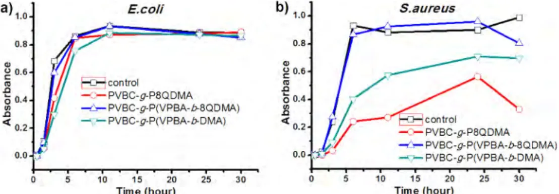
To further clarify the situation, additional antibacterial kinetic studies were performed in nutrient-free PBS with the selected particles (Figure 6) i.e. P(VPBA-b-DMA) while PVBC-g-P8QDMA SPBs were chosen for comparison and PVBC-g-P(VPBA-b-8QDMA) SPB to explain the unexpected weak antibacterial activity of S. aureus in nutrient containing PBS those are expected to exhibit high antibacterial activity.
As Figure 6(a) and (b) shows, bacterial growth reduction capability of particles were observed in the order of PVBC-g-P8QDMA>PVBC-g-P(VPBA-b-8QDMA)>PVBC-g-P(VPBA-b-DMA) for E. coli and S. aureus. PVBC-g-P8QDMA particles were able to kill all 1.0×10⁶ CFU/mL initial E. coli population in almost 5 min;
even faster than reported for the same moiety2 while other
parti-cles performed this task in 15 min.
For S. aureus, the time for 100% inhibition of 1×10⁶ CFU/mL
initial bacteria with PVBC-g-P8QDMA and PVBC-g-P(VPBA-b-8QDMA) SPBs are 5 and 15 min, respectively (Figure 6(b)). However, PVBC-g-P(VPBA-b-DMA) SPB was able to kill 99.0% initial bacteria concentration in 60 min. These results are inconsis-tent as compared to the ones obtained from the nutrient-con-taining PBS (Table 1). PVBC-g-P(VPBA-b-DMA) SPB showed better antibacterial activity in nutrient-containing PBS when compared to PVBC-g-P8QDMA and PVBC-g-P(VPBA-b-8QDMA) SPBs.
To solve this contradiction, antibacterial kinetic studies were performed under high initial bacteria population 4.0×10⁷ CFU/
mL in nutrient-containing PBS (Figure 7). The growth of E. coli in the nutrient-containing PBS slowed down more effectively by g-P(VPBA-b-DMA) SPB than g-P8QDMA and PVBC-g-P(VPBA-b-8QDMA) SPB (Figure 7(a)) probably due to the
anti-Table 1. Antibacterial activities of SPBs against S. aureus and E. coli
Code Chemical structure of microbead and SPBs Bead concentration (mg/mL) for 100% inhibition
S. aureus E. coli PVBC PVBC >4 >32 Type APVBC-g-PDMA<1 16-32 Type B1 PVBC-g-P1QDMA <1 16-32 Type B2 PVBC-g-P5QDMA 2-4 16-32 Type B3 PVBC-g-P8QDMA <1 16-32 Type C PVBC-g-PVPBA>4 >32 Type D PVBC-g-P(VPBA-co-DMA) 1-2 16-32 Type E PVBC-g-P(VPBA-b-DMA) <1 8-16 Type F PVBC-g-P(VPBA-b-8QDMA) >4 16-32
biofilm, anti-QS and antibacterial properties of its VPBA and DMA contents. Although DMA containing particles shows anti-QS capability, quaternized DMA-containing particles did not showed it.41 Owing to these properties, the PVBC-g-P8QDMA SPB can
partially stop the reproduction of the bacteria so that antibac-terial surface can be available after cleaning the dead bacteria from the surface.
On the other hand, inhibition effect of PVBC-g-P(VPBA-b-DMA) SPB that possesses less antibacterial and anti-QS activity, on S. aureus growth was found as weaker than PVBC-g-P8QDMA (Figure 7(b)). Additionally, Table 1 also shows that these two SPBs have the highest antibacterial effect in nutrient-containing PBS against S. aureus.
PVBC-g-P(VPBA-b-8QDMA) SPB (Figure 7) provided least antibacterial activities against both bacteria in the nutrient containing PBS, similar to the results obtained in the medium containing nutrients (Table 1). In the absence of nutrients, bet-ter antibacbet-terial results were obtained because of no bacbet-terial replication occurs (Figure 6). In this environment, quaternized nitrogens directly kills the bacteria, thus anti-QS property is not required. In this case, the high antibacterial activities of the P8QDMA segments in PVBC-g-P(VPBA-b-8QDMA) SPB is more pronounced as compared with the PVBC-g-P(VPBA-b-DMA) SPB that carries anti-QS properties.
Evaluation of these results suggests that if the nutrients are present in the medium, it will effectively prevent the bacterial reproduction with microbeads carrying biofilm and anti-QS properties. However, in nutrient-free environments, since anti-QS is insignificant, microbeads with only bactericidal groups
are more effective antibacterials.
3.3. Repeatability of antibacterial activity
The antibacterial activity of P(VPBA-co-DMA), PVBC-g-P(VPBA-co-8QDMA), and PVBC-g-P8QDMA slightly decreased after repeated uses. PVBC-g-P(VPBA-co-8QDMA), on the other hand, reflected less activity reduction as compared to others.
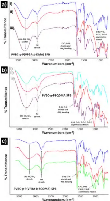
ATR-FTIR results of SPBs after 3rd and 10th consecutive use against both bacteria are given in Figure 8. Since the results of SPBs were almost similar after 3rd use against both bacteria, only
one spectrum is given in Figure 8(a-i, b-i, and c-i). The bands appeared at ~3300 (stretching; -NH2, >NH and -OH), ~2960
(stretching; C-H), ~1600 (bending; -NH2 and stretching; C=O, C=N),
~1400 (bending and stretch; aliphatic), and ~1100 (asymmet-ric stretching; C=O, P=O, P-O-C, P-O-P) confirms the adherence of E. coli and S. aureus on particle surface.54,55 Although there is
a substantial change in the intensity of these peaks for PVBC-g-P8QDMA SPB (Figure 8(b-ii and b-iii)), the change is very slight in case of PVBC-g-P(VPBA-b-DMA) SPB against E.coli (Figure 8(a-ii)) and negligible against both bacteria with PVBC-g-P(VPBA-b-8QDMA) SPB (Figure 8(c-ii and c-iii)) usage.
After 10th usage, the SEM photographs of
PVBC-g-P(VPBA-b-DMA) and PVBC-g-P8QDMA SPBs before and after interaction with E. coli and S. aureus were obtained and are given in Figure 9. It can be seen that S. aureus are embedded on the PVBC-g-P8QDMA SPB surface (Figure 9(c)), while few on the surface of PVBC-g-P(VPBA-b-DMA) SPB (Figure 9(a) and (b)). These images support the idea that PVPBA has a repulsive effect against the
Figure 7. Growth kinetics of E. coli and S.aureus in nutrient containing PBS in the present of the selected particles. Initial bacteria concentration is 4×10⁷ CFU/mL.
Figure 6. Antibacterial reduction capability of the selected particles for E. coli and S. aureus at initial bacteria population of 1.0×10⁶ CFU/mL.
a) 6 ~ Q) 5 .c E ~ 4 C: Q) 3 u Q) ::0 2 .!!! ~ Cl 1 0 ....I 0 0.8 Q) g 06
"'
.c 0 0.4 (/) .c <( 0.2 0 0.0 v 5 E.coli - 0 - PVBC-g-PSQDMA82:3
-
PVBC-g-P(VPBA-b-DMA) - 6 - PVBC-g-P(VPBA-b-8QDMA) 5 10Contacttime (min)
E.coli
f o ,control
~_.._ PVBC-g-P8Q OMA
-ic.- PVBC-g-P(VPBA-b-8QD IIIIA)
- PVBC-g-P(VPBA-b-DMA) 15 10 15 20 25 30 Time (hour)
-
;;; 5 .c5
4 C: ai 3 u Q) ::0 2 .!!!8'
....I 0 b) 1.0 0.8.,
g 0.6"'
.c 0 0.4 1l <( 0.2 0.0 0 0 S.aureus8B
PVBC-g-P8QDMA -:>- PVBC-g-P(VPBA-b-8QDMA) .../:,,-PVBC-g-P(VPBA-b-DMA) 10 20 30 40 50Contact time (min)
S.aureus
10 15 20 25 30
Time (hour)
bacteria. The surface image after purifying PVBC-g-P(VPBA-b-DMA) SPB (Figure 9(d)) after interaction with the E. coli is very close to the original surface (Figure 9(g)). However, the purified surface (Figure 9(e)) in case of S. aureus appears to have less pores than the original surface (Figure 9(g)). The results sup-port our interpretations that the PVPBA chains against S.aureus are less effective. For the PVBC-g-P8QDMA SPB, it is clear from the pores that the purified surface (Figure 9(f)) after interaction with S. aureus greatly differs from the original surface (Figure 9(h)). The results also supports the interpretations of ATR-FTIR spectra of PVBC-g-P(VPBA-b-8QDMA) SPB containing PVPBA as expressed in Figure 8(c-iii).
Although P(VPBA-b-DMA) and especially PVBC-g-P(VPBA-b-8QDMA) SPBs can be used repeatedly after purifica-tion with proposed method, PVBC-g-P8QDMA SPB may not be recycled for antibacterial activity that is against the reported findings.2,56 These results suggest that PVBC-g-P(VPBA-b-8QDMA)
SPB may be more suitable for long-term use and reproducible results, although they show somewhat lower antibacterial activ-ity as compared to PVBC-g-P8QDMA SPB.
4. Conclusions
The main objective of this study is to obtain polymeric brushes with antibacterial effect in nutrient and nutrient free medium. For this purpose, PVBC microbeads were synthesized in different constructs containing DMA and VPBA copolymeric structures. One or more repeated antibacterial activities of these SPBs were tested against E. coli and S. aureus bacteria in the indicated media. Effective antibacterial activity against both bacteria were obtained with PVBC-g-P(VPBA-b-DMA) SPB in the medium containing nutrient due to its antibacterial, anti-biofilm and anti-QS prop-erties. In addition, antibacterial kinetic studies of selected par-ticles were performed in nutrient free phosphate buffer and bacterial growth reduction capability of particles were observed in the order of PVBC-g-P8QDMA>PVBC-g-P(VPBA-b-8QDMA)> PVBC-g-P(VPBA-b-DMA) for E. coli and S. aureus. Growth kinetic studies of selected SPBs in phosphate buffer containing nutri-ent revealed that PVBC-g-P8QDMA had an inhibition effect on the growth against S.aureus in particular. In nutrients
contain-Figure 8. ATR-FTIR spectra of various microbeads after use in the antibacte-rial tests; (a) PVBC-g-P(VPBA-b-DMA) SPB, (b) PVBC-g-P8QDMA SPB, and (c) PVBC-g-P(VPBA-b-8QDMA) SPB ((i) E. coli and S.aureus after 3th, (ii) E. coli 10th and (iii) S.aureus 10th usage).
Figure 9. SEM images of particle surfaces after interactions with bacteria; (a) PVBC-g-P(VPBA-b-DMA) with E.coli, (b) PVBC-g-P(VPBA-b-DMA) with S. aureus, (c) PVBC-g-P8QDMA with S. aureus before purifying; cleaned forms are: (d) PVBC-g-P(VPBA-b-DMA), (e) PVBC-g-P(VPBA-b-DMA), (f) PVBC-g-P8QDMA; original surfaces: (g) PVBC-g-P(VPBA-co-DMA), (h) PVBC-g-P8QDMA.
CII
'
stretch C.=O, C=N sl.tel~h and -NHi bontlr~ -... PVBC-g-P(VPBA- • ..:_ 2000 b OMA) SSIP~8~--20(W~ll5C50)00-- l0010 L--:3;;-5;;;;001- 33000000 "Wave2500 numbers c ( m·') ~ i)LW
iii 01-l,NH,N~ Slti!'tCll C-0, C"'N ~ ,s.tr(Jlch~nd ~l-ll bending / --;3~501170 ,3,000 2000 2500 5 (cm·•) 1500 Wavenumber~
\fr
·
~ 7 (
-CH stretch P(VPBA-b-SQDMA) SPB PVBC-g- _ 2000 2soo (cm·') 3000 Wavenumbers 3500:i~~t
ric
stretch1000
ing medium, antibacterial, anti-QS and anti-biofilm properties gain importance. It was observed that the antibacterial effects of the SPBs obtained in the study continued with a decrease after reuse. The SPB having the best antibacterial effect was PVBC-g-P(VPBA-co-8QDMA). The continuity of antibacterial and anti-biofilm effects after re-use of SBPs and their interaction against bacteria have been successfully demonstrated by ATR-FTIR spec-tra and SEM images. As can be seen from the ATR-FTIR specspec-tra, SPBs continue their antibacterial effect even though they are decreased against both bacteria even after the 3rd and 10th use. PVBPA chains have been shown to exert a repellent effect against E. coli. It has also been found that PVBC-g-P(VPBA-b-8QDMA) SPB may be more suitable for long-term use and reproducibility. By considering all results, the PVBC-g-P(VPBA-b-8QDMA) SPB has advantageous over PVBC-g-P8QDMA SPB for long-term and repeated applications. PVBC-g-P(VPBA-b-8QDMA) SPB has good potentials for wastewater treatment in fluidized bed reactors due to their long-lasting surface properties.
Supporting Information: Information is available regarding the experimental procedure for the preparation of different type of SPBs and their ATR-FTIR spectra. The materials are available via the Internet at http://www.springer.com/13233.
References
(1) S. K. Singh, J. Anamika, S. Dipak, and D. Arti, Der Chemica Sinica, 2, 111 (2011).
(2) Z. P. Cheng, X. L. Zhu, Z. L. Shi, K. G. Neoh, and E. T. Kang, Ind. Eng. Chem. Res., 44, 7098 (2005).
(3) Z. P. Cheng, X. L. Zhu, Z. L. Shi, K. G. Neoh, and E. T. Kang, Surf. Rev. Lett., 13, 313 (2006).
(4) M. Z. Elsabee and E. S. Abdou, Mater. Sci. Eng. C-Mater., 33, 1819 (2013). (5) Y. Liu, Y. Liu, X. H. Ren, and T. S. Huang, Appl. Surf. Sci., 296, 231 (2014). (6) H. P. Yu, Y. C. Fu, G. Li, and Y. X. Liu, Holzforschung, 67, 455 (2013). (7) D. Roy, J. S. Knapp, J. T. Guthrie, and S. Perrier, Biomacromolecules, 9,
91 (2008).
(8) F. Tang, L. F. Zhang, Z. B. Zhang, Z. P. Cheng, and X. L. Zhu, J. Macromol. Sci. A, 46, 989 (2009).
(9) P. Limpiteeprakan and S. Babel, Environ. Monit. Assess, 188 (2016). (10) F. Siedenbiedel and J. C. Tiller, Polymers, 4, 46 (2012).
(11) L. L. Maharaj, M. M. Gupta, and A. K. Gadad, World J. Pharm. Pharm. Sci., 4, 126 (2015).
(12) S. Edmondson and S. P. Armes, Polym. Int., 58, 307 (2009). (13) V. Mittal, Polymers, 2, 40 (2010).
(14) H. Suzuki, M. Murou, H. Kitano, K. Ohno, and Y. Saruwatari, Colloids Surf. B: Biointerfaces, 84, 111 (2011).
(15) F. J. Xu, S. J. Yuan, S. O. Pehkonen, E. T. Kang, and K. G. Neoh, NanoBio-technology, 2, 123 (2006).
(16) A. Khabibullin, E. Mastan, K. Matyjaszewski, and S. P. Zhu, in Controlled Radical Polymerization at and from Solid Surfaces, P. Vana, Ed. 2016, Vol. 270, pp 29-76.
(17) C. J. Fristrup, K. Jankova, and S. Hvilsted, Soft Matter, 5, 4623 (2009). (18) B. W. Brooks, Chem. Eng. Technol., 33, 1737 (2010).
(19) M. Charnley, M. Textor, and C. Acikgoz, React. Funct. Polym., 71, 329 (2011).
(20) M. T. Gokmen and F. E. Du Prez, Prog. Polym. Sci., 37, 365 (2012). (21) F. X. Hu, K. G. Neoh, L. Cen, and E. T. Kang, Biotechnol. Bioeng., 89, 474
(2005).
(22) A. J. Kugel, S. M. Ebert, S. J. Stafslien, I. Hevus, A. Kohut, A. Voronov,
and B. J. Chisholm, React. Funct. Polym., 72, 69 (2012).
(23) S. Senel, H. Cicek, and A. Tuncel, J. Appl. Polym. Sci., 67, 1319 (1998). (24) S. Slomkowski and T. Basinska, Macromol. Symp., 295, 13 (2010). (25) R. Tomovska, J. C. de la Cal, and J. M. Asua, in Monitoring
Polymeriza-tion ReacPolymeriza-tions, John Wiley & Sons, 2013, pp 59-77.
(26) S. B. Lee, R. R. Koepsel, S. W. Morley, K. Matyjaszewski, Y. J. Sun, and A. J. Russell, Biomacromolecules, 5, 877 (2004).
(27) G. Q. Lu, D. C. Wu, and R. W. Fu, React. Funct. Polym., 67, 355 (2007). (28) H. Murata, R. R. Koepsel, K. Matyjaszewski, and A. J. Russell,
Biomater-ials, 28, 4870 (2007).
(29) L. Timofeeva and N. Kleshcheva, Appl. Microbiol. Biot., 89, 475 (2011). (30) F. Q. Zeng, Y. Q. Shen, S. P. Zhu, and R. Pelton, J. Polym. Sci. Pol. Chem.,
38, 3821 (2000).
(31) S. J. Baker, C. Z. Ding, T. Akama, Y.-K. Zhang, V. Hernandez, and Y. Xia, Future Medicinal Chemistry, 1, 1275 (2009).
(32) Y. J. Chang, X. Z. Liu, Q. Zhao, X. H. Yang, K. M. Wang, Q. Wang, M. Lin, and M. Yang, Chinese Chem. Lett., 26, 1203 (2015).
(33) B. Elmas, M. A. Onur, S. Senel, and A. Tuncel, Colloid Polym. Sci., 280, 1137 (2002).
(34) X. B. Li, J. Pennington, J. F. Stobaugh, and C. Schoneich, Anal. Biochem., 372, 227 (2008).
(35) Z. Lin, H. Huang, S. H. Li, J. Wang, X. Q. Tan, L. Zhang, and G. N. Chen, J. Chromatogr. A, 1271, 115 (2013).
(36) S. Senel, Colloid Surf. A, 219, 17 (2003).
(37) J. Zhang, Y. L. Ni, and X. L. Zheng, J. Sep. Sci., 38, 81 (2015).
(38) A. Adamczyk-Wozniak, O. Komarovska-Porokhnyavets, B. Misterk-iewicz, V. P. Novikov, and A. Sporzynski, Appl. Organomet. Chem., 26, 390 (2012).
(39) N. T. Ni, H. T. Chou, J. F. Wang, M. Y. Li, C. D. Lu, P. C. Tai, and B. H. Wang, Biochem. Biophys. Res. Commun., 369, 590 (2008).
(40) G. Kocak, H. Cicek, Ö. Ceylan, and V. Bütün, J. Appl. Polym. Sci., 136, 46907 (2019).
(41) H. Cicek, G. Kocak, O. Ceylan, E. A. Kutluca, Z. Dikmen, and V. Butun, J. Appl. Polym. Sci., 135, 46245 (2018).
(42) E. Yavuz, G. Bayramoglu, B. F. Senkal, and M. Y. Arica, J. Appl. Polym. Sci., 113, 2661 (2009).
(43) Y. Man, G. Peng, X. F. Lv, Y. L. Liang, Y. Wang, Y. Chen, and Y. L. Deng, Chromatographia, 78, 157 (2015).
(44) E. Yancheva, D. Paneva, V. Maximova, L. Mespouille, P. Dubois, N. Manolova, and I. Rashkov, Biomacromolecules, 8, 976 (2007).
(45) B. Suart, Infrared Spectroscopy: Fundamental and Applications, John Wiley & Sons, Ltd, 2004.
(46) G. Kahraman, O. Beskardes, Z. M. O. Rzaev, and E. Piskin, Polymer, 45, 5813 (2004).
(47) O. Cetinkaya, M. E. Duru, and H. Cicek, J. Chromatogr. B, 909, 51 (2012). (48) W. T. Lu, Z. G. Shao, G. Zhang, Y. Zhao, and B. L. Yi, J. Power Sources,
248, 905 (2014).
(49) J. M. Song, S. Y. Lee, H. S. Woo, J. Y. Sohn, and J. Shin, J. Polym. Sci. Pol. Phys., 52, 517 (2014).
(50) M. Karamitrou, E. Sarpaki, and G. Bokias, J. Appl. Polym. Sci., 133 (2016). (51) D. Roy, J. T. Guthrie, and S. Perrier, Soft Matter, 4, 145 (2008). (52) M. Wiacek, D. Wesolek, S. Rojewski, K. Bujnowicz, and E.
Schab-Bal-cerzak, Polym. Adv. Technol., 26, 49 (2015).
(53) A. King, S. Chakrabarty, W. Zhang, X. M. Zeng, D. E. Ohman, L. F. Wood, S. Abraham, R. Rao, and K. J. Wynne, Biomacromolecules, 15, 456 (2014). (54) Z. Filip, S. Hermann, and K. Demnerova, Czech J. Food Sci., 26, 458
(2008).
(55) L. D'Souza, P. Devi, T. Kamat, and C. G. Naik, Indian J. Mar. Sci., 38, 45 (2009).
(56) M. Ashfaq, S. Khan, and N. Verma, Biochem. Eng. J., 90, 79 (2014). Publisher’s Note Springer Nature remains neutral with regard to juris-dictional claims in published maps and institutional affiliations.