https://doi.org/10.1007/s00404-020-05855-1
GYNECOLOGIC ENDOCRINOLOGY AND REPRODUCTIVE MEDICINE
Oxytocin and cabergoline alleviate ovarian hyperstimulation
syndrome (OHSS) by suppressing vascular endothelial growth factor
(VEGF) in an experimental model
Ismet Hortu1,2 · Elif Karadadas3 · Gokay Ozceltik1 · Erol Tavmergen1,4 · Ege Nazan Tavmergen Goker1,4 · Gurkan Yigitturk5 · Oytun Erbas6
Received: 31 May 2020 / Accepted: 20 October 2020
© Springer-Verlag GmbH Germany, part of Springer Nature 2020
Abstract
Purpose Ovarian hyperstimulation syndrome (OHSS) is a life-threatening complication of ovarian stimulation in reproduc-tive medicine. Here, we aimed to investigate the role of oxytocin (OT) and cabergoline in the prevention and alleviation of the OHSS in an animal model.
Methods Thirty-five female immature Wistar rats were randomly assigned to five groups. The control group (n = 7) received saline only for five consecutive days. Remaining twenty-eight rats received 10 IU of pregnant mare serum gonadotropin (PMSG) followed by 30 IU of human chorionic gonadotropin (hCG) to induce OHSS. Group 2 (n = 7) was managed with no additional intervention after the induction of OHSS. Group 3 (n = 7) received 100 μg/kg cabergoline 2 h before the PMSG injection for four consecutive days and 2 h before the hCG injection on the fifth day. Group 4 (n = 7) and group 5 (n = 7) received 80 μg/kg and 160 μg/kg OT after induction of OHSS, respectively. Oxytocin was administered 2 h before the PMSG injection for four consecutive days and 2 h before the hCG injection on the fifth day. Body and ovary weight, vascular per-meability (VP), VEGF expression in the ovaries, and levels of VEGF in the peritoneal fluids were examined in all animals.
Results Cabergoline and OT reduced body weight, ovary weight, and VP compared to that of the OHSS group (p < 0.05). VEGF expressions in ovaries and peritoneal VEGF levels were decreased in cabergoline and OT groups compared to that of the OHSS groups (p < 0.001 for cabergoline and OT—80 μg/kg; p < 0.00001 for OT—160 μg/kg). However, there was no statistically significant difference in these parameters between the OT and cabergoline groups.
Conclusion Both OT and cabergoline were active in the alleviation of OHSS through suppression of VEGF and VP. Overall, we conclude that OT is effective for downregulation for VEGF and improvement in vascular permeability in OHSS.
Keywords Ovarian hyperstimulation syndrome · VEGF · Oxytocin · Cabergoline · Animal model
Introduction
Ovarian hyperstimulation syndrome (OHSS) is the most cru-cial and sometimes life-threatening iatrogenic complication of controlled ovarian hyperstimulation (COH) by exogenous gonadotropins for assisted reproductive techniques (ART) [1, 2]. It is characterized by cystic enlargement of the ova-ries and rapid transudation of protein-rich fluid from the vascular space into the peritoneal cavity. The excess fluid produces weight gain, abdominal distension, and intravas-cular depletion. If the OHSS is severe, this protein-rich fluid may also be found in pleural and pericardial cavities [3, 4]. The incidence of mild, moderate, and severe forms of OHSS is 3–10% of all ART cycles. However, it can reach 20% in high-risk women [4]. The pathophysiology of OHSS is not
* Ismet Hortu
1 Department of Obstetrics and Gynecology, Ege University
School of Medicine, Bornova, 35100 Izmir, Turkey
2 Department of Stem Cell, Ege University Institute of Health
Sciences, Bornova, 35100 Izmir, Turkey
3 Department of Biochemistry, Ege University School
of Medicine, Izmir, Turkey
4 Department of IVF Research Center, Ege University School
of Medicine, Izmir, Turkey
5 Department of Histology and Embryology, Mugla Sıtkı
Kocman University School of Medicine, Mugla, Turkey
6 Department of Physiology, Demiroglu Bilim University
clearly understood. The major pathophysiological mecha-nism underlying OHSS is an acute vascular permeability (VP) due to exposure to exogenous and/or endogenous human chorionic gonadotropin (hCG) resulting in a fluid shift from intravascular space to third space compartments. OHSS has a wide pathophysiological spectrum ranging from mild illness to severe disease. It is usually self-limited and resolves spontaneously within several days; however, it may persist longer in conception cycles [5]. Hypovolemia in the vascular system may cause declined organ perfusion, hydrothorax, ascites, electrolyte imbalance, hemoconcentra-tion, disseminated intravascular coagulation (DIC), and even renal and hepatic failure. In addition, venous thrombosis is a major cause of morbidity and mortality related to OHSS [6].
hCG-dependent increase in vascular endothelial growth factor (VEGF), the main regulator of vascular permeabil-ity (VP), is the cornerstone of the main pathophysiological processes in OHSS. Hence, targeting VEGF is one of the key points for the prevention of complications of OHSS and attenuating its clinical challenges. The high level of serum hCG induces mRNA expression of VEGF in the granulosa cells of the ovary. VP and capillary leakage develop after VEGF binds to its receptor (VEGFR2) on endothelial cells
[7]. The recruitment and development of a great number of antral follicles increase the production of VEGF during ART cycles. There is a notable link between the VEGF level and certain biological characteristics of capillary leakage and hemoconcentration [8]. Several other mediators, e.g., IL-2, IL-6, IL-8, histamine, elements of the ovarian renin–angio-tensin system, and prostaglandins, have been suggested to be involved in the pathophysiological process of the OHSS [9, 10].
Cabergoline, an ergot derivative molecule, is potent dopa-mine (DA) receptor agonist on D2 receptors. It has been used to prevent OHSS in women at high risk of OHSS undergoing ART treatments. Cabergoline reduces vascular permeabil-ity via inhibiting both VEGFR2 phosphorylation and VEGF
production [11]. In a Cochrane review, dopamine agonists were useful in the prevention of mild to severe OHSS, with-out reducing the live birth rates, clinical pregnancy, and abortion rates [12]. Currently, dopamine agonists are well-established agents that appear to be useful for the prevention of OHSS [13].
Oxytocin (OT) is a peptide hormone synthesized in the paraventricular nuclei of the hypothalamus and transported through the axons to the posterior pituitary. OT is released from the pituitary gland into the bloodstream to act as a neurotransmitter and a hormone on many organs. OT is the most important stimulant of the uterine contraction and is used primarily to induce or reinforce labor in obstetrics [14]. Another well-established action of OT is milk ejection dur-ing lactation. OT regulates the hypothalamic–pituitary–adre-nal axis in response to stress, luteal function, pregnancy, cell
proliferation, behavioral status, emotional regulation, cardio-vascular function, and neuropsychiatric disorders [15, 16]. OT is also produced by peripheral tissues, including skin, placenta, ovary, pancreas, heart, and blood vessels. OT acts through its specific receptors (OTRs) in peripheral organs. Furthermore, OT’s anti-inflammatory, anti-apoptotic, and antioxidant functions have been demonstrated previously [17–19]. The potential role of VEGF, a potent angiogenic mediator, has been shown in these studies on different patho-logical conditions such as endometriosis, cell injury, and oxidative stress. Also, recently, Ji et al. demonstrated that OT inhibited ovarian cancer metastasis by suppressing the expression of VEGF [20]. Although several studies impli-cate the role of OT in inflammatory, apoptotic, anti-oxidative pathways in various pathophysiological processes, there is a lack of data with regard to the possible amelio-rative effects of OT in OHSS which is a proinflammatory devastating process.
Many strategies and drugs have been described for the prevention and attenuation of OHSS complications [21,
22]. However, there is no exact method and/or drug to pre-vent OHSS entirely. To our knowledge, no study examined the OT for the prevention of OHSS to date. In this context, we investigated whether OT may be useful as an alterna-tive agent for the prevention and/or alleviation of OHSS by modulating VEGF. To investigate and compare the effective-ness of OT, we used animals treated with cabergoline, which is a widely known OHSS preventive drug.
Materials and methods
Animals
In this study, 35 immature female Sprague–Dawley albino rats were used. All rats were 22 days-old and weighed 45–50 g. Animals were maintained in pairs in steel cages within a temperature-controlled room (22 ± 2 ℃) and 12-h light/dark cycles. All animals were fed ad libitum. The study was approved by the Committee for Animal Research of Ege University. All animal studies strictly conform to the animal experiment guidelines of the Committee for Human Care.
Experimental design
Twenty-two-day-old female rats (weighing 45–50 g) were randomly divided into five groups: Group 1 (control, n = 7) received 0.1 ml of saline intraperitoneally (i.p.) on five con-secutive days (22–26); Group 2 (OHSS, n = 7) was given 10 IU of pregnant mare serum gonadotropin (PMSG) (Folligon®-Intervet; Schering-Plough Animal Health, Pune,
India) subcutaneously for four consecutive days and 30 IU hCG (Chorulon®-Intervet; Schering-Plough Animal Health,
Boxmeer, The Netherlands) on the fifth day to induce OHSS; Group 3 (OHSS and cabergoline; n = 7) was treated as the OHSS group, followed by oral gavage with cabergoline (100 µg/kg, Dostinex®-Pharmacia SpA, Ascoli Piceno, Italy)
dissolved in 1 ml of tap water. The drug was administered 2 h before the PMSG injection for four consecutive days and 2 h before the hCG injection on the fifth day. Group 4 (OHSS and oxytocin; n = 7) was treated as the OHSS group, followed by i.p. injection of OT (80 µg/kg, Pituisan®, Ege
Vet, Alfasan International BV, Holland) 2 h before the PMSG injection for four consecutive days and 2 h before the hCG injection on the fifth day. Group 5 (OHSS and oxy-tocin; n = 7) was treated as the OHSS group, followed by i.p. injection of OT 160 µg/kg 2 h before the PMSG injection for four consecutive days and 2 h before the hCG injection on the fifth day [23]. A previously published protocol was used for the OHSS model [22].
Permeability assay
All rats were anesthetized by i.p. injection of 80 mg/kg keta-mine hydrochloride (Alfaketa-mine®, Ege Vet, Alfasan
Interna-tional B.V., Woerden, Holland) and 4 mg/kg xylazine hydro-chloride (Alfazyme®, Ege Vet, Alfasan International B.V.,
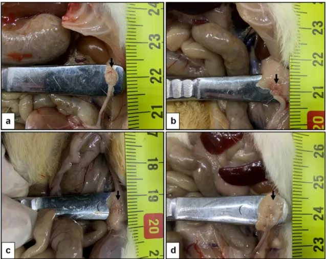
Woerden, Holland) 48 h after hCG administration to evalu-ate vascular permeability (VP) after weighing and number-ing. VP was measured as previously described [24]. Next, 0.2 ml of 5 mM Evans Blue (EB) (Sigma-Aldrich, St. Louis, MO) dye in distilled water was injected intravenously (i.v.) through an insulin injector via the femoral vein. The perito-neal cavity was filled with 5 ml saline solution (0.9% NaCl) at a 21 ℃ and was shaken for 30 s after a 30-min waiting period. Then, the peritoneal washing liquid was gently aspi-rated via a vascular catheter without damage to the tissues. Collected washing fluids were divided into two tubes with 0.05 ml of 0.1 N NaOH for VP evaluation. After centrifuga-tion (900 × g, 12 min), EB concentracentrifuga-tion was evaluated at 600 nm using the Multiskan GO Microplate Spectrophotom-eter (Thermo Fisher Scientific Inc., Waltham, MA, USA). The level of extravasated dye in the collected fluid was expressed as micrograms per 100 g body weight. Finally, all the animals were killed, and ovaries harvested bilaterally and weighed on precision scales. The macroscopic images of the ovaries in rats are shown in Fig. 1.
Histopathological evaluation of VEGF expression
Each ovarian tissue was immunohistochemically stained for VEGF expression within an isolated box with formal-dehyde. Ovaries were fixed in 10% buffered formaldehyde and embedded in paraffin blocks. Four-micron sections were cut from the blocks using a microtome and deparaffinized. Each section was incubated with H2O2 (10%) for 30 min
to remove endogenous peroxidase activity. Next, the sec-tions were blocked with 10% normal goat serum (Invitrogen, Camarillo, CA, USA) for 1 h at room temperature and incu-bated with primary antibodies against VEGF (Bioss Inc., Woburn, MA, USA; dilution 1/100) for 24 h at 4 ℃ and stained with the Histostain-Plus Bulk Kit anti-rabbit IgG (Bioss Inc., Woburn, MA, USA) and 3,3′-diaminobenzidine (DAB).
All sections were washed in PBS and photographed with an Olympus C-5050 digital camera connected to an Olympus BX51 microscope (Olympus, Hamburg, Germany). Brown cytoplasmic staining was scored positive for VEGF. The number of VEGF-positive cells was assessed systematically by scoring at least 100 granulosa and theca cells per 10 fields of tissue sections at × 100 magnification.
Biochemical evaluation
Peritoneal fluids were centrifuged (3000 rpm, 10 min at room temperature) and stored at − 20 ℃ for VEGF measure-ment. The level of cytokines was determined using enzyme-linked immunosorbent assay (ELISA) kits for VEGF (Inv-itrogen, Camarillo, CA, USA). Samples from each animal were run in duplicates. Assays were done as recommended by the manufacturer.
Statistical analysis
Statistical analysis of the data was performed with Graph-Pad Prism 8.1.1 (GraphGraph-Pad Software, La Jolla, CA, USA). Values were expressed as mean ± standard deviation (SD). Kolmogorov–Smirnov test was used to determine whether variables were normally distributed, and a one-way analysis of variance (ANOVA) was used. Bonferroni’s corrections were used for post hoc analysis. A value of p < 0.05 was considered statistically significant.
Results
Histopathological evaluation of ovaries for VEGF immunoexpression
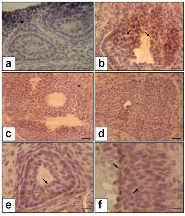
VEGF localization in the ovaries is shown in Fig. 2 for all groups. The immunohistological examination of ova-ries in the control group was found to be normal, with no staining of VEGF in granulosa cells in the antral follicles (Fig. 2a). Strong staining was observed in the granulosa cells in the OHSS group (Fig. 2b). A remarkable decrease in VEGF staining was observed in both cabergoline and OT (80 µg/kg and 160 µg/kg) groups (Fig. 2c–f). VEGF immunoexpression percent was found to be increased in the OHSS group compared to the control group (35.9 ± 6.4%
vs. 1.2 ± 0.3%; p < 0.0001). VEGF immunoexpression percent was significantly lower in both OT groups (80 µg/ kg and 160 µg/kg) than in the OHSS group (12.5 ± 2.03% vs 1.2 ± 0.3%, p < 0.001 for 80 µg/kg OT; 8.6 ± 1.5% vs 1.2 ± 0.3%, p < 0.00001 for 160 µg/kg OT). Additionally, VEGF immunoexpression percent was significantly lower in the cabergoline group compared to the OHSS group (10.2 ± 1.9% vs.1.2 ± 0.3%, p < 0.001). However, there was no statistically significant difference between cabergoline and oxytocin groups (p: 0.66).
Measurement of ovarian weight and body weight
Ovarian weight was notably higher in OHSS groups com-pared to the control group (49.2 ± 8.4 mg vs 21.15 ± 2.2 mg;
p < 0.005). Also, ovaries of rats treated with 160 µg/
kg and 80 µg/kg of OT had significantly lower weight than ovaries of the OHSS groups (29.4 ± 6.07 mg and 33.6 ± 5.2 mg vs 49.2 ± 8.4 mg; p < 0.001 and p < 0.01, respectively). Similarly, ovaries of cabergoline-treated rats
had significantly lower weight than ovaries of the OHSS groups (38.5 ± 3.1 mg vs49.2 ± 8.4 mg; p < 0.05).
There were significant alterations in body weight among all groups. OHSS caused significant weight gain com-pared to the control group (69.6 ± 7.2 g vs 46.6 ± 8.4 g;
p < 0.005). Body weight of rats treated with cabergoline
and OT (160 µg/kg and 80 µg/kg) was significantly lower than of the OHSS groups (49.05 ± 4.4 g and 55.9 ± 8.5 g for OT, 54.5 ± 4.09 g for cabergoline vs 69.6 ± 7.2 g; p < 0.05, respectively). However, the differences between cabergoline and OT groups were not statistically significant (p = 0.11).
The effects of hyperstimulation, cabergoline, and OT on the weight of ovaries and body and on VP in OHSS are presented in Table 1.
Biochemical analysis
Peritoneal VEGF levels were found to be significantly increased in the OHSS group compared to the control group (56.5 ± 8.2 pg/ml vs 4.1 ± 0.8 pg/ml; p < 0.0001). The level of VEGF in the peritoneal fluid was significantly
Fig. 1 Macroscopic images of the ovaries in different groups. a Control group, b OHSS group, c OHSS + cabergoline group, d OHSS + OT—160 µg/kg group; arrows indicate ovary
lower in both OT groups (80 µg/kg and 160 µg/kg) compared to the OHSS group (26.5 ± 4.4 pg/ml vs. 56.5 ± 8.2 pg/ml, p < 0.001 for 80 µg/kg OT; 18.5 ± 3.2 pg/ ml vs. 56.5 ± 8.2 pg/ml, p < 0.00001 for 160 µg/kg OT). Furthermore, VEGF levels in the peritoneal fluid were sig-nificantly decreased in the cabergoline group compared
to the OHSS group (25.06 ± 9.05 pg/ml vs 56.5 ± 8.2 pg/ ml; p < 0.001).
The VP was significantly increased in the OHSS group compared to the control group (34.5 ± 3.07 mM/100 g vs 15.06 ± 5.1 mM/100 g; p < 0.005). When high-dose OT (160 µg/kg) group was compared with the OHSS group,
Fig. 2 VEGF immunoexpression in the ovaries (the a–e figure is × 40 magnification, and f figure is × 100 magnification). a No staining in the control group, b increased expression of VEGF in granulosa cells in the OHSS group; arrow indicates increased staining of VEGF, c decreased VEGF expression in cabergoline group, d decreased VEGF
expression in the group of OT—80 µg/kg; arrow indicates declined staining of VEGF, e and f decreased VEGF expression in the group of OT—160 µg/kg; arrow indicates declined staining of VEGF. Scale bars indicate 125 µm for a, b, c, d, and e; 250 µm for f
a significant reduction was found for the high-dose OT group (16.9 ± 2.05 mM/100 g vs34.5 ± 3.07 mM/100 g;
p < 0.001). Furthermore, in rats treated with 80 µg/
kg OT we observed a remarkable decline in VP com-pared to the OHSS group (22.04 ± 6.3 mM/100 g vs 34.5 ± 3.07 mM/100 g; p < 0.01). VP also decreased significantly in the group of cabergoline com-pared to the OHSS group (24.3 ± 6.01 mM/100 g vs 34.5 ± 3.07 mM/100 g; p < 0.05). However, the VP and peritoneal VEGF levels were not significantly differ-ent between the OT groups and the cabergoline group (p = 0.18). The VEGF immunoexpression percent and the levels of peritoneal VEGF are shown in Table 2 for all groups. Also, comparisons of all biochemical and immu-nohistochemical parameters evaluated in this study are shown in Figs. 3 and 4 as well.
Discussion
OHSS is a prominent serious complication of COH. Besides its potential detrimental complications that affect the qual-ity of life, e.g., thromboembolic-related events, clinically many hemodynamic issues can occur. Prevention and prompt treatment of OHSS might be lifesaving. In the current study, we established a rat model of OHSS using a combination of PMSG and hCG. VEGF expressions in the ovaries and VEGF levels in the peritoneal fluid were significantly reduced in OT- and cabergoline-treated groups in compari-son with OHSS groups.
Furthermore, body and ovary weight and VP decreased strikingly in the OT- and cabergoline-treated groups com-pared to the OHSS group. Therefore, OT is a promising agent that could potentially prevent and alleviate the poten-tially harmful effects of OHSS. Known main risk factors of OHSS include a history of OHSS, low body mass index,
Table 1 Comparison of ovarian weight, body weight, and VP among groups
The experimental groups (n = 7) were analyzed for ovary weight, body weight, and vascular permeability. Values were expressed as mean ± standard deviation (SD). The groups were evaluated with Kolmogorov–Smirnov for normal distribution and analyzed with ANOVA test. Bonferroni correction was applied as the post hoc test
VP vascular permeability, OT oxytocin, OHSS ovarian hyperstimulation syndrome p value < 0.05 was considered as statistically significant
*p < 0.005, OHSS group compared to control group
**p < 0.001, OT of 160 µg/kg group compared to OHSS group
# p < 0.05, OHSS + cabergoline or OHSS + OT group compared to OHSS group
## p < 0.01, OHSS + cabergoline or OHSS + OT of 80 µg/kg group compared to OHSS group
Control
(n = 7) OHSS(n = 7) OHSS + Cabergoline(n = 7) OHSS + 80 µg/kg OT(n = 7) OHSS + 160 µg/kg OT(n = 7)
Ovary weight (mg) 21.15 ± 2.2 49.2 ± 8.4* 38.5 ± 3.1# 33.6 ± 5.2## 29.4 ± 6.07**
Body weight (g) 46.6 ± 8.4 69.6 ± 7.2* 54.5 ± 4.09# 55.9 ± 8.5# 49.05 ± 4.4#
Vascular permeability
(Evans Blue mM/100 g) 15.06 ± 5.1 34.5 ± 3.07* 24.3 ± 6.01
# 22.04 ± 6.3## 16.9 ± 2.05**
Table 2 Comparison of ovarian expression of VEGF and peritoneal VEGF levels among groups
The experimental groups (n = 7) were analyzed for percentage of VEGF immunoexpression and peritoneal VEGF. Values were expressed as mean ± standard deviation (SD). The groups were evaluated with Kolmogorov–Smirnov test for normal distribution. The analysis was performed with ANOVA followed by Bonferroni correction as post hoc test
VEGF vascular endothelial growth factor, OT oxytocin, OHSS ovarian hyperstimulation syndrome p value < 0.05 was considered as statistically significant
*p < 0.0001, OHSS group compared to control group
**p < 0.00001, OT of 160 µg/kg group compared to OHSS group
# p < 0.001, OHSS + cabergoline or OHSS + OT of 80 µg/kg group compared to OHSS group
Control
(n = 7) OHSS(n = 7) OHSS + Cabergoline(n = 7) OHSS + 80 µg/kg OT(n = 7) OHSS + 160 µg/kg OT(n = 7) VEGF immunoexpression
percent (%) 1.2 ± 0.3 35.9 ± 6.4* 10.2 ± 1.9
# 12.5 ± 2.03# 8.6 ± 1.5**
Fig. 3 a, b, c Graphical comparison of ovary weight, body weight, and VP in each group. The experimental groups for each parameter were analyzed with ANOVA, and Bonferroni correction was applied as the post hoc test. A p value < 0.05 was considered as statistically significant. *p < 0.005, OHSS group compared to control group.
**p < 0.001, OT of 160 µg/kg group compared to OHSS group.
#p < 0.05, OHSS + cabergoline or OHSS + OT group compared to
OHSS group. ##p < 0.01, OHSS + cabergoline or OHSS + OT group
compared to OHSS group. VP vascular permeability, OT oxytocin, OHSS ovarian hyperstimulation syndrome
Fig. 4 a, b Graphical comparison of VEGF expression and peritoneal VEGF levels in each group. The percent of VEGF immunoexpres-sion (%) and peritoneal VEGF was evaluated. Each parameter was evaluated by comparing with the control group and by comparisons between experimental groups via using ANOVA test. Bonferroni cor-rection was applied as the post hoc test. A p value < 0.05 was
consid-ered as statistically significant. *p < 0.0001, OHSS group compared to control group. **p < 0.00001, OT of 160 µg/kg group compared to OHSS group. #p < 0.001, OHSS + cabergoline or OHSS + OT of
80 µg/kg group compared to OHSS group. VEGF vascular endothe-lial growth factor, OT oxytocin, OHSS ovarian hyperstimulation syn-drome
young age, having polycystic ovaries, increased antral fol-licle count, or high anti-Müllerian hormone (AMH) levels, and high-dose hCG application [3]. The management of OHSS remains a subject of great importance not only for IVF providers but also for physicians who deal with affected cases in emergency departments. So far, to prevent OHSS in ART cycles, several strategies have been used in infertil-ity practice. These include cycle cancellation, withholding exogenous gonadotropins and delaying hCG application until the serum estradiol (E2) levels decrease to a secure
level (coasting), GnRH agonist usage to launch final oocyte maturation, albumin infusion during oocyte retrieval, in vitro oocyte maturation, cryopreservation of all embryos, and use of dopamine agonists, e.g., cabergoline after oocyte pickup [25, 26]. Despite these preventive methods, no clear mecha-nisms have fully explained how these factors contribute to OHSS. However, it is well established that vasoactive sub-stances released from ovaries into the circulatory system under hCG induction play the key role in this syndrome [27].
Dopamine agonists have long been used in reproductive endocrinology and infertility practice to reduce the OHSS manifestations without any maternal and/or fetal adverse effects in early periods of pregnancy [12]. In our study, we found that 100 µg/kg cabergoline 2 h before the gonadotro-pin injection for four consecutive days and 2 h before the hCG injection on the fifth day significantly reduced ovary weight, body weight, VP, VEGF expressions in the ovary, and VEGF levels in peritoneal fluid. A recent randomized clinical study by Tehraninejad et al. [28] demonstrated that for women at high risk for developing OHSS cabergoline administration before oocyte pickup was superior to intrave-nous human albumin in the prevention of OHSS. Previously, the effectiveness of dopamine agonists, e.g., cabergoline in the treatment of OHSS, has been attributed to its ability to decrease VP related to VEGF receptor 2 (VEGFR2) [29,
30]. Not only the treatment but also the OHSS prevention by cabergoline without compromising pregnancy has been emphasized [31]. We investigated the repression of OHSS by both cabergoline and OT in the experimental model and observed that our findings were consistent with other studies in the literature. Recently, many different agents have been tested as OHSS agents in animal models. Akman et al. [32] compared montelukast and cabergoline in the rat model of OHSS. They demonstrated a reduction in VP and VEGF expression in the cabergoline- and montelukast-treated rats in comparison with non-treated (only OHSS) controls. Also, they found that body weight, ovary weight, and peritoneal VEGF levels were significantly lower in the drug-treated groups than in the control (only OHSS) group. In another study, Sahin et al. [33] reported the effectiveness of both letrozole and cabergoline in the prevention of the OHSS through decreased VP and expression of VEGF in ovarian tissue of experimental animals. Unlike us, this team also
examined pigment epithelium-derived growth factor (PEDF) known as an antioxidant, anti-thrombogenic, and anti-angi-ogenic agent and an inhibitor of vascular permeability in blood vessels of animals. Levels of VP and PEDF were significantly lower in the letrozole and cabergoline group compared to the OHSS group.
In obstetrics, OT is a well-known and widely used drug. Besides, it is considered a safe drug for clinical use [34]. However, numerous potential effects of OT have been documented only in obstetrics. It is worth mentioning that besides its endocrine and paracrine activities, OT also has anti-angiogenic properties, alters immune responses, and suppresses VEGF, known as a powerful vasoactive mediator [15], which plays a crucial role in normal (e.g., embryogen-esis, healing of injured tissues) or pathological angiogenesis (e.g., carcinogenesis, metastasis of tumor) and inflamma-tion [35]. In addition, VEGF receptors are commonly found on endothelial cells where it is believed they are responsi-ble for the transition of capillary leakage in OHSS [36]. In this regard, we have designed this experimental study on the OHSS model. To the best of our knowledge, there is no study regarding OT’s possible effects on OHSS both clini-cal and experimental yet. We demonstrated that OT signifi-cantly reduced VEGF expression and VEGF levels in the peritoneal fluid after the induction of OHSS. In this context in our study, OT may have exerted its effects through the mechanisms as mentioned earlier. In the present study, VP and peritoneal VEGF levels were not significantly different between the OT groups and the cabergoline group. Hence, it is worth to express other properties and common side effects of both agents. The main adverse effects of oxytocin include hypersensitivity, anaphylactic reactions, flushing, cardiac arrhythmia, nausea, and vomiting. On the other hand, the main side effects of cabergoline include headache, dizzi-ness, weakness or lack of energy, nausea, and constipation. If the consumption is higher in cabergoline, some serious adverse events (shortness of breath, chest pain, cough, and heart valve problems) might occur [37, 38]. Indeed, this was an animal model study, and therefore, before results can be generalized to human beings, clinical studies are necessary to confirm the findings. Moreover, the long-term effects of OT on pregnancy and women need to be further studied.
The study has some limitations. First, we established an animal model to investigate the effects of both OT and cabergoline on OHSS; thus, our results may not be adapted immediately to human beings without large-scale, proven clinical studies. Second, we did not measure plasma estrogen levels of the rats. Further studies are required to support and elucidate the exact mechanisms underlying the protective role of OT both before and during the COH to support our results. To the best of our knowledge, this is the first study to investigate the potential effects of the OT in an experimen-tally induced OHSS model. As a consequence, OT appears
to be as beneficial as cabergoline both in treating and in alle-viating OHSS by modulating VEGF. The long-term effects of both OT and cabergoline, way of administration protocol, and appropriate drug dosage should be explored in further randomized clinical studies.
Author contributions IH contributed to conception and design, data extraction, and writing manuscript; EK and GO collected the data and performed interpretation and statistical analysis; GY done the his-tological analysis; ET and ENTG helped in supervising and critical revision; OE contributed to conception and design and performed the experiment.
Compliance with ethical standards
Conflict of interest All the authors declare that there is no conflict of interest.
Ethical approval The present study was approved by the Committee for Animal Research of Ege University, Izmir, Turkey. All procedures performed in studies involving animals were in accordance with the recommendations in the Guide for the Care and Use of Laboratory Animals of the National Institutes of Health.
Informed consent The informed consent is not applicable for the cur-rent study.
References
1. Petrenko AP, Castelo-Branco C, Marshalov DV, Salov IA, Shif-man EM (2019) Ovarian hyperstimulation syndrome. A new look at an old problem. Gynecol Endocrinol 35:651–656
2. Humaidan P, Nelson SM, Devroey P, Coddington CC, Schwartz LB, Gordon K et al (2016) Ovarian hyperstimulation syndrome: review and new classification criteria for reporting in clinical tri-als. Hum Reprod 31:1997–2004
3. Practice Committee of the American Society for Reproductive Medicine (2016) Prevention and treatment of moderate and severe ovarian hyperstimulation syndrome: a guideline. Fertil Steril 106:1634–1647
4. Zohav E, Almog B, Cohen A, Levin I, Deutsch V, Many A et al (2017) A New perspective on the risk of hypercoagulopathy in ovarian hyperstimulation syndrome using thromboelastography. Reprod Sci 24:1600–1606
5. Nastri CO, Teixeira DM, Moroni RM, Leitao VM, Martins WP (2015) Ovarian hyperstimulation syndrome: pathophysiology, staging, prediction and prevention. Ultrasound Obstet Gynecol 45:377–393
6. Taylor HS, Pal L, Seli E (2020) Speroff’s Clinical Gynecologic Endocrinology and Infertility. In: Hugh ST, Lubna P, Emre S (eds) Induction of Ovulation 9th ed. Wolters Kluwer, New York pp 2784-2888
7. Soares SR (2012) Etiology of OHSS and use of dopamine ago-nists. Fertil Steril 97:517–522
8. Gómez R, Ferrero H, Delgado-Rosas F, Gaytan M, Morales C, Zimmermenn RC et al (2011) Evidences for the existence of a low dopaminergic tone in polycystic ovarian syndrome: implications for OHSS development and treatment. J Clin Endocrinol Metab 96:2484–2492
9. Soares SR, Gómez R, Simón C, García-Velasco JA, Pellicer A (2008) Targeting the vascular endothelial growth factor system to prevent ovarian hyperstimulation syndrome. Hum Reprod Update 14:321–333
10. Nelson SM (2017) Prevention and management of ovarian hyper-stimulation syndrome. Thromb Res 151:S61–S64
11. Orvieto R, Dratvinman-Storobinsky O, Lantsberg D, Haas J, Mashiach R, Cohen Y (2014) Interleukin-2 and SOCS-1 proteins involvement in the pathophysiology of severe ovarian hyperstimu-lation syndrome– a preliminary proof of concept. J Ovarian Res 7:106
12. Tang H, Mourad S, Zhai SD, Hart RJ (2016) Dopamine agonists for preventing ovarian hyperstimulation syndrome. Cochrane Database Syst Rev 11:CD008605
13. Kasum M, Oreśković S, Franulić D, Čheić E, Lila A, Vujić G et al (2017) Current medical strategies in the prevention of ovarian hyperstimulation syndrome. Acta Clin Croat 56:133–142 14. Pergialiotis V, Frountzas M, Prodromidou A, Prapa S, Perrea DN,
Vlachos GD (2016) Propranolol and oxytocin versus oxytocin alone for induction and augmentation of labor: a meta-analysis of randomized trials. Arch Gynecol Obstet 293:721–729
15. Viero C, Shibuya I, Kitamura N, Verkhratsky A, Fujihara H, Katoh A et al (2010) REVIEW: oxytocin: crossing the bridge between basic science and pharmacotherapy. CNS NeurosciTher 16:e138–e156
16. Schaller F, Watrin F, Sturny R, Massacrier A, Szepetowski P, Muscatelli F (2010) A single postnatal injection of oxytocin res-cues the lethal feeding behaviour in mouse newborns deficient for the imprinted Magel2 gene. Hum Mol Gen 19:4895–4905 17. Ragy MM, Aziz NM (2017) Prevention of renal
ischemia/reper-fusion-induced renal and hepatic injury in adult male Albino rats by oxytocin: role of nitricoxide. J Basic Clin Physiol Pharmacol 28:615–621
18. Hortu I, Ozceltik G, Ergenoglu AM, Yigitturk G, Atasoy O, Erbas O (2020) Protective effect of oxytocin on a methotrexate-induced ovarian toxicity model. Arch Gynecol Obstet 301:1317–1324 19. Hortu I, Ozceltik G, Karadadas E, Erbas O, Yigitturk G, Ulukus
M (2020) The role of ankaferd blood stopper and oxytocin as potential therapeutic agents in endometriosis: a rat model. Curr Med Sci 40:556–562
20. Ji H, Liu N, Yin Y, Wang X, Chen X, Li J et al (2018) Oxyto-cininhibitsovariancancermetastasisbyrepressingtheexpression of MMP-2 and VEGF. J Cancer 9:1379–1384
21. Kasap E, Turan GA, Eskicioğlu F, Cengiz H, Gur EB, Sivrikoz ON et al (2016) Comparison between resveratrol and cabergoline in preventing ovarian hyper stimulation syndrome in a rat model. Gynecol Endocrinol 32:634–640
22. Saylan A, Arioz DT, Koken T, Dilek H, Saylan F, Yilmazer M (2010) Prevention of ovarian hyperstimulation syndrome in a rat model: efficacy comparison between cabergoline and meloxicam. Acta Obstet Gynecol Scand 89:692–699
23. Petersson M, Lundeberg T, Sohlstrom A, Wiberg U, UvnasMoberg K (1998) Oxytocin increases the survival of musculocutaneous flaps. Naunyn Schmiedebergs Arch Pharmacol 357:701–704 24. Ferrero H, Garcia-Pascual CM, Gaytán M, Morales C, Simón C,
Gaytán F et al (2014) Dopamine receptor 2 activation inhibits ovarian vascular endothelial growth factor secretion in an ovarian hyperstimulation syndrome (OHSS) animal model: implications for treatment of OHSS with dopamine receptor 2 agonists. Fertil Steril 102:1468–1476
25. D’Angelo A, Amso NN, Hassan R (2017) Coasting (withholding gonadotrophins) for preventing ovarian hyperstimulation syn-drome. Cochrane Database Syst Rev 5:CD002811
26. El Tokhy O, Kopeika J, El-Toukhy T (2016) An update on the prevention of ovarian hyperstimulation syndrome. Womens Health (Lond) 12:496–503
27. Guo JL, Zhang DD, Zhao Y, Zhang D, Zhang XM, Zhou CQ et al (2016) Pharmacologic interventions in preventing ovarian hyperstimulation syndrome: a systematic review and network meta-analysis. Sci Rep 6:19093
28. Tehraninejad ES, Hafezi M, Arabipoor A, Aziminekoo E, Cheh-razi M, Bahmanabadi A (2012) Comparison of cabergoline and intravenous albumin in the prevention of ovarian hyperstimula-tion syndrome: a randomized clinical trial. J Assist Reprod Gen 29:259–264
29. Ferrero H, García-Pascual CM, Pellicer N, Simón C, Pellicer A, Gómez R (2015) Dopamine agonist inhibits vascular endothelial growth factor protein production and secretion in granulosa cells. Reprod Biol Endocrinol 13:104
30. Gomez R, Soares SR, Busso C, Garcia-Velasco JA, Simón C, Pel-licer A (2010) Physiology and pathology of ovarian hyperstimula-tion syndrome. Semin Reprod Med 28:448–457
31. Gaafar S, El-Gezary D, El Maghraby HA (2019) Early onset of cabergoline therapy for prophylaxis from ovarian hyperstimulation syndrome (OHSS): a potentially safer and more effective protocol. Reprod Biol 19:145–148
32. Akman L, Sahin G, Erbas O, Aktug H, Akdogan A, Goker EN et al (2015) Comparison of montelukast and cabergoline for pre-vention of ovarian hyperstimulation syndrome: in an experimental rat model. Gynecol Endocrinol 31:369–373
33. Şahin N, Apaydın N, Töz E, Sivrikoz ON, Genç M, Turan GA et al (2016) Comparison of the effects of letrozole and cabergoline
on vascular permeability, ovarian diameter, ovarian tissue VEGF levels, and blood PEDF levels, in a rat model of ovarian hyper-stimulation syndrome. Arch Gynecol Obstet 293:1101–1106 34. Alfirevic Z, Keeney E, Dowswell T, Welton NJ, Medley N, Dias S
et al (2016) Which method is the best for the induction of labour? A systematic review, network meta-analysis and cost-effectiveness analysis. HealthTechnol Assess 20:1–584
35. Matsumoto K, Ema M (2014) Roles of VEGF-A signalling in development, regeneration, and tumours. J Biochem 156:1–10 36. Kasum M (2010) New insights in mechanisms for development of
ovarian hyperstimulation syndrome. Coll Antropol 34:1139–1143 37. Laurence L. Brunton, John S. Lazo, Keith L. Parker (2006) Good-man & GillGood-man’s The Pharmacological Basis of Therapeutics. In: Laurence LB, Randa H-D, Björn CK (eds) Hormones and Hor-mone Antagonists 11th ed. McGraw-HILL, New York pp 1500. 38. Laurence L. Brunton, John S. Lazo, Keith L. Parker (2006)
Good-man & GillGood-man’s The Pharmacological Basis of Therapeutics. In: Laurence LB, Randa H-D, Björn CK (eds) Hormones and Hor-mone Antagonists 11th ed. McGraw-HILL, New York pp 1508. Publisher’s Note Springer Nature remains neutral with regard to jurisdictional claims in published maps and institutional affiliations.