Cem Simsek1,2
&Aylin Karalezli2&Murat Dogru1&Takashi Kojima1
Published online: 9 July 2019
# Springer Science+Business Media, LLC, part of Springer Nature 2019
Abstract
Purpose of Review We reviewed recent findings on in vivo confocal microscopy (IVCM) of the ocular surface in dry eye and related diseases.
Recent Findings In dry eye disease, IVCM allows for corneal structure evaluation at the cellular level and is frequently used in diagnosis, disease course follow-up, and management. IVCM also enables a detailed examination of variations, such as abnormal hyperreflexia keratocytes and inflammatory cells, altered corneal superficial cell density, and basal cell density. In addition, several cellular alterations in ocular surface diseases have been detected using IVCM. Many studies have used IVCM to evaluate qualitative and quantitative changes in the corneal nerves associated with dry eye disease, enabling characterization of the morphology, density, and disease or surgically induced alterations of the subbasal nerve plexus.
Summary IVCM is a valuable and promising complementary method for clinical diagnosis and follow-up in dry eye and related diseases.
Keywords In vivo confocal microscopy . Dry eye diseases . Ocular surface . Corneal subbasal nerves
Introduction
Dry eye disease (DED) is one of the most frequently encoun-tered diseases that affect hundreds of millions of people worldwide. Typical clinical presentation of severe DED is a limitation of daily activities, foreign body sensation, burning, itchiness, redness, pain, ocular fatigue, and visual disturbance. While the prevalence of DED in adults ranges from 10 to 20%, this rate is up to 33% in patients over 50 years old [1]. In the USA alone, approximately 7–10 million Americans require artificial tear preparations, with patients spending over $100 million/year [2].
The 2017 Dry Eye WorkShop (DEWS) report defined DED as a multifactorial disease of the ocular surface char-acterized by loss of the homeostasis of the tear film,
accompanied by ocular symptoms, in which tear film in-stability and hyperosmolarity, ocular surface inflammation and damage, and neurosensory abnormalities play etiolog-ical roles [3]. Despite the wide range of dry eye research described in the DEWS report, there are still many areas that require investigation at the pathophysiological level and in vivo confocal microscopy (IVCM) is a useful tool to help with these investigations.
In Vivo Confocal Scanning Laser Microscopy
IVCM is a noninvasive imaging method that allows for the study of corneal structures at the cellular level and is fre-quently used in the differential diagnosis and follow-up of healthy corneas as well as eye diseases. The Heidelberg Retina Tomograph (HRT) with the Rostock Cornea Module (Heidelberg Engineering, Heidelberg, Germany) is most frequently used for corneal surface layer examina-tion. HRT uses a 670-nm red wavelength diode laser source, provides 400 × 400 μm real-time images of the cornea with a lateral resolution of 1μm/pixel, and the total period of IVCM assessment is approximately 5–10 min per eye. Through IVCM, high-resolution images of epithelial cells, keratocytes, endothelial cells, corneal subbasalThis article is part of the Topical Collection on Cornea * Takashi Kojima
1
Department of Ophthalmology, Keio University School of Medicine, Shinanomachi 35, Shinjuku-ku, Tokyo 160-8582, Japan
2 Department of Ophthalmology, Sitki Kocman University,
Mugla, Turkey
CORNEA (T YAMAGUCHI, SECTION EDITOR)
In Vivo Confocal Microscopy Evaluation in Dry Eye
and Related Diseases
nerves, immune/inflammatory cells, and meibomian gland structures can be acquired, while also changes in these structures may also be monitored. With the increasing use of IVCM in clinics, many studies have begun to evaluate the structures of healthy and pathological corneas [4,5••].
IVCM Evaluation of DED
IVCM enables the detailed examination of variations in the corneal layers associated with dry eyes and other ocular sur-face diseases, as well as the subbasal nerve plexus associated with corneal neuralgia [6].
In recent IVCM studies, many changes in meibomian glands in Sjögren syndrome [7], graft versus host disease [8], ocular demodicosis, superior limbic keratoconjunctivitis, contact lens use [9], and aging [10] have been reported. Studies using IVCM have shown changes in the corneal epi-thelium such as decreased superficial cell density and in-creased basal cell density in DED [11–13]. Abnormal hyperreflexia keratocytes in the stroma were also detected with IVCM [6,14]. It was thought that these changes occurred as a result of pro-inflammatory mediators causing metabolic activation [6,14].
Sjögren Syndrome
Sjögren syndrome (SS) is a chronic, systemic, autoimmune disease characterized by lymphocytic infiltration in all exo-crine glands, especially in the lacrimal and salivary glands. The main symptoms of this disease, which is basically an autoimmune exocrinopathy, are mouth and eye dryness (sicca symptoms), but SS can also affect many organs and systems. In SS, the most prominent eye finding is keratoconjuncti-vitis sicca (KCS), in other words, xerophthalmia or dry eye, which develops in the cornea and conjunctival epithelium, with symptoms such as burning, stinging, itching, and sensa-tion of the presence of a foreign body. Photophobia, redness, mucous discharge in the morning, eyestrain, and blurred vi-sion may also occur in the patients. Secondary infections, and rarely corneal perforations, can be seen and may lead to vision and even eye loss [15]. During diagnosis, the lack of tears is determined quantitatively with the most commonly used Schirmer test in practice. Ocular surface damage can be inves-tigated with fluorescence in the cornea and with Rose Bengal and lissamine green vital staining in the conjunctiva.
During the examination of SS patients with the cornea, irregular corneal epithelium, decreased superficial corneal ep-ithelial density, decreased subbasal cell count, increased tor-tuosity, bend-like formation in increased subbasal nerves, ac-tivated keratocytes, and decreased corneal thickness can be observed with IVCM [7,16]. Furthermore, in patients with
SS, subbasal nerve density was reported to be correlated with vital staining score and/or negative Schirmer test results [16]. In our previous study on patients with SS, we showed a sig-nificant increase in inflammatory cell density with IVCM, which is a sign of inflammation in the cornea and conjunctiva. These inflammatory cells were mostly polymorphonuclear cells, dendritic cells, and/or lymphocytes [17••].
Dendritic cells around conjunctiva and the limbus can be observed in many conditions, such as keratoconjunctivitis, ocular injury, use of contact lens, Sjögren syndrome DED (SSDE), non-Sjögren syndrome DED (NSSDE), atopic kera-toconjunctivitis, and herpes keratosis. Another considerable finding was the notable reduction in the density of the super-ficial, intermediate, and deeper conjunctival epithelial cells, possibly due to the increase of the ocular surface inflammatory aspect and impairments in the overall turnover of epithelial cells. When we examined the positive Rose Bengal stained areas in the conjunctiva with IVCM, we clearly observed round, dark spots likely corresponding to micro cysts (Fig. 1). These epithelial micro cysts were significantly in-creased in patients with SS. These studies have shown that IVCM is a useful method for diagnosis and follow-up in SS, especially in the evaluation of inflammation and epithelial status.
Superior Limbic Keratoconjunctivitis
Although the cause of superior limbic keratoconjunctivitis (SLK) is not precisely known, it is thought to be caused by mechanical shear stress, which is caused by the friction between the upper eyelid and the superior bulbar conjunctiva. SLK was found to be associated with autoimmune thyroid disease [18]. SLK is a chronic and recurrent disease which causes ocular irritation and redness. This typically occurs in women between 20 and 70 years old and may act up between 1 and 10 years. This disease is usually bilateral; mild papillary reaction in the tarsal conjunctiva, thickening and injection in the upper bulbar conjunctiva, hypertrophy in the upper limbus, squamous meta-plasia and above the limbus in the upper bulbar conjunctiva, and fluorescein and Rose Bengal staining on the superior area of the cornea is frequently observed [18–20].
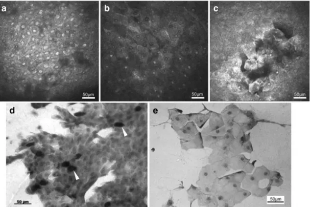
As we have already mentioned, IVCM is a highly effective method for demonstrating corneal and conjunctival changes in patients with SSDE and non-SSDE disease. In a previous study, the mean individual epithelial cell area (MEICA), nucleocytoplasmic ratio (N/C), and inflammatory cell density in patients with SLK were investigated with IVCM and im-pression cytology (Fig.2) [21]. Morphological changes such as cellular expansion, cell dropout, reduced cell cohesion, and shrunken nuclei were observed in both impression cytology and IVCM. Evaluation with IVCM showed that the inflam-matory cell density was significantly higher in SLK patients
than in the control group. Based on IVCM, MEICA values were significantly higher and N/C ratio was significantly low-er in SLK patients compared to the control group. Confocal microscopy in patients with SLK revealed that superficial ep-ithelial cells were enlarged and had pyknotic nuclei. Additionally, round, white bodies, presumably goblet cells, were observed in normal control eyes. Such cells were not found in SLK patients [21].
Laser scanning confocal microscopy seems to be an effi-cient noninvasive tool in the evaluation of phenotypic alter-ations of the conjunctival epithelium in SLK and may serve as an alternative for impression cytology. N/C ratio and inflam-matory cell density appear to be two new promising parame-ters of in vivo confocal microscopy in the assessment of ocular surface disease in SLK.
Examination of Corneal Nerves with IVCM
The 2017 DEWS report extensively discussed the role of neuronal involvement and neurosensory abnormalities in DED [3]. The cornea is the most densely innervatedtissue in the body and it is supplied primarily by the ophthalmic terminal branch of the trigeminal nerve [22]. CSN fibers have a substantial function in corneal ho-meostasis and have a primary role in the maintenance of ocular surface sensation and epithelial integrity via regulation of epithelial cell proliferation and wound healing [23].
Many studies have used IVCM to evaluate DED-dependent qualitative and quantitative changes in the cor-neal nerves [24,25]. These studies usually were focusing on the density of the corneal subbasal nerve. Although a decrease in corneal nerve density was detected in the ma-jority of studies [10,26–29], there are studies reporting an increase in nerve density in patients with SS [30]. However, Hoşal et al. [31] and Tuominen et al. [11] com-pared DED patients with control groups in terms of cor-neal subbasal nerve density and did not detect any chang-es. The results of these studies are thought to depend on the severity and the different stages of DED, the difference in neural regeneration/degeneration patterns, the level of inflammation, and the change in paralgesia and allodynia as a result of repetitive effects on the corneal nerves.
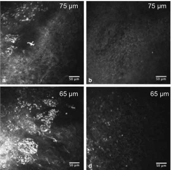
Fig. 1 a, c Widespread inflammation including dendritic cells, polymorphonuclear cells, and/or lymphocytes can be observed in the conjunctival epithelium in a 52-year-old woman with SS. b, d After 1-month dry eye treatment, significant improvement of conjunctival inflammatory infiltrates at two conjunctival epithelial depths (65 and 75 m) was observed [48]. Reprinted with permission from Wakamatsu TH, Sato EA, Matsumoto Y, Ibrahim OM, Dogru M, Kaido M, Ishida R, Tsubota K. Conjunctival in vivo confocal scanning laser microscopy in patients with Sjögren syndrome. Invest Ophthalmol Vis Sci. 2010 Jan;51(1):144–50
Corneal sensitivity is very important for maintaining a healthy ocular surface. Corneal epithelium becomes more susceptible to external factors, when there is a decrease in blink reflex and tears due to any reason. As a result of excessive evaporation and cooling, tear osmolarity in-creases. Local inflammation and peripheral nerve damage occur as a result of stress in the ocular mucosal epithe-lium depending on a decrease in the amount of tears and an increase in osmolarity [32]. Local inflammation and nerve damage can cause short- and long-term genetic and molecular changes in primary sensorial neurons [33]. Sensorial nerve terminals are located in a densely and superficially between the epithelial cells on the corneal surface. Therefore, corneal superficial nerves are vulner-able to environmental factors (air pollution, low humidi-ty), trauma (cataract and refractive surgery), and ocular surface diseases (pterygium, conjunctivochalasis, keratoconus) [14, 34, 35]. However, changes in the den-sity and structure of subbasal nerves and epithelial nerve terminals depend on changes in tear secretion, according to studies in animal models [36, 37]. Similarly, studies on patients with tear deficiency due to various etiological
reasons have shown changes in the number, tortuosity, branching pattern, and reflectivity of subbasal nerve fi-bers [7, 38, 39].
Other morphological parameters associated with cor-neal subbasal nerve density with IVCM were tortuosity, reflectivity, and beading pattern [7, 28, 30, 38, 40, 41]. These studies have shown an increase in these parame-ters and this increase is thought to be caused by neural regeneration after damage in the subbasal corneal nerves. In patients with dry eyes, an increase in the density of other immune cells along with DCs was determined in studies with IVCM [24, 25, 28, 42]. In these patients, clinical symptoms were parallel with the increase in DC density [43]. Therefore, IVCM can be considered as an important tool that can help diagnosis and treatment of DED. In our recent study, we evaluated the changes in tortuosity, reflective, and DC cell density in mice with dry eyes exposed to scopolamine for 1 month and re-vealed significant changes in these parameters. We thought that these changes occurred due to regeneration as a result of dry eye stress in the cells as a consequence of scopolamine application [44•].
Fig. 2 Comparison of conjunctival epithelial cell differentiation between SLK patients and the control group. a–c Representative conjunctival epithelial cell images in confocal microscopy. a, d Eyes of a healthy individual. b, c, e Representative IVCM and impression cytology images of eyes in SLK patients. b, e Remarkable expansion of cell size with pyknotic nuclei was demonstrated in patients with SLK. c In some areas, sloughing of the superficial conjunctival epithelium was observed.
White arrowheads indicate the presumable goblet cells with glycogen overload [21]. Reprinted with permission from Kojima T, Matsumoto Y, Ibrahim OM, Sato EA, Dogru M, Tsubota K. In vivo evaluation of superior limbic keratoconjunctivitis using laser scanning confocal microscopy and conjunctival impression cytology. Invest Ophthalmol Vis Sci. 2010 Aug;51(8):3986–92
Meibomian Gland Disease
Recently, IVCM has also been frequently used in the exami-nation of meibomian gland dysfunction (MGD). Meibomian gland disorders are common in daily ophthalmology practice [45]. MGD is a term commonly used for obstructive meibomian gland disease. A decrease in meibomian gland lipid secretion, increase in tear evaporation, and decrease in tear stability can lead to deterioration in the lubrication of the ocular surface and damage to the corneal epithelium. In the etiology of DED, MGD is a significant factor [46].
IVCM was useful in characterizing phenotypic alterations in MGD such as subepithelial fibrosis and obstruction of meibomian gland (MG) orifices [47]. Additionally, our group previously reported that IVCM was efficient in describing phenotypic alterations in MGD by devising new diagnostic parameters such as acinar unit density and acinar unit diameter reflecting histopathological changes such as glandular atrophy and acinar/ductal dilatation [48].
IVCM can be used to measure morphological changes in the meibomian glands, the diameters and densities of the MG acinar unit, diameter of the MG orifices, and the density of periglandular inflammatory cells [7,9,48, 49]. IVCM also enables semi-quantitative evaluation of meibum secretion re-flectivity and the heterogeneity of the glandular interstices and acinar wall [7,9]. In patients with MG disease, a decrease in acinar cell density, an increase in acinar unit diameters, and an increase of reflectance in meibomian secretions were observed [7,9,49]. However, in patients with SS, small acinar units, increased inflammatory cell density, and decreased homoge-neity in periglandular interstitium were demonstrated [7].
IVCM parameters showed acceptable sensitivity and specificity, and the cutoff parameters certainly helped cli-nicians in MGD diagnosis [49]. Similarly, another IVCM parameter described by our group in a recent study is periglandular inflammatory cell density, which suggested the potential of this novel technology in differentiating inflammatory obstructive MGD from non-inflammatory subtypes as well as the potential for evaluating the out-come of different treatment protocols [7].
Graft Versus Host Disease
Graft versus host disease (GVHD) is the most important com-plication of allogeneic hematopoietic stem cell transplanta-tion, as a result of donor cell recognition by the host immune system as foreign antigens and subsequent attack. GVHD is a condition that affects many organs and systems, is character-ized by high morbidity, and can lead to a wide variety of clinical outcomes [50,51]. The eye is mostly affected by GVHD with an incidence between 60 and 90% [52, 53]. GVHD can cause various ocular conditions such as corneal
epitheliopathy, MGD, ocular surface inflammation, conjunc-tival cicatricial disease, lacrimal gland dysfunction, hyper-emia, uveitis, scleritis, and retinal microvasculopathy in the eye [54]. Additionally, the most common pathology in GVHD is DED, which has been reported to occur in 40–70% of GVHD patients [52,53,55].
In patients receiving hematopoietic stem cell therapy (HSCT) by our group, GVHD and non-GVHD patients were compared using IVCM, and significant differences were ob-served [56]. Subbasal corneal nerve density was significantly lower in patients with GVHD compared to individuals with normal eyes. These findings were consistent with Kheirkhah et al. [57]. This change in subbasal corneal nerve cell density was thought to be related not only to GVHD-related DED, but also to the immune reaction that occurred. In the same study, the tortuosity and branching patterns of the subbasal nerves were evaluated and a significant increase was observed in both. It is thought that this increase is caused by a regeneration process initiated by subcutaneous nerves by nerve growth factors and cytokines released during inflammation due to ocular surface damage [56]. Another change in the ocular surface of patients with GVHD is the density changes in den-dritic cells, which played an important role in the primary immune response. These cells primarily play a role in the initiation of immune responses as antigen presenting cells. As a result, in the same study, the density of DCs in both the central cornea and limbal epithelium was found to be signifi-cantly high in patients with DED due to GVHD. In addition, significant morphological changes were observed in distinct subbasal nerves in patients with dry eye associated with GVHD receiving HSCT [56]. In light of all these findings, IVCM is considered as a potentially helpful technique in the evaluation of ocular surface changes in the eyes especially during inflammation in GVHD.
Ocular Demodicosis
Demodex folliculorum (DF) and Demodex brevis (DB) are mites that are parasites only in humans and settle in human hair follicles and pilosebaceous units [58]. It is widely be-lieved that there may be a potential risk for skin diseases [59,60]. DF and DB can be found in humans, especially in the face, nose, eyelashes, ears, and genital area [59]. Although they can remain in the intact skin, in the hair follicles, and in the meibomian glands without any pathogenic effects, they can be pathogenic in certain cases that skin cleaning is not performed well and the immune system is suppressed, causing inflammatory dermatitis, contributing to the development of keratosis and epithelioma, cuasing acne and acne rosacea [60–64]. Occasionally, they can cause strong skin reactions and prominent pigmentation [60]. Moreover, a large number
of mites may be located in the eyelid follicles and may cause keratosis, hyperplasia, and melanocyte aggregation [65].
Demodex mites can cause pathogenic conditions such as papulopustular rosacea, pityriasis folliculorum, rosacea-like demodicosis, demodicosis gravis (granulomatous rosacea-like demodicosis), and blepharitis when they over-proliferate or penetrate the dermis [66]. Previous studies have shown that
the number of DFs in eyelashes was higher in blepharitis patients than in the control group, and it was determined that pruritus occurred parallelly with the ovulation period of Demodex in itchy blepharitis cases [62,67–70].
In our previous study, we evaluated the feasibility and efficacy of IVCM in diagnosis and follow-up in patients with blepharitis-related cylindrical dandruff. According to
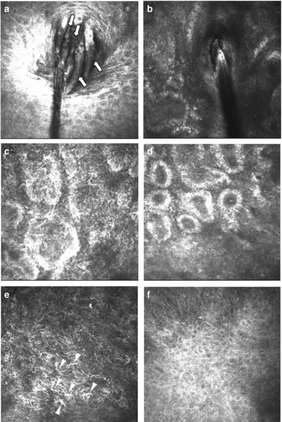
Fig. 3 a Heavy demodicosis infestation of eyelash follicles in a 72-year-old female patient shown by in vivo confocal microscopy. Note the presence of several Demodex colonies. b
Representative IVCM images of eyelash bulbs in a healthy female control individual; note the absence of mites. c–f IVCM images of meibomian gland acinar units and palpebral conjunctiva before and after tea tree oil treatment of the patient in panel a. c Heterogeneous reflectivity of the gland lumen and dilatation of meibomian gland acinar units can be seen. e Prominent inflammatory infiltrates can be observed in the palpebral conjunctiva near the eyelid margin. d, f After treatment with tea tree oil, a significant improvement was observed in acinar dilatation and conjunctival cell infiltration [66]. Reprinted with permission from Kojima T, Ishida R, Sato EA, Kawakita T, Ibrahim OM, Matsumoto Y, Kaido M, Dogru M, Tsubota K. In vivo evaluation of ocular demodicosis using laser scanning confocal microscopy. Invest Ophthalmol Vis Sci. 2011 Feb 1;52(1):565–9
our results, IVCM was a highly effective method for the evaluation of meibomian glands/conjunctival diseases, ac-inar dilatation, periglandular inflammation, and conjuncti-val inflammation as well as the demonstration of embed-ded mites in the bulb. In the evaluation of patients with demodicosis with IVCM, dilution in meibomian gland ac-inar units and periglandular inflammatory infiltrates with DCs were observed (Fig.3). Similarly, prominent inflam-matory infiltrates were found in the palpebral conjunctiva adjacent to the eyelids. In addition, IVCM has been shown to be a very useful technology in the follow-up of changes in the eyelids, meibomian, and conjunctival diseases fol-lowing tea tree oil treatment [66]. As a consequence, laser scanning confocal microscopy is an efficient noninvasive tool in the diagnosis and follow-up of ocular demodicosis infestation.
Conclusions
Recently, the number of studies involving IVCM has increased dramatically [71••]. In the light of all these
studies, IVCM is thought to be an extremely promising method, easy to implement, cost-effective, and minimally invasive providing rapid outcomes for the evaluation of ocular surface structures in dry eye and related diseases at the cellular level. Although it is a very practical meth-od, there are some difficulties in evaluation with IVCM. IVCM can display a small area of the entire cornea. In order to see the entire cornea, it is necessary to acquire many shots from different regions and these areas may sometimes overlap. In addition, it is very difficult to reevaluate the same area which has been previously im-aged. In addition, stable contact with the patient’s eye sometimes can be difficult, even if local anesthesia is used. Additional concerns include the standardization of image acquisition, interpretation, and quantification [72]. Despite these difficulties, IVCM is a valuable and prom-ising complementary method in clinical diagnosis and follow-up in DED.
Compliance with Ethical Standards
Conflict of Interest Simsek C, Karalezli A, and Dogru M each declare no potential conflicts of interest.
Kojima T has received personal fees from Santen pharmaceutical, Otsuka pharmaceutical, Alcon, Eye Lens, Carl Zeiss Meditec, and Echo electricity.
Human and Animal Rights and Informed Consent All reported studies/ experiments with human or animal subjects performed by the authors have been previously published and complied with all applicable ethical standards (including the Helsinki declaration and its amendments, institutional/national research committee standards, and international/na-tional/institutional guidelines).
References
Papers of particular interest, published recently, have been highlighted as:
• Of importance •• Of major importance
1. Moss SE, Klein R, Klein BE. Long-term incidence of dry eye in an older population. Optom Vis Sci. 2008;85:668–74.https://doi.org/ 10.1097/OPX.0b013e318181a947.
2. Lemp MA. Epidemiology and classification of dry eye. Adv Exp Med Biol. 1998;438:791–803.
3. Craig JP, Nelson JD, Azar DT, Belmonte C, Bron AJ, Chauhan SK, et al. TFOS DEWS II report executive summary. Ocul Surf. 2017;15:802–12.https://doi.org/10.1016/j.jtos.2017.08.003. 4. Patel SV, Bourne WM. Corneal endothelial cell loss 9 years after
excimer laser keratorefractive surgery. Arch Ophthalmol. 2009;127:1423–7.https://doi.org/10.1001/archophthalmol.2009. 192.
5.•• Villani E, Magnani F, Viola F, Santaniello A, Scorza R, Nucci P, et al. In vivo confocal evaluation of the ocular surface morpho-functional unit in dry eye. Optom Vis Sci. 2013;90:576–86.
https://doi.org/10.1097/OPX.0b013e318294c184. A clinical study indicating the evaluation of the whole ocular surface morpho-functional unit in patients with dry eye.
6. Efron N. Contact lens-induced changes in the anterior eye as ob-served in vivo with the confocal microscope. Prog Retin Eye Res. 2007;26:398–436.https://doi.org/10.1016/j.preteyeres.2007.03. 003.
7. Villani E, Beretta S, De Capitani M, Galimberti D, Viola F, Ratiglia R. In vivo confocal microscopy of meibomian glands in Sjogren’s syndrome. Invest Ophthalmol Vis Sci. 2011;52:933–9.https://doi. org/10.1167/iovs.10-5995.
8. Ban Y, Ogawa Y, Ibrahim OM, Tatematsu Y, Kamoi M, Uchino M, et al. Morphologic evaluation of meibomian glands in chronic graft-versus-host disease using in vivo laser confocal microscopy. Mol Vis. 2011;17:2533–43.
9. Villani E, Ceresara G, Beretta S, Magnani F, Viola F, Ratiglia R. In vivo confocal microscopy of meibomian glands in contact lens wearers. Invest Ophthalmol Vis Sci. 2011;52:5215–9.https://doi. org/10.1167/iovs.11-7427.
10. Villani E, Canton V, Magnani F, Viola F, Nucci P, Ratiglia R. The aging Meibomian gland: an in vivo confocal study. Invest Ophthalmol Vis Sci. 2013;54:4735–40.https://doi.org/10.1167/ iovs.13-11914.
11. Tuominen IS, Konttinen YT, Vesaluoma MH, Moilanen JA, Helinto M, Tervo TM. Corneal innervation and morphology in primary Sjogren’s syndrome. Invest Ophthalmol Vis Sci. 2003;44:2545–9. 12. Villani E, Galimberti D, Viola F, Mapelli C, Del Papa N, Ratiglia R.
Corneal involvement in rheumatoid arthritis: an in vivo confocal study. Invest Ophthalmol Vis Sci. 2008;49:560–4.https://doi.org/ 10.1167/iovs.07-0893.
13. Villani E, Galimberti D, Viola F, Mapelli C, Ratiglia R. The cornea in Sjogren’s syndrome: an in vivo confocal study. Invest Ophthalmol Vis Sci. 2007;48:2017–22.https://doi.org/10.1167/ iovs.06-1129.
14. Villani E, Viola F, Sala R, Salvi M, Mapelli C, Curro N, et al. Corneal involvement in Graves’ orbitopathy: an in vivo confocal study. Invest Ophthalmol Vis Sci. 2010;51:4574–8.https://doi.org/ 10.1167/iovs.10-5380.
15. Akpek EK, Lindsley KB, Adyanthaya RS, Swamy R, Baer AN, McDonnell PJ. Treatment of Sjogren’s syndrome-associated dry eye an evidence-based review. Ophthalmology. 2011;118:1242– 52.https://doi.org/10.1016/j.ophtha.2010.12.016.
16. Zhang M, Chen J, Luo L, Xiao Q, Sun M, Liu Z. Altered corneal nerves in aqueous tear deficiency viewed by in vivo confocal mi-croscopy. Cornea. 2005;24:818–24.
17.•• Wakamatsu TH, Sato EA, Matsumoto Y, Ibrahim OM, Dogru M, Kaido M, et al. Conjunctival in vivo confocal scanning laser mi-croscopy in patients with Sjogren syndrome. Invest Ophthalmol Vis Sci. 2010;51:144–50.https://doi.org/10.1167/iovs.08-2722. A clinical study shows the alterations of conjunctival morphology in Sjögren syndrome by describing new confocal microscopy parameters such as conjunctival epithelial cell and microcyst densities and levels of inflammatory infiltrate. 18. Udell IJ, Kenyon KR, Sawa M, Dohlman CH. Treatment of
rior limbic keratoconjunctivitis by thermocauterization of the supe-rior bulbar conjunctiva. Ophthalmology. 1986;93:162–6. 19. Wander AH, Masukawa T. Unusual appearance of condensed
chro-matin in conjunctival cells in superior limbic keratoconjunctivitis. Lancet. 1981;2(8236):42–3.
20. Collin HB, Donshik PC, Foster CS, Boruchoff SA, Cavanagh HD. Keratinization of the bulbar conjunctival epithelium in superior limbic keratoconjunctivitis in humans. An electron microscopic study. Acta Ophthalmol. 1978;56:531–43.
21. Kojima T, Matsumoto Y, Ibrahim OM, Sato EA, Dogru M, Tsubota K. In vivo evaluation of superior limbic keratoconjunctivitis using laser scanning confocal microscopy and conjunctival impression cytology. Invest Ophthalmol Vis Sci. 2010;51:3986–92.https:// doi.org/10.1167/iovs.09-4932.
22. Muller LJ, Pels L, Vrensen GF. Ultrastructural organization of hu-man corneal nerves. Invest Ophthalmol Vis Sci. 1996;37:476–88. 23. Oliveira-Soto L, Charman WN. Some possible longer-term ocular
changes following excimer laser refractive surgery. Ophthalmic Physiol Opt. 2002;22:274–88.
24. McGowan DP, Lawrenson JG, Ruskell GL. Touch sensitivity of the eyelid margin and palpebral conjunctiva. Acta Ophthalmol. 1994;72:57–60.
25. Norn MS. Conjunctival sensitivity in pathological cases, with si-multaneous measurement of corneal and lid margin sensitivity. Acta Ophthalmol. 1975;53:450–7.
26. McMonnies CW. Incomplete blinking: exposure keratopathy, lid wiper epitheliopathy, dry eye, refractive surgery, and dry contact lenses. Cont Lens Anterior Eye. 2007;30:37–51.https://doi.org/10. 1016/j.clae.2006.12.002.
27. Qazi Y, Kheirkhah A, Blackie C, Cruzat A, Trinidad M, Williams C, et al. In vivo detection of clinically non-apparent ocular surface inflammation in patients with meibomian gland dysfunction-associated refractory dry eye symptoms: a pilot study. Eye (Lond). 2015;29:1099–110.https://doi.org/10.1038/eye.2015.103. 28. Tomlinson A, Bron AJ, Korb DR, Amano S, Paugh JR, Pearce EI,
et al. The international workshop on meibomian gland dysfunction: report of the diagnosis subcommittee. Invest Ophthalmol Vis Sci. 2011;52:2006–49.https://doi.org/10.1167/iovs.10-6997f. 29. Simsek C, Kojima T, Dogru M, Tsubota K. Alterations of murine
subbasal corneal nerves after environmental dry eye stress. Invest Ophthalmol Vis Sci. 2018;59:1986–95.https://doi.org/10.1167/ iovs.17-23743.
30. Lienert JP, Tarko L, Uchino M, Christen WG, Schaumberg DA. Long-term natural history of dry eye disease from the patient’s perspective. Ophthalmology. 2016;123:425–33.https://doi.org/10. 1016/j.ophtha.2015.10.011.
31. Hosal BM, Ornek N, Zilelioglu G, Elhan AH. Morphology of cor-neal nerves and corcor-neal sensation in dry eye: a preliminary study. Eye (Lond). 2005;19:1276–9.https://doi.org/10.1038/sj.eye. 6701760.
32. Blodi BA, Byrne KA, Tabbara KF. Goblet cell population among patients with inactive trachoma. Int Ophthalmol. 1988;12:41–5.
33. Papas EB. Key factors in the subjective and objective assessment of conjunctival erythema. Invest Ophthalmol Vis Sci. 2000;41:687– 91.
34. Wakamatsu TH, Okada N, Kojima T, Matsumoto Y, Ibrahim OM, Dogru M, et al. Evaluation of conjunctival inflammatory status by confocal scanning laser microscopy and conjunctival brush cytolo-gy in patients with atopic keratoconjunctivitis (AKC). Mol Vis. 2009;15:1611–9.
35. Zhivov A, Stachs O, Kraak R, Stave J, Guthoff RF. In vivo confocal microscopy of the ocular surface. Ocul Surf. 2006;4:81–93. 36. Cocho L, Fernandez I, Calonge M, Martinez V, Gonzalez-Garcia
MJ, Caballero D, et al. Biomarkers in ocular chronic graft versus host disease: tear cytokine- and chemokine-based predictive model. Invest Ophthalmol Vis Sci. 2016;57:746–58.https://doi.org/10. 1167/iovs.15-18615.
37. Hara S, Kojima T, Ishida R, Goto E, Matsumoto Y, Kaido M, et al. Evaluation of tear stability after surgery for conjunctivochalasis. Optom Vis Sci. 2011;88:1112–8.https://doi.org/10.1097/OPX. 0b013e3182223573.
38. Villani E, Galimberti D, Del Papa N, Nucci P, Ratiglia R. Inflammation in dry eye associated with rheumatoid arthritis: cyto-kine and in vivo confocal microscopy study. Innate Immun. 2013;19:420–7.https://doi.org/10.1177/1753425912471692. 39. Villani E, Garoli E, Termine V, Pichi F, Ratiglia R, Nucci P. Corneal
confocal microscopy in dry eye treated with corticosteroids. Optom Vis Sci. 2015;92:e290–5. https://doi.org/10.1097/OPX. 0000000000000600.
40. Efron N, Brennan NA, Morgan PB, Wilson T. Lid wiper epitheliopathy. Prog Retin Eye Res. 2016;53:140–74.https://doi. org/10.1016/j.preteyeres.2016.04.004.
41. Walker PM, Lane KJ, Ousler GW 3rd, Abelson MB. Diurnal vari-ation of visual function and the signs and symptoms of dry eye. Cornea. 2010;29:607–12. https://doi.org/10.1097/ICO. 0b013e3181c11e45.
42. Golebiowski B, Chim K, So J, Jalbert I. Lid margins: sensitivity, staining, meibomian gland dysfunction, and symptoms. Optom Vis S c i . 2 0 1 2 ; 8 9 : 1 4 4 3–9. h t t p s : / / d o i . o r g / 1 0 . 1 0 9 7 / O P X . 0b013e3182693cef.
43. Cornish KS, Gregory ME, Ramaesh K. Systemic cyclosporine A in severe atopic keratoconjunctivitis. Eur J Ophthalmol. 2010;20:844– 51.
44.• Simsek C, Kojima T, Nagata T, Dogru M, Tsubota K. Changes in murine subbasal corneal nerves after scopolamine-induced dry eye stress exposure. Invest Ophthalmol Vis Sci. 2019;60:615–23.
https://doi.org/10.1167/iovs.18-26318. This animal study indicates that prolonged exposure to dry eye conditions resulted in morphologic alterations and branch patterns of corneal subbasal nerves in wild type mice.
45. Driver PJ, Lemp MA. Meibomian gland dysfunction. Surv Ophthalmol. 1996;40(5):343–67.
46. McCulley JP, Shine WE. Meibomian gland function and the tear lipid layer. Ocul Surf. 2003;1:97–106.
47. Messmer EM, Torres Suarez E, Mackert MI, Zapp DM, Kampik A. In vivo confocal microscopy in blepharitis. Klin Monatsbl Augenheilkd. 2005;222:894–900. https://doi.org/10.1055/s-2005-858798.
48. Matsumoto Y, Sato EA, Ibrahim OM, Dogru M, Tsubota K. The application of in vivo laser confocal microscopy to the diagnosis and evaluation of meibomian gland dysfunction. Mol Vis. 2008;14: 1263–71.
49. Ibrahim OM, Matsumoto Y, Dogru M, Adan ES, Wakamatsu TH, Goto T, et al. The efficacy, sensitivity, and specificity of in vivo laser confocal microscopy in the diagnosis of meibomian gland dysfunction. Ophthalmology. 2010;117:665–72.https://doi.org/10. 1016/j.ophtha.2009.12.029.
50. Jagasia MH, Greinix HT, Arora M, Williams KM, Wolff D, Cowen EW, Palmer J, Weisdorf D, Treister NS, Cheng GS, Kerr H, Stratton P, Duarte RF, McDonald GB, Inamoto Y, Vigorito A, Arai S, Datiles MB, Jacobsohn D, Heller T, Kitko CL, Mitchell SA, Martin PJ, Shulman H, Wu RS, Cutler CS, Vogelsang GB, Lee SJ, Pavletic SZ, Flowers MED National Institutes of Health Consensus development project on criteria for clinical trials in chronic graft-versus-host disease: I. The 2014 diagnosis and staging working group report. Biol Blood Marrow Transplant 2015:21: 389–401 e1. doi:https://doi.org/10.1016/j.bbmt.2014.12.001. 51. Jamil MO, Mineishi S. State-of-the-art acute and chronic GVHD
treatment. Int J Hematol. 2015;101:452–66.https://doi.org/10. 1007/s12185-015-1785-1.
52. Shikari H, Antin JH, Dana R. Ocular graft-versus-host disease: a review. Surv Ophthalmol. 2013;58(3):233–51.https://doi.org/10. 1016/j.survophthal.2012.08.004.
53. Hessen M, Akpek EK. Ocular graft-versus-host disease. Curr Opin Allergy Clin Immunol. 2012;12:540–7.https://doi.org/10.1097/ ACI.0b013e328357b4b9.
54. Saboo US, Amparo F, Abud TB, Schaumberg DA, Dana R. Vision-related quality of life in patients with ocular graft-versus-host dis-ease. Ophthalmology. 2015;122:1669–74.https://doi.org/10.1016/ j.ophtha.2015.04.011.
55. Anderson NG, Regillo C. Ocular manifestations of graft versus host disease. Curr Opin Ophthalmol. 2004;15:503–7.
56. He J, Ogawa Y, Mukai S, Saijo-Ban Y, Kamoi M, Uchino M, et al. In vivo confocal microscopy evaluation of ocular surface with graft-versus-host disease-related dry eye disease. Sci Rep. 2017;7:10720.
https://doi.org/10.1038/s41598-017-10237-w.
57. Kheirkhah A, Rahimi Darabad R, Cruzat A, Hajrasouliha AR, Witkin D, Wong N, et al. Corneal epithelial immune dendritic cell alterations in subtypes of dry eye disease: a pilot in vivo confocal microscopic study. Invest Ophthalmol Vis Sci. 2015;56:7179–85.
https://doi.org/10.1167/iovs.15-17433.
58. Nutting WB. Hair follicle mites (Acari: Demodicidae) of man. Int J Dermatol. 1976;15:79–98.
59. Desch C, Nutting WB. Demodex folliculorum (Simon) and D. brevis akbulatova of man: redescription and reevaluation. J Parasitol. 1972;58:169–77.
60. Norn MS. Demodex folliculorum. Incidence, regional distribution, pathogenicity. Dan Med Bull. 1971;18:14–7.
61. Rufli T, Mumcuoglu Y. The hair follicle mites Demodex folliculorum and Demodex brevis: biology and medical impor-tance. Rev Dermatol. 1981;162:1–11.
62. Roth AM. Demodex folliculorum in hair follicles of eyelid skin. Ann Ophthalmol. 1979;11:37–40.
63. Erbagci Z, Ozgoztasi O. The significance of Demodex folliculorum density in rosacea. Int J Dermatol. 1998;37:421–5.
64. Georgala S, Katoulis AC, Kylafis GD, Koumantaki-Mathioudaki E, Georgala C, Aroni K. Increased density of Demodex folliculorum and evidence of delayed hypersensitivity reaction in subjects with papulopustular rosacea. J Eur Acad Dermatol Venereol. 2001;15: 441–4.
65. Rodriguez AE, Ferrer C, Alio JL. Chronic blepharitis and Demodex. Arch Soc Esp Oftalmol. 2005;80:635–42.
66. Kojima T, Ishida R, Sato EA, Kawakita T, Ibrahim OM, Matsumoto Y, et al. In vivo evaluation of ocular demodicosis using laser scan-ning confocal microscopy. Invest Ophthalmol Vis Sci. 2011;52: 565–9.https://doi.org/10.1167/iovs.10-5477.
67. Kheirkhah A, Casas V, Li W, Raju VK, Tseng SC. Corneal mani-festations of ocular demodex infestation. Am J Ophthalmol. 2007;143:743–9.https://doi.org/10.1016/j.ajo.2007.01.054. 68. Kemal M, Sumer Z, Toker MI, Erdogan H, Topalkara A, Akbulut
M. The prevalence of Demodex folliculorum in blepharitis patients and the normal population. Ophthalmic Epidemiol. 2005;12:287– 90.https://doi.org/10.1080/092865805910057.
69. Gao YY, Di Pascuale MA, Li W, Liu DT, Baradaran-Rafii A, Elizondo A, et al. High prevalence of Demodex in eyelashes with cylindrical dandruff. Invest Ophthalmol Vis Sci. 2005;46:3089–94.
https://doi.org/10.1167/iovs.05-0275.
70. Clifford CW, Fulk GW. Association of diabetes, lash loss, and Staphylococcus aureus with infestation of eyelids by Demodex folliculorum (Acari: Demodicidae). J Med Entomol. 1990;27: 467–70.
71.•• Villani E, Baudouin C, Efron N, Hamrah P, Kojima T, Patel SV, et al. In vivo confocal microscopy of the ocular surface: from bench to bedside. Curr Eye Res. 2014;39:213–31.https://doi.org/10.3109/ 02713683.2013.842592. A comprehensive review illustrating the advances in confocal microscopy imaging technique for clinical diagnosis and management of the ocular surface and focusing on recent and promising attempts.
72. Niederer RL, McGhee CN. Clinical in vivo confocal microscopy of the human cornea in health and disease. Prog Retin Eye Res. 2010;29:30–58.https://doi.org/10.1016/j.preteyeres.2009.11.001. Publisher’s Note Springer Nature remains neutral with regard to jurisdictional claims in published maps and institutional affiliations.