GENETIC AND EPIGENETIC EFFECT OF
ESTROGEN ON MESENCHYMAL STEM CELL
MAINTENANCE AND DIFFERENTIATION
A THESIS
SUBMITTED TO THE DEPARTMENT
OF MOLECULAR BIOLOGY AND GENETICS AND THE
GRADUATE SCHOOL OF ENGINEERING AND SCIENCE
OF BILKENT UNIVERSITY
IN PARTIAL FULFILLMENT OF THE REQUIREMENTS
FOR THE DEGREE OF
DOCTOR OF PHILOSOPHY
BY
CEYLAN VERDA BİTİRİM
OCTOBER, 2013
ii
I certify that I have read this thesis and that in my opinion it is fully adequate, in scope and in quality, as a thesis for the degree of Doctor of Philosophy.
Assoc. Prof.Dr. Işık YULUĞ (Advisor) Prof. Dr. K. Can AKÇALI (Co-Advisor) I certify that I have read this thesis and that in my opinion it is fully adequate, in scope
and in quality, as a thesis for the degree of Doctor of Philosophy.
Assist. Prof. Dr.ÖzlenKONU
I certify that I have read this thesis and that in my opinion it is fully adequate, in scope and in quality, as a thesis for the degree of Doctor of Philosophy.
Assoc. Prof. Dr. Rengül Çetin ATALAY I certify that I have read this thesis and that in my opinion it is fully adequate, in scope and in quality, as a thesis for the degree of Doctor of Philosophy.
Assoc. Prof. Dr. Mayda GÜRSEL I certify that I have read this thesis and that in my opinion it is fully adequate, in scope and in quality, as a thesis for the degree of Doctor of Philosophy.
. Prof.Dr.Mehmet Yakup ARICA
Approved for the Graduate School of Engineering and Science
Prof. Dr. Levent Onural
iii
ABSTRACT
GENETIC AND EPIGENETIC EFFECT OF ESTROGEN ON
MESENCHYMAL STEM CELL MAINTENANCE AND
DIFFERENTIATION
Ceylan Verda Bitirim
Ph.D. in Molecular Biology and Genetics Supervisor: Assoc. Prof. Dr. Işık Yuluğ
October 2013, Pages 151
Mesenchymal stem cells (MSCs) have the potential to differentiate into multiple cell types and immune privileged characteristics. These features make MSCs a hope in tissue engineering and cell based treatment applications. Tremendous amount of studies were carried out in order to produce an ideal biomaterial as a scaffold for cell transplantation. In recent studies, carbon nanotubes (CNT) were identified as a novel scaffold array due to their unique physical, chemical and electrical properties among the other biomaterials.The effect of estrogen hormone on the regulation of MSC maintenance, proliferation and differentiation was reported. However, its role in maintenance of MSCs on scaffold materials such as CNTs and the genetic and epigenetic regulation of MSC differentiation have not fully been elucidated. Therefore our aim was to examine the possible role of estrogen in the MSCs’ maintenance seeded on CNT surfaces and genetic and epigenetic regulation of the key transcription
iv
factors involved in adipogenic, osteogenic and chondrogenic differentiaton of MSCs. Our results revealed the enhanced effect of estrogen on the viability of MCSs which were seeded and incubated on multiwalled carbon nanotubes (MWCNT). In addition we demonstrated that passaging causes decrease of cell viability and the number of attached cells on CNT materials. We have also shown the effect of estrogen on the epigenetic and genetic regulation of MSC differentiation. Estrogen treatment decreased the expression of major adipogenic transcription factors; C/EBPα, FABP4, PPARγ, Adipsin and increased key osteogenic transcription factor RUNX2 in MSCs from both normal female and ovariectomized rats, suggesting inhibitory and stimulatory effect of estrogen on adipogenesis and osteogenesis respectively. We have also shown that the subcellular localization of PPARγ and ETS1 is changed in response to estrogen deficiency. Among modified histones, we found that H3K27me2, H3K27me3 and H3K36me2 protein levels were reduced after estrogen treatment both in female and ovariectomized animals. In addition, ChIP analysis showed that estrogen treatment caused an increase in H3K27me2, H3K27me3 and ERα levels at the promoters of C/EBPα, FABP4, PPARγ, Adipsin and RUNX2. Bisulfite sequencing analysis revealed that in the absence of estrogen, DNA hypermethylation was established in C/EBPα and PPARγ promoters whereas in ERα promoters CpG hypomethylation was observed after estrogen treatment. In conclusion, estrogen causes epigenetic and genetic changes in maintenace and differentiation of MSCs. Understanding the
v
effect of estrogen on the genetic and epigenetic regulation of the major transcription factors may lead to clues for new treatment in chronic diseases such as obesity, osteoporosis and ostearthiritis.
vi
ÖZET
ÖSTROJEN HORMONUNUN MEZENKİMAL KÖK
HÜCRELERİN MUHAFAZASINDAKİ VE
FARKLILAŞMASINDAKİ GENETİK VE EPİGENETİK
ETKİSİ
Ceylan Verda Bitirim
Moleküler Biyoloji ve Genetik Doktorası Tez Yöneticisi: Doç. Dr. Işık Yuluğ
Ekim 2013, Sayfa 151
Mezenkimal kök hücreler (MKH) kemik, yağ, kıkırdak, sinir ve karaciğer hücrelerine dönüşebilme potansiyeli olan, immün reaksiyon oluşturma özelliği olmayan hücrelerdir. Bu özellikler kök hücreleri doku mühendisliği ve hücre tedavisi alanları için yeni bir umut haline getirdi. Hücre transplantasyonunda kullanılmak üzere ideal yüzey üretmek için pek çok çalışma yürütüldü. Son yıllarda farklı fiziksel, kimyasal ve elektriksel özelliklerinden dolayı karbon nanotüpler (KNT) yeni biyomateryal yüzeyler olarak tanımlandı. Östrojen hormonunun mezenkimal kök hücre devamlılığı, çoğalması ve farklılaşması üzerine etkileri rapor edilmiştir. Fakat östrojenin biyomateryal yüzey üzerindeki mezenkimal kök hücrenin devamlılıgındaki ve mezenkimal kök hücre farklılaşmasındaki genetik ve epigenetik rolü tam olarak araştırılmamıştır. Bu nedenle, bu çalışmada östrojenin karbon nantüp üzerine ekilen kök hücrelerin devamlılığındaki ve adipojenik, östeojenik ve kondrojek farklılaşmaya katılan
vii
anahtar transkripsiyon faktörlerinin genetik ve epigenetik regülasyonundaki rolünü araştrmak amaçlanmıştır. Sonuçlarımız östrojenin çok duvarlı karbon nanotüpler üzerindeki MKHlerin canlılıgını arttırıcı etkisini göstermiştir. Ayrıca hücre canlılıgı ve KNT materyalin üzerine yapışan MKH sayısının passajlama ile azaldıgını gösterdik. Bu çalışmada östrojenin MKH farklılaşmasının genetik ve epigenetik regülasyonuna etkisini gösterebildik. Östrojen tedavisi, normal dişi ve overi alınmış dişiden alınan MKH’de temel adipojenik transkripsiyon faktörler olan C/EBPα, FABP4, PPARγ, Adipsinin gen ifadesini azaltırken, anahtar transkripsiyon faktörü olan RUNX2 nin ifadesini arttırır. Bu sonucun östrojenin adipojenik farklılaşma üzerine baskılayıcı etkisini, östeojenik farklılaşma üzerine arttırıcı etkisini gösterdiği söylenebilir. PPARγ ve diğer transkripsiyon faktörü ETS1 proteinlerinin hücresel lokalizasyonunun östrojen yokluguna bağlı değişimi ayrıca gösterilmiştir. Diğer modifiye histonlar arasında, H3K27me2, H3K27me3 ve H3K36me2 protein düzeylerinin östrojen tedavisinden sonra azaldığını bulduk. Ek olarak, kromatin immünopresipitasyon analizleri H3K27me2, H3K27me3 ve ERα düzeylerinin C/EBPα, FABP4, PPARγ, Adipsin ve RUNX2 promotörleri üzerinde östrojen tedavisi sonucu arttıgını gösterdi. Bisülfit sekanslama analizleri östrojen yoklugunda C/EBPα ve PPARγ promotörleri üzerinde DNA hipermetilasyonu görülürken, ERα promotöründe CpG hipometilsayonu görüldü. Sonuç olarak, östrojen MKH devamlılıgı ve farklılaşmasında epigenetik ve genetik değişimlere yol açar. Östrojenin temel transkripsiyon faktölerinin regülasyonundaki etkisinin
viii
anlaşılması obezite, osteoporoz ve osteoartirit gibi kronik hastalıkların tedavisi için yeni ipuçları yaratır.
ix
ACKNOWLEDGEMENT
First and foremost, from my heart, I would like to express my gratitute to my supervisor Prof. Dr. K. Can Akçalı for giving me the opportunity to work in his lab and providing his continuous support, endless patience and knowledge during my PhD study. His valuable guidance opened a door to a new life for me. It has been a big honor for me to work with him.
I want to thank Assoc. Prof. Dr. Özlen Konu, Assoc. Prof. Dr. Rengül Çetin Atalay, Prof. Dr. Yakup Arıca, Assoc. Prof. Dr. Mayda Gürsel and especially Assist. Prof. Dr. Işık Yuluğ for being members of my thesis committee and for their contribution in this thesis through their helpful suggestions.
I would like to express my heartful thanks to the past and present members of Akçalı Group; Merve Aydın, Ece Akhan, Damla Gözen, Şahika Çıngır, Fatma Ayaloğlu-Bütün, Zeynep Tokçaer-Keskin, Sumru Bayın, Sinan Gültekin and to present members of Yuluğ Grup; Nilüfer Sayar and Gurbet Karahan for their patience, tremendous help and understanding during my research. I would also like to thank to the senior students; Fatma Rabia Ürün, Ezgi Kaya and Gizem Gökçe Dinç who helped me and took part in this project with me. Working with
x
you was great! I also would like to thank Abdullah Ünnü, Füsun Elvan, Bilge Kılıç and Turan Daştandır for being always there for us.
I am very grateful to Assist. Prof. Dr. Erman Bengü for his supervision and to the members of Bengü group, especially Gökçe Küçükayan for her help during the carbon nanotube studies.
I would like to express my sincere appreciation to Assist. Prof. İhsan Gürsel for his support, guidance and encouragement.
I would like to thank The Scientific and Technological Research Council of Turkey (TÜBİTAK) for their financial support throughout my thesis work.
I am truly indebted to all of my beloved friends in MBG family, especially Büşra Yağabasan, Gülşah Dal, Özlem Tufanlı, Dilek Çevik, Pelin Telkoparan-Akıllılar, Füsun Doldur-Ballı, Nilüfer Sayar and Gökhan Yıldız for their support, incredible friendship and help whenever I needed during this long and arduous journey. They were always there to help whenever I had moments of crisis with my PhD. Without them this journey would be very difficult and boring. I would like to dedicate my special thankfulness to my dear friend Gözde Güçlüler for her invaluable friendship and being there for me whenever I needed them the most.
xi
I would like to also dedicate my special thankfulness to my beloved friend Şeyma Demirsoy. Being your friend and sharing the same lab with you was gorgeous.
I would like to express my deepest love and gratitude to Defne Bayık, for everything she has added to my life and to teach me looking the world through different perspectives. She gave more meaning not just to this journey but also to my life with her invaluable friendship, endless patience and encouragement. She will be a part of my family forever.
Once for all, I would also like to extend my sincere gratitude to my family, especially to my dearest mother and father for their unconditional and endless love and support throughout my life; and to my cousin Berk Zafer for his academic guidance and his priceless support. Without his guidance I couldn’t be here.
xii
CONTENT
CHAPTER 1 INTRODUCTION...………..………....1
1.1 EMBRYONIC STEMCELLS AND INDUCED PLURIPOTENT STEM CELLS……….………...5
1.2 ADULT STEM CELLS………...…………10
1.2.1 MESENCHYMALSTEM CELLS………..……...11
1.3 USE OF CARBON NANATUBES AS THREE-DIMENSIONAL SCAFFOLD MATERIAL.………....16
1.4 ESTROGEN AND ESTROGEN RECEPTOR PATHWAY…………...…18
1.5 EPIGENETICS……….………...……22
1.5.1 DNA METHYLATION……….…………....24
1.5.2 HISTONE MODIFICATIONS…….….………....26
1.6 EPIGENETIC ALTERATIONS IN STEM CELLS……….………...31
1.7 ESTROGEN AND EPIGENETIC REGULATION……….………..34
1.7.1 RELATIONSHIP BETWEEN THE ROLE OF ESTROGEN IN EPIGENETIC REGULATION AND MESENCHYMAL STEM CELL...36
CHAPTER 2 AIM OF THE STUDY...39
CHAPTER 3 MATERIALS AND METHODS………...…..42
3.1 ANIMALS………...42
3.1.1 OVARIECTOMIZATION……….……….42
3.2 SYNTHESIS, PATERNING CHARACTERIZATION OF CARBON NANOTUBES...43
3.3 ISOLATION OF THE CELLS FROM RAT BONE MARROW AND CULTURING OF MSC………...….……...….44
xiii
3.4.1 CALCULATION THE NUMBER OF ATTACHED MSCS ON
CNT………...46
3.4.2 CELL PROLIFERATION ASSAY FOR THE MESENCYHMAL STEM CELLS ON CNT SURFACES……….47
3.5 ADIPOGENIC AND OSTEOGENIC DIFFERENTIATION………….…47
3.6 DETECTION OF ADIPOGENIC AND OSTEOGENIC INDUCTION...48
3.6.1 OIL RED O STAINING………..………..48
3.6.2 ALIZARIN RED STAINING………….………...49
3.7 TOTAL RNA ISOLATION FROM MSCs AND DIFFERENTIATED CELLS………...49
3.8 cDNA SYNTHESIS………....50
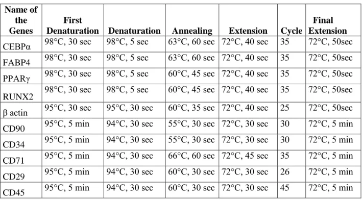
3.9 PRIMER DESIGN FOR QUANTITATIVE REAL TIME PCR AND REVERSE TRANSCRIPTASE PCR………....51
3.10 QUANTITATIVE REAL TIME PCR………...………....51
3.11 REVERSE TRANSCRIPTASE PCR………..…..52
3.12 AGAROSE GEL ELECTROPHORESIS………...…………...55
3.13 SEMI QUANTITATIVE ANALYSIS ………...56
3.14 IMMUNOFLUORESCENCE STAINING FOR TRANSCRIPTION FACTORS……….56
3.15 PROTEIN ISOLATION AND QUANTIFICATION………….……...57
3.15.1 TOTAL PROTEIN ISOLATION AND QUANTIFICATION……58
3.15.2 HISTONE PROTEIN EXTRACTION FROM MSCs…….…...58
3.15.3 CYTOPLASMIC AND NUCLEAR PROTEIN EXTRACTION FROM MSCs ……….……...59
3.17 WESTERN BLOTTING………...61
3.17.1 SDS- POLYACRYLAMIDE GEL ELECTROPHORESIS……...61
3.17.2 TRANSFER OF THE PROTEINS TO PVDF MEMBRANE……..61
3.17.2.1 SEMI-DRY TRANSFER ……….………....61
xiv
3.17.2.3 IMMUNOLOGICAL DTECETION OF THE
PROTEINS ……….……..63 3.18 CHROMATIN IMMUNOPRECIPITATION (ChIP) ASSAY…………..64
3.18.1 IN VIVO CROSSLINKING AND DIGESTION OF
CHROMATIN……….64 3.18.2 ANALYSIS OF CHROMATIN DIGESTION AND
CONCENTRATION………..……….66 3.18.3 CHROMATIN IMMUNOPRECIPITATION AND DNA
PURIFICATION……….………....66 3.18.4 QUANTIFICATION OF IMMUNOPRECIPITATED DNA BY qPCR………...67 3.19 BISULFITE SEQUENCING ANALYSIS……….………...68 3.19.1 BISULFITE CONVERSION………...68 3.19.2 PREPARING OF DH5α COMPETENT CELLS…………..……...68 3.19.3 TA CLONING AND PLASMID DNA ISOLATION…...………..69 3.20 STATISTICAL ANALYSIS………...72 CHAPTER 4 RESULTS..………...73 4.1 CHARACTERIZATION AND EXPERIMENTAL DESIGN OF CNT ARRAYS………....75 4.2 CHARACTERIZATION OF MESENCHYMAL STEM CELLS…...76 4.3 SEEDING OF MESENCHYMAL STEM CELLS ON THE CNT
ARRAYS……...78 4.3.1 CELL VIABILITY OF MSCS ON NON-COATED CNTs IN THE
PRESENCE AND ABSENCE OF ESTROGEN………78 4.3.2 CALCULATION OF THE NUMBER OF ATTACHED MSCS ON
COLLAGEN COATED AND NON-COATED CNTs………...79 4.3.3 CELL VIABILITY OF PASSAGED MSCs ON
xv
4.4 EXPRESSION OF TRANSCRIPTION FACTORS INVOLVED IN ADIPOGENIC, OSTEOGENIC AND CHONDROGENIC
DIFFERENTITATION IN MSCs………..83
4.5 EXPRESSION OF ADIPOGENIC AND OSTEOGENIC TRANSCRIPTION FACTORS IN DIFFERENTIATED MSC………….…..87
4.6 TRANSCRIPTION FACTOR LOCALIZATION………..………...91
4.7 EFFECT OF ESTROGEN ON THE EXPRESSION OF HISTONE MODIFICATION PROTEINS AND ENZYMES IN MSCs..………...…96
4.8 ChIP ASSAY……….…..….99
4.8.1 EZH2, H3K27me2 AND H3K27me3 BINDING STATUS OF ADIPOGENIC GENES………...99
4.8.2 H3K27me2 AND H3K27me3 BINDING STATUS OF RUNX2 GENE………....104
4.8.3 ER ALPHA BINDING STATUS OF ADIPOGENIC GENES………...105
4.9 CpG METHYLATION STATUS OF ADIPOGENIC GENE PROMOTERS………..…106
CHAPTER 5 DISCUSSION………...109
CHAPTER 6 FUTURE PERSPECTIVES………..….123
REFERENCES………...126
xvi
LIST OF FIGURES
FIGURE 1.1 TYPE OF STEM CELLS AND THEIR ORIGIN ... 2 FIGURE 1.2 GENERATION AND APPLICATIONS OF IPS CELLS ... 9
FIGURE 1.3 TYPES OF CELLS DIFFERENTIATED FROM BONE MARROW MSC AND THEIR GERM LAYERS ... 14
FIGURE 1.4 ESTROGEN RECEPTOR SIGNALING PATHWAYS ... 22 FIGURE 1.5 CORE HISTONE STRUCTURE ... 27 FIGURE 1.6 POST-TRANSLATIONAL COVALENT HISTONE MODIFICATIONS ... 29 FIGURE 1.7 DEMONSTRATION OF REPRESSION OF EARLY TARGET GENES MEDIATED BY ER ALPHA ... 35
FIGURE 3.1 DISTRUBITION OF CPG RESIDUES IN PROMOTER REGIONS ... 70
FIGURE 4.1 PREPARATION OF CNT ... 76 FIGURE 4.2 CHARACTERIZATION OF MSC ... 77
FIGURE 4.3.1 EVALUATION OF THE PERCENT VIABLE MSCS ON NON-COATED PATTERNED CNT ARRAYS BY MTT ASSAY AT P0 ... 78
FIGURE 4.3.2 SEM IMAGES AND AVERAGE AREA DENSITY OF MSCS ON CNT ... 81
FIGURE 4.3.3 THE PERNCET OF VIABLE OF PASSAGED MSCS ON NON-COATED AND COLLAGEN PATTERNED CNT ARRAYS ... 82
FIGURE 4.4 EFFECTOFESTROGENONTHEEXPRESSIONPROFILEOF TRANSCRIPTIONFACTORS ... 86
xvii
FIGURE 4.5 DETECTION OF ADIPOGENIC DIFFERENTIATION………88 FIGURE 4.6 EFFECT OF ESTROGEN ON THE EXPRESSION OF THE ADIPOGENIC TRANSCRIPTION FACTORS IN ADIPOCYTE…………...89 FIGURE4.7DETECTIONOFOSTEOGENICDIFFERENTIATIOAN. ... 90
FIGURE4.8EFFECTOFESTROGENONTHEEXPRESSIONOF RUNX2INOSTEOCYTES ... 91
FIGURE4.9CONFIRMATIONOFPROCEDUREPERFORMEDTO
SEPARATENUCLEARANDCYTOPLASMICFRACTIONINMSCS. ... 92 FIGURE4.10SUBCELLULARLOCALIZATIONOFPPARGAMMABY IMMUNOFLUORESCENCE ... 93 FIGURE 4.11 SUBCELLULAR LOCALIZATION OF PPAR GAMMA BY WESTERN BLOTTING ... 94 FIGURE 4.12 SUBCELLULAR LOCALIZATION OF ETS1 BY
IMMUNOFLUORESCENCE ... 95
FIGURE 4.13 SUBCELLULAR LOCALIZATION OF ETS1 BY BY WESTERN BLOTTING ... 96
FIGURE 4.14 EVALUATION OF TOTAL PROTEIN LEVEL OF HISTONE MODIFICATION PROTEINS………..98 FIGURE 4.15 EVALUATION OF TOTAL PROTEIN LEVEL OF HISTONE MODIFIER ENZYME PROTEINS.. ... 99 FIGURE4.16CONFIRMATIONOFTHEDIGESTIONOFCHROMATIN SAMPLES ... 100
FIGURE4.17CHIPANALAYSISOFHISTONEMODIFICATIONSAT THE
THEADIPOGENICGENES………...102
FIGURE4.18CHIPANALAYSISOFEZH2AT THE
xviii
FIGURE 4.19 CHIP ANALAYSIS OF HISTONE MODIFICATIONS AT RUNX2……….………104 FIGURE 4.20 CHIP ANALYSIS OF ER ALPHA AT TRANSCRIPTION FACTORS ... 106 FIGURE 4.21 DETERMINATION OF THE LEVEL OF CPG
METHYLATIONS AT PPAR GAMMA, C/EBP ALPHA AND ER ALPHA PROMOTERS………..107 FIGURE 4.22 BISULFITE ANALYSIS OF PPAR GAMMA, C/EBP ALPHA AND ER ALPHA ………..…….108
xix
LIST OF TABLES
TABLE 1.1 DIFFERENT TYPES OF ADULT STEM CELLS AND THEIR LOCATION IN THE BODY………....12 TABLE 1.2 MAMMALIAN HISTONE MODIFIERS………....30 TABLE 3.1 THE PRODUCT SIZE AND SEQUENCES OF PRIMERS USED FOR QRT-PCR………..………53 TABLE 3.2 QRT-PCR CONDITIONS……..………...54 TABLE 3.3 EFFICIENCIES OF PRIMERS USED FOR QRT-PCR..……….55 TABLE 3.4 RT-PCR CONDITIONS………...….55 TABLE 3.5 DILUTION RATES OF ANTIBODIES USED FOR
IMMUNOFLUORESCENCE………...……....57 TABLE 3.6 PRIMARY AND SECONDARY ANTIBODIES USED IN
WESTERN BLOTTING AND THEIR DILUTION RATES IN BLOCKING SOLUTION………...………60 TABLE 3.7 LIST OF PRIMERS USED FOR BISULFITE SPECIFIC PCR AND THE THEIR PRODUCT SIZE………....70 TABLE 3.8 BISULFITE SPECIFIC PCR CONDITIONS……….……..70 TABLE 3.9 AMOUNTS OF REACTION COMPONENTS FOR
LIGATION……….………...……71 TABLE 4.1 FOLD CHANGES AND P VALUES OF TRANSCRIPTON FACTORS QRT-PCR RESULTS………..………87
xx
ABBREVIATIONS
ALP Alkaline PhosphataseATF3 Cyclic AMP-dependent Transcription Factor 3 AP-1 Activator Protein 1
ATP Adenosine Triphosphate ASC Adult Stem Cell
BMP4 Bone Morpohogenetic Protein 4 bp Base pairs
BSA Bovine Serum Albumin BSPCR Bisulfite Specific PCR CD Cluster of Differentiation
cDNA Complementary Deoxyribonucleic Acid C/EBPα CCAAT-Enhancer-Binding Protein alpha
CoREST Co-Repressor repressor Element 1 Silencing Transcription factor CNT Carbon Nanotubes
CpG Unmethylated Cytosine-Guaniosine motifs CHD1 Chromodomain Helicase DNA-binding 1
Dax-1 Dosage-sensitive sex reversal, adrenal hypoplasia critical region, on chromosome X, gene 1
ddH2O Double distilled water
xxi
DMEM Dulbecco's Modified Eagle's Medium Low glucose DMSO Dimethyl Sulfoxide
DNA Deoxyribonucleic acid DNMT DNA Methyl transferase dNTP Deoxy nucleotide triposphate ECM Extra Cellular matrix
EDTA Ethylenediaminetetraacetic acid ER Endoplasmic Reticullum ERE Estrogen Receptor Element ERα/β Estrogen Receptor alpha/beta ESC Embryonic Stem Cells FABP 4 Fatty Acid Binding Protein 4 FBS Fetal Bovine Serum
Fgf2 Fibroblast growth factor 2
G Grams
GVHD Graft-Versus-Host Disease H3 Histone 3
HAT Histone Acetyl Transferease HDAC Histone Deacetylase
HKMT Histone Lysine Methyltransferase Hsp Heat shock protein
xxii
H3K Histone-3 lysine
HLA Human Leukocyte Antigen HRP Horse Raddish Peroxidase IBMX Isobutyl Methyl Xanthine
ICAM-1 Intercellular Adhesion Molecule 1 ICM Inner Cell Mass
iPS Induced Pluripotent Stem KLF4 Kruppel-Like Factor 4
Jmj Jumonji
LB Luria-Bertani Broth
LIF Leukemia Inhibitory Factor LSD1 Lysine Specific Demethylase 1
M Molar
Mg Mili Gram
ml Mili liter
mM Mili molar
mm Mili meter
MAPK Mitogen-Activated Kinase MBD Methly-CpG Binding Proteins MSC Mesenchymal Stem Cell MWCNT Multiwalled Carbon Nanotubes MHC Major Histocompatibility Complex
xxiii
NR Nuclear Receptors
NuRD Nucleosome Remodeling Deacetylase Oct Octamer-binding transcription factor 3 Ovex Ovariectomized
PAGE Polyacrylamide Gel Electrophoresis PBS Phosphate Buffered Saline
PCR Polymerase Chain Reaction PGC Primordial Germ Cell PLA Poly-L-lactide
PLGA Poly Lactic-co-Glycolic Acid
PPARγ Peroxisome Proliferator-Activated Receptor gamma PRC Polycomb Repressor Complex
PTM Post-Translational Modifications PVDF Polyvinlidenefluoride
RNA Ribonucleic Acid
RUNX2 Runt-related transcription factor 2 rpm Revolution per minute
RT-PCR Reverse Transcriptase PCR qRT-PCR Quantitative Real Time PCR SEM Scanning Electron Microscope SDS Sodium Dodecyl Sulfate
xxiv
TAE Tris Acetate EDTA TB Transformation Buffer TBS Tris Buffered Saline
TBS-T Tris Buffered Saline Tween
TEM Transmission Electron Microscope TERT Telomerase Reverse Transcriptase TGF-β Transforming Growth Factor beta
V Volt
1
CHAPTER 1
INTRODUCTION
In the last several decades researchers have been trying to understand and explain complex processes involved in cellular development and molecular factors which regulate cell differentiation and formation of multi-cellular organisms. However several questions remained unresolved until 1998. Isolation of human pluripotent stem cells in 1998 has provided new opportunities to answer the questions about human development and fundamentals of life.
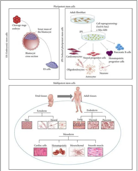
Stem cells are unspecialized cells that have self-renewal ability and can differentiate into specialized cells of the body (http://stemcells.nih.gov). They can be categorized according to their timing or potency. According to their potency abilities and origin, different types of stem cells were shown in Figure 1.1.
2
Figure 1.1 Type of stem cells and their origin; pluripotent embryonic stem cells (ES), induced pluripotent stem cells (iPS), and multipotent fetal and adult stem cells (Figure is adopted from 1)
3
Based on their potency, stem cells can be classified as totipotent, pluripotent, multipotent and unipotent. Totipotent stem cells differentiate into the cells of three embryonic germ layers; ectoderm, endoderm, mesoderm and extra embryonic tissues, whereas pluripotent stem cells differentiate into the cells of three embryonic germ layers, but cannot give rise to the cells of the extra embryonic tissue. Multipotent stem cells can differentiate into the cell types from other different tissues that they originated. Unipotent stem cells can give rise only one specific cell lineage (http://stemcells.nih.gov). Zygote or fertilized egg is a single totipotent cell and generates all the cells in the body, whereas embryonic stem cells (ESC) are pluripotent cells and derived from the inner cell mass of an early mammalian embryo2,3. Adult stem cells, are usually multipotent and reside in several specific organs of adults4,5.
ESCs are undoubtfully key source to understand molecular basis of cellular development as well as cellular threapeutic applications. However due to ethical reasons regarding to the use of human embryo, the scientists focused on the usage of induced pluripotent stem cells (iPSC) and adult stem cells (ASC). iPSCs were first isolated by transfecting four key transcription factors; Oct 4, c-MYC, Sox 2, Klf 4 into mouse fibroblast to differentiate them into embryonic- like stem cells in 20066. This invention seemed to overcome the ethical obstacles against the using of embryo. However the methods of induction with retro viral vectors and their ability to form teratomas are still some of the
4
factors which may limit their usage.
Among ASCs, mesenchymal stem cells (MSC), also referred to as bone marrow derived stromal cells, have became a potential cell source for regenerative medicine, stem cell-based therapy and tissue engineering applications due to their unique features like differentiation potential, nonimmunogenic characteristics and homing ability7,8.
In this study, we aimed to investigate the role of estrogen in the maintenance and differentiation of MSCs. In addition to normal culture conditions, we also examined estrogen’s effect on MSCs when cultured on carbon nanotube arrays (CNTs). We further investigated the genetic and epigenetic changes during the differentiation of MSCs by estrogen hormone since our previous studies showed that estrogen enhances proliferation and inhibits apoptosis of MSCs.
In the introduction section first I will briefly explain stem cells, ESCs, iPS cells, ASCs. Then the importance of MSCs in tissue engineering applications will be defined. The functions and regulation mechanisms of estrogen hormone and the basic concepts of epigenetics will be explained afterwards. Finally, the introduction chapter will end with the explanation of the epigenetic and genetic regulation of MSCs differentiation upon estrogen treatment.
5
1.1 EMBRYONIC STEM CELLS AND INDUCED
PLURIPOTENT STEM CELLS
Fertilized oocyte undergoes very rapid cellular proliferation which is defined as cleavage stage of early development. ES cells are formed in the inner cell mass (ICM) at this stage. Following cleavage hollow of cells are distributed to the appropriate parts of the developing embryo and this process is called gastrulation. Formation of three germ layers, endoderm, ectoderm and mesoderm, is the final products of gastrulation.
ESCs were first isolated by Evans and Kaufman in 1981 from ICM of mouse embryo and from human embryo by Thomson in 19982. They have indefinite self-renewal capacity and broad differentiation potential to develop cells representative of three germ layers9.
When they were injected into mouse blastocysts, they fulfill all developmental requirements differentiating into the cells of three embryonic germ layers and transmitting to the germline of chimeric animals9. In addition, teratoma and embryoid body formations are evidences of pluripotency properties of these cells in vivo and in vitro respectively10. Maintenance of pluripotency capability of ESCs is provided by expression of key pluripotency genes; Oct4, Sox2, and Nanog11,12.
6
Two X chromosomes cytologically or functionally are active in undifferentiated ESCs13. During cell cycle, ESCs undergo symmetric division and generates two daughter cells both of which maintain their stem cell identity. The cell cycle structure of ESCs can be characterized by short G1 phase which can account for the rapid rate of cell division14 . Gı phase activate the cylin dependent kinases (Cdk) in order to begin new round of replication. When ES cells undergo the differentiation, cell cycle structure changes in order to extend significantly G1 phase15.
Both human and mouse ESCs require a feeder layer from irradiated mouse fibroblasts. It was well established that leukemia inhibitory factor (LIF) is an important mediator which keeps mouse ESCs in undifferentiate state through activation of JAK/Stat3 signaling pathway16,17. When LIF is added into the cell culture medium, mouse ESCs can also grow in the absence of feeder layer17,18. On the contrary, LIF addition does not support undifferentiated proliferation of human ES cells without feeder layer. On the other hand, self renewal capacity of human ESCs is controlled by different factors like Fgf2, TGF-β, noggin, activin, and Wnt18,19.
Thanks to their ability to self-renewal and differentiaton potential into the cell types from all germ layers, ESCs have been considered as a promising source
7
for cell-based therapy in treatment of several degenerative diseases such as diabetes mellitus, Parkinson’s disease, Alzheimer, spinal cord injury20 and osteogenesis imperfecta21,22.
Despite the advantages of ESCs as a promising tool for cell based therapies, there are also some medical and ethical pitfalls23. Since their isolation from blastocyst requires the destruction of the embryo, different political and religious groups are opposed to ESC research. In addition, ESCs cause immune reactions in the host and as mentioned above have ability to form teratomas22. Therfore, due to these concerns, researchers sought alternative pluripotent stem cell sources. The reprogramming of fibroblasts to ESC-like cells, which was accomplished by Takahashi and Yamanaka in 2006, was a new hope for stem cell researches6. The invention of induced pluripotent stem cells is considered as another major breakthrough in stem cell research field.
Induced pluripotent cells were directly generated from mouse embryonic or adult fibroblast cells by introducing combining of four selected factors; Oct3/4, Sox 2, Klf 4, c-Myc. These transcription factors play key roles in the induction of pluripotency of somatic cells. The factors were chosen among 24 candidate genes. These critical four genes, which are called as Yamanaka factors, were introduced to fibroblasts by retroviral transfection6, 24.
8
The iPS cells and ESCs share lots of similarities in many respects. First of all, iPS cells can give rise to cells from three embryonic germ layers; ectoderm, endoderm and mesoderm as exactly similar to ESCs. Moreover, the pluripotency ability of iPS cells was demonstrated by teratoma formation after the subcutaneous injection into nude mice and embryoid body formation in vitro like ESCs. Also the cells are expressing ES cell markers like Nanog, Sox2, Eras, Cripto and Dax1. Furthermore, analyses of genome-wide expression patterns and global histone modifications have shown a high degree of similarity between ESCs and iPSCs. Finally, iPSC clones exhibit similar morphology to ES cells such as round shape, large nucleoli and scant cytoplasm25,26.
In light of these considerations, iPSCs are seems to be promising tools for cell based therapy, regenerative medicine and drug development overcoming the difficulties regarding the use of human embryos and tissue rejection problem following transplantation in patients. iPSCs enable patient-specific iPS cell based therapy and minimize rejection. In a previous study, it was reported that generation of iPS cells from patients with different genetic diseases including Parkinson disease, adenosine deaminase deficiency-related severe combined immunodeficiency, Huntington disease, Down syndrome and juvenile-onset diabetes mellitus offers opportunity to investigate the causes of these diseases and drug development27. Alternatively, if the mutation which causes a disease
9
was known, it could be repaired in iPS cells by gene targeting. Afterwards, the healthy tissue-specific cells can be transplanted into the patients following differentiation24,28. Generation and threapeutic applications of iPS cells was shown in Figure 1.2.
Figure 1.2 Generation and applications of iPS cells (Figure is adopted from28)
On the other hand, a major limitation of this technology is the use of viruses that integrate into the genome. Using viruses is also associated with the risk of tumor formation due to reactivation of the viral trans-genes. Thereby, after invention of the iPSCs in 2006, transient transfection methods were extensively studied. To improve the efficiency and to overcome the viral transfection issue, different cell sources such as human dermal fibroblasts, foreskin fibroblasts, fetal liver cells and hepatocytes have been used with different delivery techniques such as inducible lentiviral vectors29, adenovectors35, plasmids26 Cre/LoxP system after single plasmid transfection36. PiggyBac transposons31,32, chemical compounds33, purified recombinant proteins without using any virus or plasmid40 and synthetic modified mRNA molecules which encode the
10
Yamanaka Factors and Lin2841 for reprogramming. However, the efficiency of all these approaches is extremely low. Furthermore, the epigenetic memory problem of iPS cells has not been already solved. Although, somatic cells were reprogrammed to pluripotent cells resembling ESCs, these generated iPS cells keep the epigenetic memory of their tissue of origin 34.
1.2 ADULT STEM CELLS
Adult stem cells (ASC) are undifferentiated cells which reside in a specific organ or tissue. The major role of ASCs is to maintain the homeostasis and to repair the tissue in which they are found. Most types of adult stem cells are multipotent. They have long term, but not unlimited, self-renewal ability and give rise to mature specialized cells35. Typically, adult stem cells divide to generate an intermediate cell; progenitor or precursor cell. Progenitor cells develop into terminally differentiated cells36,37.
ASCs can remain quiescent for a long time in tissue. The specific area in which stem cells reside is called as “niche” (http://stemcells.nih.gov). The ASC reservoir in tissues or organs is protected by asymmetric cell division. Asymmetric cell division is regulated by a niche-derived signals38. After the cell division, one of the two daughter cell contacts the niche retaining their stem cell identity, whereas the other daughter cell leaves the niche and committed to
11
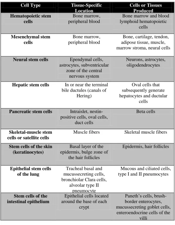
differentiate38. Under normal conditions, in case of the tissue injury or when then the maintenance of the tissue is required ASCs are triggered in order to divide and recruit41. ASCs show clonal growth. Thanks to the asymmetric cell division, the clones contain many cells that have proliferation potential of progenitor cell37. Until now, the existence of ASCs was identified in many tissues and organs and such as blood, bone marrow, liver, skin, testis, ovarian epithelium, muscle, adipose tissue, heart, teeth and brain. Different types of adult stem cells and their location in the body in human was listed in Table 1.1.
Adult bone marrow and peripheral blood contain hematopoietic stem cells (HSC) and MSCs. The first identified ASCs population was HSCs. These cells were first mentioned by Till and McCulloch in 196149. HSCs give rise to all lymphocyte and blood cell types41,42.
1.2.1 MESENCHYMAL STEM CELLS
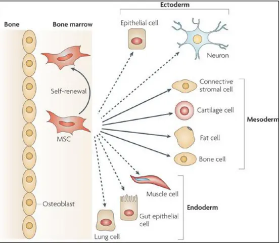
MSCs were first identified in stormal part of bone marrow in 1966 by Friedenstein43 and further characterization was accomplished by Pittenger in 199944. It was demonstrated that human MCSs can give rise to several different cell types including osteocytes, myoctes, adipocytes45,46, chondrocytes47, cardiomyocytes48 and neurons49. The types of cells that are derived form MSC
12
Cell Type Tissue-Specific Location Cells or Tissues Produced Hematopoietic stem cells Bone marrow, peripheral blood
Bone marrow and blood lymphoid hematopoietic cells Mesenchymal stem cells Bone marrow, peripheral blood
Bone, cartilage, tendon, adipose tissue, muscle, marrow stroma, neural cells
Neural stem cells Ependymal cells, astrocytes, subventricular
zone of the central nervous system
Neurons, astrocytes, oligodendrocytes
Hepatic stem cells In or near the terminal bile ductules (canals of
Hering)
Oval cells that subsequently generate hepatocytes and ductular
cells
Pancreatic stem cells Intraislet, nestin-positive cells, oval cells,
duct cells
Beta cells
Skeletal-muscle stem cells or satellite cells
Muscle fibers Skeletal muscle fibers
Stem cells of the skin (keratinocytes)
Basal layer of the epidermis, bulge zone of
the hair follicles
Epidermis, hair follicles
Epithelial stem cells of the lung
Tracheal basal and mucussecreting cells, bronchiolar Clara cells,
alveolar type II pneumocyte
Mucous and ciliated cells, type I and II pneumocytes
Stem cells of the intestinal epithelium
Epithelial cells located around the base of each
crypt
Paneth’s cells, brush-border enterocytes, mucussecreting goblet cells,
enteroendocrine cells of the villi
Table 1.1 Different types of adult stem cells and their location in the body
(Reproduced with permission from Körbling, M et al.41, Copyright Massachusetts Medical Society)
13
was shown in Figure 1.3. In addition, MSCs support the long-term hematopoiesis and expansion of embryonic stem cells via expressing cytokines and growth factors48. MSCs have been isolated from different tissues other than bone marrow, such as adipose tissue, amniotic fluids, periosteum, fetal tissues, dental pulp, peripheral blood, liver and spleen. Morphologically MSCs show heterogeneity, ranging from narrow spindle shaped to cuboidal, packed cells
51,52
.
Phenotypically, adult human MSCs are positive for a number of markers, such as CD90 (Thy-1), CD117 (c-kit), CD71, CD105, CD73, CD44, and adhesion molecules like VCAM-1 (vascular cell adhesion molecule), ICAM-1 (intercellular adhesion molecule), CD166 (activated leukocyte cell adhesion molecule) and CD29 (integrin beta-1). They do not express hematopoietic markers; CD34, CD45, CD14, co-stimulatory molecules CD40, CD86, CD80 48,51,52,53
.
Adult human MSCs express major histocompatibility complex (MHC) class I, whereas human leukocyte antigen (HLA) class II antigens and MHC class II expressions are absent. In addition MSCs have an immunosuppressive function. They inhibit T cell and B cell proliferation and differentiation of B cells into plasma cells. This phenotype shows the non-immunogenic character of the
14
Figure 1.3 Types of cells differentiated from bone marrow derived MSC and their germ layers (Figure is adopted from 54)
MSC51,55. It has been shown by in vivo models that MSCs can prevent and treat graft-versus-host disease (GVHD) seen in allogeneic stem cell transplantation50. Also MSCs may be used in the treatment of autoimmune disorders55-57.
While MSCs are differentiating into different cell lineages under appropriate conditions, expression of pluripotency genes such as Oct4, Sox2, TERT (telomerase reverse transcriptase) decrease. The expression of transcription factors, which are involved in MSC differentiation such as RUNX2, ALP,
15
PPARγ, C/EBPα, are also regulated by epigenetic mechanisms. Histone modifications and CpG methylayion states on these genes are changed upon differentiation induction. Thus, the presence of these epigenetic regulation mechanisms is crucial for MSC differentiation58,59.
MSCs may follow different ways during repair of an injured tissue or organ. First, host MSCs can enter into peripheral blood circulation and migrate to target the ischemic tissue. Secondly, MSCs that were injected exogenously can enter the blood circulation and home to injured tissue or inflammation site. As third mechanism, MSCs can be locally injected at the site of injury. The injected MSCs may differentiate into tissue specific stem cells to regenerate. On the other hand, the methods using in MSC homing/trafficking are still inadequate to determine the exact location7,52,60 .
Due to their differentiation potential and immune-privileged characteristic, MSCs derived from bone marrow is considered as promising cell source for tissue engineering applications in recent years61. However, the adhesion of stem cells on ECM is the major concern for particular growth and differentiation. Different types of synthetic and natural biomaterials were demonstrated as scaffold material for MSC transplantation especially for therapeutic applications of cartilage and bone defects61-63. Porous ceramics cylinders and biodegradable polymers; poly-L-lactide (PLA) and poly-Llactide-co-glycolide
16
(PLGA), hydrogel alginate and collagen type gels were demonstrated as alternative scaffolds60-66. Adhesion, proliferation and differentiaton of bone marrow derived MSCs on these constructs were demonstrated through in vivo and in vitro experiments60,64,66,67. For instance, repairing of full-thickness articular cartilage defects in knees of rabbits and bone defects in dogs successfully accomplished through the autologous-MSC transplantation on collagen-type-1 gel and porous ceramics cylinders respectively66,69.
1.3 USE OF CARBON NANOTUBES AS THREE-
DIMENSIONAL SCAFFOLD MATERIAL
The fundamental principle of tissue engineering approach is to repair and restore the diseased and damaged tissue with different strategies70. Over the past decades, using of biodegradable and biocompatible scaffolds in tissue engineering field have developed.
The major purpose is producing a construct, which can structurally and functionally mimic the extra cellular matrix (ECM) of cells67,71 , to transplant into diseased or defective tissue after seeding the autologous cells onto these scaffolds. That’s why, producing an ideal scaffold protein, which can support different cell types and capable of stimulating cell growth, adhesion and proliferation mimicking the native tissue, is a critical point in tissue
17
engineering.
In last decade, tremendous amount of attempt have been applied to produce better scaffold construct which can closely mimic the ECM of native tissues67,72. For this purpose different kinds of biomaterials were used as scaffold such as porous ceramics cylinders, biodegradable polymers like poly-L-lactide (PLA) and poly-poly-L-lactide-co-glycolide (PLGA), hydrogel alginate and collagen type gels60-66. In addition, the importance of topographical pattern on cell viability was also demonstrated in a number of studies. Especially it was shown that dimension and roughness features of surface induce cell growth and support attachment of the cells72. Thereby, scaffold proteins are promising tools in tissue engineering due to their patterned surface and nanometer scale dimension73,74. It was reported that the rougher surface induced gene expression of fibroblast cells67. Moreover, surface properties of scaffolds have potential to develope new environment which is able to stimulate cell proliferation and adhesion72. Due to their patterned surface and nanometer scale dimension, scaffold proteins are considered as promising tools in tissue engineering73-75.
In recent years, carbon nanotubes (CNTs) have became an attractive alternative scaffold material in tissue regeneration field due to their dimension, chemical stability, their high tensile mechanical strength, electrical conductivites, biocompatible feature and chemical properties74. Recently, CNT have been
18
proposed for biomedical applications such as cell tracking and labeling, tissue engineering scaffolds, nanosensors, and vehicles for controlled release of drugs or delivery of bioactive agents65.
As mentioned, due to their multi-lineage differentiaton potentials and immune-privileged characteristics, MSCs have become a feasible and potential source for tissue engineering applications76. It was also reported that CNT–derived patterned surfaces provides an ideal and suitable scaffolds for MSCs and neural stem cells77, 78.
In addition to topographical features of scaffold material, the growth factors or small molecules which added into cell culture media are also important for tisseue engineering. Because, these factors support the attachment, proliferation and differentiaton of cells seeded onto biomaterial.
1.4 ESTROGEN AND ESTROGEN RECEPTOR PATHWAY
Estrogen, one of the steroid hormones, shows a wide variety of effects on growth, differentiation, sexual behavior, apoptosis, development, reproductive and cardiovascular functions and many physiological processes in female and male79, 80. Estrogen shows its effect in a broad range of tissues such as liver,
19
brain, bone, cardiovascular and reproductive systems81. The most dominant estrogen that found in human is 17β estradiol compared to estrone and estriol82
.
Estrogen does not only influence the differentiation, maturation and function of stem cells, but also have a proliferative and antiapoptotic effect by changing the transcriptional activity of related genes79,83 .
Nuclear receptors (NR) coordinately regulate the expression of genes that have a role in numerous of biological processes such as development, reproduction and homeostasis. NRs activate or repress gene transcription usually by binding to NR-specific ligands such as hormones, vitamins, lipid metabolites and xenobiotics84.
Steroid hormone receptors are member of nuclear receptor family and act classically as transcription factors. Two major estrogen receptors; estrogen receptor alpha (ERα) and beta (ERβ), convey the regulatory signal of estrogen and control the transcription of target genes. ERs function both as signal transducer and transcriptional factor to regulate the target genes85.
ERα (NR3A1 in human) was identified by Jensen and Jacobsen in 1962 and ERβ (NR3A2 in human) was identified in 1966 by Gustafsson in human, mouse and rat. Two receptors are encoded by different genes located on different
20
chomosomes. Moreover, any splice variant was not identified until now86,87. ERs share similar structural anatomy and sequence homology. An ER protein consists of six modular domains; N-terminal domain, which is called as A and B domains, is involved in transcriptional activation of target gene; DNA binding domain (C domain) enables the dimerization and binding of receptor to estrogen response element (ERE) on DNA; the hinge (D domain) is involved in receptor dimerization; C-terminal domain (E domain) is E2-binding domain; F domain modulates transcriptional activity of ER88.
Two major ER pathways were identified so far; genomic or classical pathway and non-genomic pathway. In classical pathway, which is also called as estrogen response element (ERE) dependent pathway, estrogen hormone activates ER in the cytosol resulted in the formation of homodimer. Receptor translocates into to the nucleus after activation and homodimerization respectively and binds to estrogen response element (ERE), which is located at the promoter region of the target gene interacting with specific co-activators and co-repressors subsequently82,83,89. ERs localize in cytoplasm associating with heat shock proteins (Hsps) such as Hsp 90, Hsp 70, Hsp 56 in the absence of ligand. The Hsps maintain the estrogen receptors in inactive state. Binding of ligand to ERs trigger them to dissociate from Hsp and to dimerize90. The ER activation is generally stimulated in response to binding of 17-estradiol (E2) or other agonists; estrone (E1) and estriol (E3) in humans82.
21
In addition to classical ERE-dependent pathway, ERs can also regulate gene expression without direct binding to ERE on DNA. In this alternative pathway, ligand-activated ER associates with another transcription factor such as CRE-like element, which binds to Jun/ATF transcription factors, AP-1 site, which binds to Jun/Fos transcription factor, and CCAAAT site, which binds to SP1 and NF-Y transcription factors88,90,91, which, in turn regulates the transcription of genes that lack of ERE site.
In the third form of genomic pathway, also referred to as ligand-independent pathway, growth factors are involved in process instead of estrogen. Nuclear ERs are phosphorylated by protein kinases such as ERK 1-2, p38 MAPK, cyclin dependent kinases, protein kinase A and Akt upon growth factor stimulation. Phosphorylated and activated ERs bind to ERE site of target genes 91,92
.
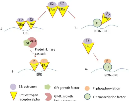
In the second major ER pathway, non-genomic pathway, numerous of signaling pathways such as mitogen-activated kinase (MAPK) pathway, PI3K/Akt pathway and JAK/STAT pathway are induced by estrogen-activated membrane bound ER86. The estrogen receptor signaling pathways were summarized in Figure 1.4.
22
Figure 1.4 Estrogen receptor signaling pathways 1. ERE-dependent classical pathway 2. ERE-independent genomic pathway 3. Ligand-independent genomic pathway 4.Non-genomic pathway
1.5 EPIGENETICS
Conrad Waddington came up with the term “epigenetic” 60 years ago. It is classically defined as stable alterations in gene expression that are mitotically and/or meiotically heritable and occur on DNA or chromatin structure without changing nucleotide sequence93-95. Epigenetic modifications include reversible changes on given phenotype, but do not cause any change in genotype. Epigenetic mechanisms are master regulators of transcriptional pathways during major biological processes such as embryonic development, cellular memory,
23
cell and tissue specific gene expression, differentiation and adult tissue maintenance96,97. Gene regulation at the epigenetic level occurs through covalent modifications both on DNA and histones. These modifications regulate the gene transcription by opening chromatin structure (euchromatin), which enables the accessibility of transcription factors to activate gene expression, or facilitating condensed DNA structure (heterochromatin), which does not allow the entering of transcription factors to repress gene expression 97,98
. Loosing of proper chromatin modifications cause lethality during embryonic developmental stage99,100. Epigenetic modifications despite of their stability can be reprogrammed using different strategies such as treatment with chemical compounds or hormones, transfection of transcription factors into the cells and nuclear fusion98. For instance, generation of iPS cells from somatic parental cells provided insight into epigenetic reprogramming of cells102.
There are different kinds of epigenetic modifications such as DNA methylation, DNA hydroxymethylation, histone modifications, chromatin remodeling and microRNAs. However, DNA methylation and histone protein modifications are considered as two major epigenetic modifications involved in regulation of stem cell.
24
1.5.1 DNA METHYLATION
The important role of DNA methylation/demethylation was first suggested by Griffith and Mahler in 1969103. DNA methylation is catalyzed by DNA methyltransferases (DNMTs) that add a methyl group (CH3) on 5’position of cytosine (m5C), which is at cytosine-phosphate-guanine (CpG) dinucleotides in some fungal, invertebrates, some bacteria species, flowering plants and in all vertebrates. DNA methyltransferases are conserved proteins and establish methylation at genes during in early development and cell division 109,110. Regions, which consist of high frequency of CpG residues, are defined as CpG islands96,104. CpG islands are located near the promoter regions of genes. According to computational analysis there are almost 29.000 CpG islands in human genome105.
Epigenetic modifications can be widely reprogrammed in both germ cells and the preimplantated embryo. At two cell stages, the major genome-wide DNA methylation occurs in mammals; in primordial germ cells (PGC) while migrating to genital ridge and after fertilization in embryo. Early PGCs initially possess similar DNA methylation status to somatic cells. Upon fertilization, paternal and maternal genome is passively demethylated before replication and during replication respectively. After implantation, lineage specific de nova methylations which are catalyzed by two different DNMTs; DNMT3a,
25
DNMT3b. When PGCs enter into the genital ridge, the methylation is erased in response to signal from somatic cells in the gonadal anlagen106,107. De novo metylation occurs also in terminally differentiated somatic cells, but slower than embryonic cells and germ cells. Moreover, progressive global hypomethylation pattern on genomic DNA in somatic cells is associated with cancer and age108,109.
Cytosine methylation is also associated with genomic imprinting and X-inactivation, which is identified as completely silencing one of two identical alleles. Failure in imprinting of specific loci causes several disorders in human 110,111,112
. During X-chromosome inactivation process, one of the two X chromosomes is randomly inactivated in female through dosage compensation mechanism and cytosine methylation 109,110,113.
Methylated DNA associates with methly-CpG binding proteins (MBD) to repress gene expression. The major MBDs identified in mammalian are Mbd1, Mbd2, Mbd3, Mbd4 and MeCP2114,115. After binding to methylated CpG, MeCP2 recruits histone deactylases116, Mbd 1 recruits Suv39H1, which is a histone 3 lysine-9 (H3K9) methylase and HP1, which is a methyl lysine binding protein, respectively117 and Mbd2 and Mbd3 recruits NuRD, which is a ATP-dependent nucleosome remodeling and histone deactelyase complex118.
26
The major question is that how epigenetic modifications can be propagated to daughter cells during cell division. DNA methylation pattern is maintained thanks to semi-conservative replication. DNMT1 enzyme provides the passing of methylation mark on the parental DNA strand to daughter cells during mitosis. On the other hand, de novo methylations both on hemimethylated and unmodified cytosines are catalyzed by DNMT3A and DNMT3B111,119.
1.5.2 HISTONE MODIFICATIONS
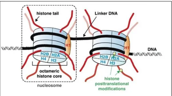
In eucaryotes, double-helix DNA is packaged by tightly wrapping 145-147 base pairs around an octomer. This DNA:protein compact structure is called as chromatin and nucleosome is the basic unit of chromatin. Nucleosome is composed of two copies of four core histone proteins; H2A, H2B, H3 and H4. In addition, histone protein H1 is a linker histone which establishes on DNA between nucleosomes and facilitates condensed nucleosome chain called as chromatin fiber121. Core histone protein stucture was shown in Figure 1.5.
27
Figure 1.5 Core histone structure (Figure is adopted from194)
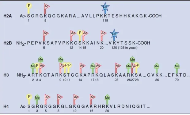
Packaging is required to establish the whole genomic DNA in nucleus. Dynamic N-terminal tails of histones, which protrude out from nucleosome structure, are subjected by post-translational modifications (PTMs). These modifications regulate gene transcription through controlling the accesibilty of transcription factors and co-regulators. There are eight major PTMs; methylation, acetylation, phosphorylation, ubiquitination, sumolaytion, deamination, ADP-ribosylation and proline isomerization. The modifications which occur on histone tails can be propagated to daughter cells during cell division like DNA methylation. Post-translational modifications occur on lysine (K), arginine (R), serine (S) and threonine residues of tails and they are regulated by specific enzymes termed chromatin modifiers. Furthermore, there are different forms of modifications that occur on these residues. For instance,
28
lysine residues can be mono-, di-, or trimethylated and one or two methyl group can be added to arginine residues 96,118,120,121.
Genes are regulated by different combinations of histone modifications at a specific locus, which is known as histone code. Histone code hypothesis was firstly suggested by Strahl and Allis in 2000 122,123.
Covalent modifications which take place on specific sites of histone tails associated with both activation and repression of transcription. Methylation of lysine 9 and lysine 27 of histone H3 (H3K9me / H3K27me) correlates with repression state, whereas methylation of lysine 4 or lysine 36 (H3K4me/ H3K36me) and acetylation of lysine 9 (H3K9ac) associates with active genes 124,125
. Post-translational covalent histone modifications on core histone proteins was shown in Figure 1.6.
First identified histone modifier enzyme was a histone acetyltransferase (HAT) Gcn 5126. HATs are considered as transcriptional activators because they provide the neuralization of positive charge of lysine via transferring acetyl group to ε amino group of lysine. Thus, they facilitated weakened DNA-histone interaction, which enables the access of transcription factors and co-activators. Histone deacetylases (HDACs) remove the acetyl groups from lysines and considered as repressors127. There are three main acetyltransferase families;
29
GNAT, MYST, and CBP/p300 and four classes of HDAC Classes I, II, IV and III, which is homologous to yeast Sir2128.
Figure 1.6 Post-translational covalent histone modifications on core histone proteins. P: Phosphorylation, Ac: Acetylation, Ub: Ubiquitinylation, Me: Methylation (Figure is adopted from118).
Histone methylation occurs on the lysine and arginine residues of tails. Methylation of lysines implies both active and repressive chromatin states118. Methylation of H3K9 and H3K27 associates with silent chromatin state, whereas methylation of H3K4 and H3K36 associates with euchromatin state129. In recent studies it was reported that, in mouse embryonic stem cells, bivalent domains are found which contain both H3K27me3 and H3K4me3 marks130.
30
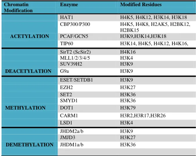
SUV39H, which is the first identified histone lysine methyltransferase (HKMT), is responsible for the methylation of H3K9. HKMTs specifically methylate lysine residues at a specific degree; mono-, di-, tri- methylation132,124. SET7/9 catalayzes demethylaion of H3K4 and EZH2 targets H3K27. Histone modifier enzymes and modified residues were given in Table 1.2.
Chromatin Modification
Enzyme Modified Residues
HAT1 H4K5, H4K12, H3K14, H3K18 CBP300/P300 H4K5, H4K8, H2AK5, H2BK12, H2BK15 ACETYLATION PCAF/GCN5 H3K9,H3K14,H3K18 TIP60 H3K14, H4K5, H4K12, H4K16, SirT2 (ScSir2) H4K16 MLL1/2/3/4/5 H3K4 SUV39H2 H3K9 DEACETYLATION G9a H3K9 ESET/SETDB1 H3K9 EZH2 H3K27 SET2 H3K36 SMYD1 H3K36 METHYLATION DOT1 H3K79 CARM1 H3R2,H3R17,H3R26 LSD1 H3K4 JHDM2a/b H3K9 JMJD3 H3K27 DEMETHYLATION JHDM1a/b H3K36
31
1.6 EPIGENETIC ALTERATIONS IN STEM CELLS
In ESCs, lineage specific genes are repressed through both DNA methylation and repressive histone modifications such as Histone-3 lysine-9 (H3K9) and Histone-3 lysine-27 (H3K27), whereas, the expressional regulation of pluripotency assocaited genes such as Oct4, Nanog and Sox2 is correlated with DNA methylation110,131. Moreover it was demonstrated that Polycomb group (PcG) protein complexes and HP1 are required for gene repression in ESCs132. Regulation of cell lineage speficic genes are mediated by bivalent histone modifications. Bivalent domains are considered as transcription-ready sites. These genes are regulated by both H3K4 and K27 trimethylation and the genes are activated upon differentiation induction133. Genome-wide distribution of DNA methylation, histone methylation and chromatin modifications regulates the changes that occur during cell differentiation. Thereby, epigenetic modification patterns including both histone modifications and DNA methylations of ESCs are clearly distinguished from differentiated somatic cells. The genome-scale DNA methylation maps and chromatin-state maps of ESCs revealed that housekeeping genes are highly expressed and enriched with H3K4me3 active mark, whereas lineage specific genes are repressed through both DNA methylation and repressive histone modifications such as H3K9 and H3K27134. DNA methylation is under the control of DNA methyltransferase Dnmt3a, Dnmt3b and Dnmt1 during embryogenesis135. A failure which changes
32
the maintenance of DNA methylation results in emryonic lethality in mice136 and several diseases such as cancer, Alpha-thalassemia, mental retardation and fragile-X syndrome112. It was also reported that the passaging also causes change in DNA methylation pattern of ESCs. In a previous study, it was demonstrated that methylation of tumor supressor genes; RASSF1, DCR1 and PTPN6 increase at late passages in human ESCs138.
Direct reprogramming of somatic cells by transcription-factor transduction yields iPSCs with remarkable similarity to embryonic stem cells. These two pluripotent stem cells share very similar epigenetic profiles137. On the other hand large-scale methylation comparisons have identified differences such as CpG methylation which is higher in iPSC than in ESCs138. In addition whole-genome screening revelaed the presence of differentially methylated regions (DMRs) within iPSC. These regionas are resistant to reprogramming139. Actually, the differences in pluriportent states and differential potential of iPCs and ESCs was not observed, whereas, retaining of the residual epignetic marks after differentiation influences cell fates in iPS cells. This problem manifests the epigenetic memory140. For instance, iPSCs derived neural progenitors and fibroblasts demonstrate decreased blood-forming potential in vitro due to the retained residual methylation at loci which is required for haematopoietic cell fate.
33
cell types are hypomethylated in MSCs. DNA hypomethylation contributes transcriptionally permissive state. But this hypomethylated state does not predict differentiaton capacity of MSCs. Since these genes are mainly regulated by post-translational histone modifications. H3K4 and H3K27 bivalency on lineage-specific genes was demonstrated. After differentiation induction, H3K27 demethylases activates the transcription of these genes5, 141. In MSCs on adipogenic gene promoters including PPARγ2, leptin, fabp4, and lipoprotein lipase (lpl) hypomethylation is established. In addition the adipogenic genes show mosaic CpG methylation patterns59. During differentiation demethylation of H3K9, acetylation of H3 and H3K4 trimethylation were also shown on adipogenic gene promoters142,143. MSC cultures from bone marrow show stable DNA methylation pattern between early and late passages. Osteocalcin is a ostegeneic linege specific gene and target of RUNX2. In the presence of osteogenic induction, on osteocalcin promoter acetylation of H3 and H4 increases, wheras DNA metyhlation decreases144. It was known that osteogenesis has a dominant effect on adipogenesis and osteoblastic inducers; bone morphogenetic protein 2 (BMP2) and Wnt ligands, suppresses transcription of PPARγ recruting repressor histone methyltransferase145
. It was also reported that JMJD2B and JMJD3B enzymes are the link between the osteogenesis and chondrogenesis. It was shown that overexperssion of these demethylases stimulates the expression of alkaline phospahatase (ALP), which is an osteoblast differentiation marker, whereas depletion of these demethylases
34
resulted in a significant increase in adipogenic differentiaton in MSCs146.
1.7 ESTROGEN AND EPIGENETIC REGULATION
Co-activators and co-repressors are directly recruited by nuclear receptors as a consequence of ligand-binding. Until now approximately 285 co-regulators associated with nuclear receptors were identified. Also it was reported that a mutation, which occurs in the expression of these co-regulators, causes diseases such as multiple cancers, Alzheimer, Parkinson and Huntington diseases84. Estrogen receptors also regulate gene transcription interacting with co-activators or co-repressors; enzymatic complexes such as HATs, SWI/SNF complexes and HDACs89.
Effect of estrogen on epigenetic mechanisms has been mostly investigated in breast cancers. In breast cancer, regulation of gene expression is mediated by methylation of DNA and acetylation of histone proteins and estrogen plays an important role in both formation of breast cancer and breast cancer treatment. It was well established that estrogen stimulates development of certain breast cancers and hormonal therapy via anti-estrogen treatment is widely used in treatment of these cancers147. H3K9me3 demethylase JMJD2B and H3K4 methyltransferase MLL2 complex directly interacts with ER. This is responsible for the cellular function of ERα in MCF-7 breast cancer cell line148
35
In the ER-negative breast cancer cells, promoter of ER gene is hypermethylated and ER mRNA is absent. Treatment of the ER-negative breast tumors with DNMT and HDAC inhibitors imposes to the reactivation of ERα149.
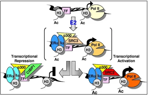
In contrast to known stimulator effect of estrogen on gene expression, estrogen occupied ERα may also repress the gene expression by recruiting corepressor/HDAC complexes150-153 and DNA methyltransferase which enables establishment of hypermethylation on specific CpG loci152. On the other hand, high level of acetylation was observed on E2-stimulated genes. The proposed model was demonstrated in Figure 1.7.
Figure 1.7 Demonstration of repression of early target genes mediated by ERα (Figure is adopted from153)