Can mean platelet volume and
neutrophil-to-lymphocyte ratio be biomarkers of acute
exacerbation of bronchiectasis in children?
Hikmet t. NaCaroglu
1, SemiHa BaHCeCi erdem
1, Sait karamaN
1, SelCuk YaziCi
2,
demet CaN
11Department of Pediatric Allergy and Immunology, Dr. Behcet Uz Children Disease and Surgery Training and Research Hospital, Izmir, Turkey
2Department of Paediatrics, Balιkesir University Medical Faculty, Balιkesir, Turkey
Abstract
Introduction: Bronchiectasis (Be) is a parenchymal lung disease evolving as a result of
recur-rent lung infections and chronic inflammation. although it has been shown in adult studies that mean platelet volume (mPV) and neutrophil-to-lymphocyte ratio (Nlr) can be used as biomarkers of airway inflammation, knowledge is limited in the paediatric age group. the aim of our study is to investigate the potential of mPV and Nlr as biomarkers that may indicate acute exacerbations of non-cystic fibrosis Be in children.
Material and methods: Children with non-cystic fibrosis Be (n = 50), who were followed in the
division of Paediatric Pulmonology of our hospital between June 2010 and July 2015, were involved in the present retrospective cross-sectional study. Haemogram values during acute exacerbations and non-exacerbation periods, and a control group were compared.
Results: in children with bronchiectasis, the average leukocyte count (p < 0.001), platelet count
(p = 0.018), absolute neutrophil count (p < 0.001), and Nlr (p < 0.001) were higher, as expected, when compared with the control group. Nlr values, in the period of acute exacerbation were signifi-cantly higher than the values of both the non-exacerbation periods (p = 0.02) and the control group (p < 0.001). in contrast, mPV values in the period of acute exacerbation did not exhibit a significant difference from those of non-exacerbation periods (p = 0.530) and the control group (p = 0.103).
Conclusions: it was concluded that leukocyte count, platelet count, absolute neutrophil count, and
Nlr can be used to show chronic inflammation in Be, but only Nlr and absolute neutrophil count can be used as biomarkers to show acute exacerbations.
Key words: mean platelet volume, neutrophil-to-lymphocyte ratio, childhood, bronchiectasis.
(Centr eur J immunol 2017; 42 (4): 358-362)
Introduction
Bronchiectasis (BE) is a chronic inflammatory lung disease that leads to persistent productive cough, recurrent lower respiratory tract infections, and obstructive lung dis-ease in children and adults [1]. The disdis-ease is characterised by abnormal and persistent dilatation of the airways, which occurs as a result of the chronic inflammation due to insuf-ficient mucosal clearance and recurrent/chronic infections of the airways [2]. The exacerbations observed in BE pa-tients, reduce lung function by increasing the damage in the lung parenchyma, impair quality of life and lead to the development of respiratory failure in patients [3]. In BE patients, identification of biomarkers of inflammation showing relapse is of great importance. Most of the present
biomarkers require cooperation of the patient or tend to be invasive, therefore they cannot be used effectively. For example, the spirometric measurements that are often used require effective cooperation and thus have limited use in small children, and methods such as bronchial biopsy and bronchoalveolar lavage (BAL) are not practical because they tend to be invasive [4, 5].
Neutrophils and platelets are known to be engaged in the inflammatory process in BE and to play a role in pro-gressive lung damage [1-3]. Mean platelet volume (MPV) is a value that indicates platelet activation, and can be used as a biomarker in inflammation [6-8]. Also in adult studies, it has been shown that the neutrophil-to-lymphocyte ratio (NLR) may be a biomarker of airway inflammation [9].
The aim of our study is to investigate the potential of MPV and NLR as biomarkers that may indicate acute ex-acerbations of non-cystic fibrosis (CF) BE in children.
Material and methods
Patient population
All patients who had a diagnosis of non-CF BE or were hospitalised due to acute exacerbation between June 2010 and July 2015, were included in the present retrospective cross-sectional study. Average age of the 50 children who were hospitalised due to acute exacerbation was detect-ed as 151 (±53 SD) months. Twenty-four (48%) were male and 26 (52%) were female. In the control group, 45 (51.7%) were male, 42 (48.3%) were female, and the average age was 150 (±36 SD) months. The diagnosis was confirmed by thorax High-Resolution Computed Tomogra-phy (HRCT) in the patients who had a persistent wet cough and abnormal chest X-ray findings that did not respond to antibiotic therapy and also maintained non-regressive symptoms for 2-4 weeks [10]. The aetiology of BE was detected as; primary immune deficiency 26%, protracted bacterial bronchitis 22%, bronchiolitis obliterans 18%, pri-mary ciliary dyskinesia 10%, lung disease secondary to gastro-oesophageal reflux 8%, foreign body aspiration 6%, tuberculosis 6%, and congenital malformation 4%. Exacer-bation criteria were defined as having more than two of the following signs and symptoms: increase in cough frequen-cy, sputum production with changes in sputum content, dyspnoea, tachypnoea, reduced exercise capacity, loss of appetite, haemoptysis, fever above 38oC, and clinically
de-termined crackles and wheezing [10]. Children who were diagnosed with sepsis, iron deficiency anaemia, obesity, hyperlipidaemia, diabetes mellitus, hypertension, chronic renal failure, nephrotic syndrome, inflammatory bowel dis-ease, connective tissue disdis-ease, or receiving systemic ste-roids within the last month were excluded from our study
because it was previously reported that these diseases af-fected MPV and NLR values. The demographic parameters of patients and haemogram findings in non-exacerbation periods were obtained from the file records. When patients were hospitalised due to acute exacerbation, full blood count and C-reactive protein (CRP) were studied. Haemo-gram parameters and CRP values in acute exacerbations (Group 1) and non-exacerbation periods (Group 2) were compared. Eighty-seven healthy children with equivalent age and gender were randomised as the control group. Then the same parameters were compared with the data of the patients in the healthy control group (Group 3). Approval by the local Ethics Committee was granted.
Counting blood samples
Whole blood count was performed via Beckman Coulter LH 780, and blood samples which were anticoagulated with K3EDTA were used. The Coulter principle is volumetric analysis. Haemoglobin level, white blood cell count (WBC), platelet count, and MPV values were recorded for each pa-tient. The reference range for MPV was between 7.0 and 11.0 fL. The neutrophil-lymphocyte ratio was calculated by dividing the percentage of neutrophils and lymphocytes in complete blood count analysis. Serum CRP level was mea-sured using an Abbott c8000 Architect (USA) device.
Statistical analyses
Statistical Package for Social Sciences (SPSS for Win-dows 15.0 Chicago, USA) software was used to analyse the data. Results were given as either mean ± standard devia-tion (SD) or median (min-max) according to the distribudevia-tion. The Chi-squared test was used to compare grouped data. The paired t-test was used to compare group-specific mea-surements, and the independent Student’s t-test to compare measurements among independent groups. Kruskal-Wallis test and Mann-Whitney u tests were used for non-normal-ly distributed variables. Pearson’s correlation ananon-normal-lysis was
Table 1. Comparison of haemogram parameters and C-reactive protein levels between acute exacerbation (Group 1),
non-exacerbation (Group 2), and controls (Group 3)
Parameter Group 1 (n = 50) Group 2 (n = 50) Group 3 (n = 87) 1-2 p 2-3 p 1-3 p WBC (× 103/µl) 10.8 ±4.1 8.5 ±3.5 7.3 ±1.4 0.83 < 0.001 < 0.001
Absolute neutrophil count (× 103/µl) 7.3 ±4.1 4.9 ±3.1 3.8 ±1.1 0.001 < 0.001 < 0.001 Absolute lymphocyte count (× 103/µl) 2.1 ±0.8 2.4 ±0.9 2.7 ±0.6 0.71 0.607 < 0.001
NLR 5.1 ±6.3 2.3 ±2.2 1.5 ±1.2 0.02 0.01 < 0.001
CRP (mg/l) 4.1 ±7.0 0.45 ±0.5 < 0.001 -
-Plt (× 103/µl) 319 ±103 336 ±106 285 ±62 0.284 0.001 0.018
MPV (fl) 8.5 ±0.9 8.6 ±1.09 8.2 ±0.7 0.530 0.03 0.103
WBC – white blood cell, NLR – neutrophil-to-lymphocyte ratio, CRP – C-reactive protein, Plt – platelet, MPV – mean platelet volume data are shown as means ± standard deviation for normally distributed variables
used to explore correlations between the measurements.
P-values < 0.05 were considered statistically significant.
Results
Leukocyte count, platelet count, MPV, neutrophil count, absolute lymphocyte count, and NLR values of patients during acute exacerbations and non-exacerbation periods and of control group are shown in Table 1. Platelet counts of both acute exacerbations and non-exacerbation periods were significantly higher than those in the con-trol group (p = 0.001, p = 0.018). Average MPV values of non-exacerbation periods were significantly higher than in the control group (p = 0.03). In contrast, the MPV values of acute exacerbation were not statistically different when compared to non-exacerbation periods or the control group (p = 0.103, p = 0.530, respectively) (Table 1).
Absolute neutrophil counts in the periods of acute ex-acerbations and non-exacerbation were significantly higher than in the control group (p < 0.001). When periods of acute exacerbations and non-exacerbation were compared, absolute neutrophil count was found to be significantly higher in the period of exacerbation (p = 0.001) (Table1). NLR revealed a statistically significant increase both in the period of acute exacerbation and during non-exacer-bation when compared with the control group (p = 0.02,
p = 0.01). Also, when compared to each other, NLR in the
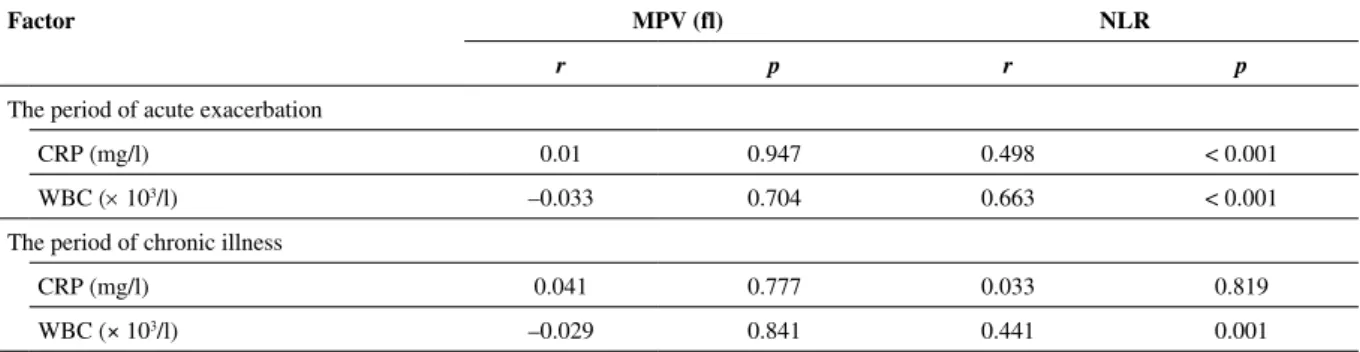
exacerbation period was significantly higher than during the period of non-exacerbation (p < 0.001) (Table 1). The CRP values in the acute exacerbation period were significantly higher than in the non-exacerbation period (p < 0.001) (Table 1). We investigated the correlation between leukocyte count and CRP values with the MPV. When acute exacerbations and non- exacerbation periods were compared, there was no correlation. A moderate-ly (r = 0.498) significant, positive correlation was found between NLR and acute exacerbation period CRP levels (p < 0.001). Similarly, a moderately (r = 0.663) significant
positive correlation was found between NLR and acute ex-acerbation-period leucocyte numbers (p < 0.001) (Table 2).
Discussion
In the present study, which evaluates the potential of haemogram parameters of children with BE (especially MPV and NLR) as biomarkers of acute exacerbation, it was detected that 1) leukocyte count, platelet count, abso-lute neutrophil count, and NLR show chronic inflamma-tion of BE, and 2) only absolute neutrophil count and NLR can be used as biomarkers of acute exacerbation. To the best of our knowledge, there is no study in the literature that evaluates the roles of both MPV and NLR in predict-ing acute exacerbation in patients with BE.
Mean platelet volume is correlated with platelet func-tion and activafunc-tion [6]. Platelet activafunc-tion that occurs in the process of inflammation can be measured indirectly through MPV. Mean platelet volume alone represents both platelet stimulation and the rate of platelet production [11]. CD62, CD63, GP IIB/IIIA, PF4, and thromboglobulin can be used as markers of platelet activation [12]. These tests are not routinely used due to their high cost and need of specialised equipment [13].Measurement of mean platelet volume is a cheap, effective, and an easy method that is closely correlated with platelet function and activation and is used in the assessment of platelet function.
Recent studies show that platelets, one of the most im-portant elements of the haemostasis process, also play a role in the development of immune response [14]. Usually in adult studies, it has been shown that there is platelet activa-tion in different inflammatory lung diseases [8, 9, 15-21].
In the study by Bansal et al. [15]involving 100 patients with chronic obstructive pulmonary disease (COPD) and 100 healthy subjects, the MPV values in COPD patients were detected as being higher than those in the control group. Erden et al. [16]found MPV values to be higher during flare-up periods in their work about exacerbation
Table 2. Correlation of neutrophil count and C-reactive protein levels and correlation of mean platelet volume and
neu-trophil-to-lymphocyte ratio in patient groups during acute exacerbation and non-exacerbation chronic disease periods
Factor MPV (fl) NLR
r p r p
The period of acute exacerbation
CRP (mg/l) 0.01 0.947 0.498 < 0.001
WBC (× 103/l) –0.033 0.704 0.663 < 0.001
The period of chronic illness
CRP (mg/l) 0.041 0.777 0.033 0.819
WBC (× 103/l) –0.029 0.841 0.441 0.001
CRP – C-reactive protein, WBC – white blood cell, MPV – mean platelet volume, NLR – neutrophil-to-lymphocyte ratio Pearson correlation analysis
of COPD. In another study with 85 COPD patients, MPV values and the number of leukocytes were found to be sig-nificantly higher in COPD patients compared to the con-trol group [17]. In contrast, in the study of Uysal et al. [18]
evaluating children with BE, MPV levels in the acute exac-erbation period were found to be significantly lower when compared to the non-exacerbation period and the control group. In asthmatic children, as another chronic airway dis-ease, Tuncel et al. [19]did not find any difference between MPV levels of acute episodes and symptom-free periods. Similarly, in our study about children with asthma, there were no difference between MPV values in acute episodes and symptom-free periods [20]. As seen in the literature, different conclusions concerning use of MPV as a marker of inflammation are reported. In our study, no statistical difference was detected between MPV levels of acute ex-acerbation periods and non-exex-acerbation periods, so it can be concluded that MPV cannot be used as a marker for ex-acerbation. Different results in various studies may result from methodological changes. Also, MPV levels may be influenced by the method of blood sampling, care of blood samples, time between sampling and analysing, and the drugs used by the cases [18]. In our study, platelet counts in the acute exacerbation and non-exacerbation periods were significantly higher than in the control group. This result leads to the idea that increased platelet count is associated with chronic inflammation in BE, but measurement of MPV is not adequate for showing inflammation via platelets in cases of acute exacerbation.
NLR has been used as a marker for inflammation in several diseases because the physiological responses of circulating leucocytes in the human body to stress are an increase in the number of neutrophils and a decrease in the number of lymphocytes [21, 22]. In various studies about NLR, many domains such as systemic oxidative stress, ac-tivation of circulating inflammatory cells, an increase in plasma cytokines and acute phase proteins, weight loss, and skeletal muscle dysfunction are observed related to the inflammatory response in the lungs [9, 23-28]. Related to this, in studies of adult patients with COPD, significant ris-es in serum CRP levels and leukocyte counts in the period of acute exacerbation of COPD have been shown [23-25]. Drews et al. [26] showed an increase in sputum eosinophil counts in the majority of children with atopic asthma, and in contrast, a significantly higher sputum neutrophil rate in nonatopic children with asthma. Zhang et al. [27], in a study evaluating the blood cell counts and the percent-ages of neutrophils and eosinophils in sputum cytology in asthmatic patients, detected an increase of NLR in neutro-philic asthma and reported this finding to be a practical pa-rameter especially in uncontrolled asthma. Furukawa et al. [28] showed that the number of neutrophils in sputum were increased in asthmatic children, and there is a small cor-relation between the NLR of the patients with the number of hospital admissions. In a study of 50 adult patients with
asthma, Fu et al. [29]found that the number of neutrophils in the sputum is higher in asthmatic patients with systemic inflammation. Gunay et al. [30], in a study on 269 patients with COPD, showed that NLR were higher than controls in patients with stable COPD, and there is a positive rela-tionship between CRP and NLR. Similarly, in our study, leukocyte count, absolute neutrophil count, NLR, and CRP values in children with BE were higher when compared to the control group. Also, a moderate positive correlation be-tween NLR and CRP, with leukocyte count, was detected. Therefore, we think that NLR could be used as a marker to show acute exacerbation.
The limitations of our study are the insufficient number of patients and its retrospective nature. Thus, the effects of respiratory function tests in acute exacerbations and non-exacerbation periods, sputum cytology, colonisation status on NRL, and MPV could not be studied. We think that correlation analysis with proinflammatory cytokines, which has been shown to play a role in the inflammation process of acute exacerbation and progressive period of BE patients, such as IL-6, IL-8, TNF-α, and ICAM, should be done in subsequent studies.
As a result of this study, it can be concluded that there may be a role played by platelets in the pathophysiology of BE, but MPV measurement is not a potential biomark-er of acute exacbiomark-erbations. In contrast, NLR and absolute neutrophil count, as a quick, cheap, and easily measurable feature, can be used as a marker for acute exacerbations of BE in children.
the authors declare no conflict of interest. References
1. Re Redding GJ (2009): Bronchiectasis in children. Pediatr Clin North Am 56: 157-171.
2. Karadag B, Karakoc F, Ersu R, et al. (2005): Non-cystic fi-brosis bronchiectasis in children: a persisting problem in de-veloping countries. Respiration 72: 233-238.
3. Wilson CB, Jones PW, O’Leary CJ, et al. (1997): Vali dation of the St. George’s respiratory questionnaire in bron chiectasis. Am J Respir Crit Care Med 156: 536-541.
4. Bloemen K, Van Den Heuvel R, Govarts E, et al. (2011): A new approach to study exhaled proteins as potential bio-markers for asthma. Clin Exp Allergy 41: 346-356.
5. van de Kant KD, Jansen MA, Klaassen EM, et al. (2012): Elevated inflammatory markers at preschool age precedeper-sistent wheezing at school age. Pediatr Allergy Immunol 23: 259-264.
6. Wiwanitkit V (2004): Plateletcrit, mean platelet volume, platelet distribution width: its expected values and correlation with parallel red blood cell parameters. Clin Appl Thromb Hemost 10: 175-178.
7. Kowal-Bielecka O, Kowal K, Lewszuk A, et al. (2005): B-thromboglobulin and platelet factor in bronchoalveolar la-vage fluid of patients with systemic sclerosis. Ann Rheum Dis 64: 484-486.
8. Yamamoto H, Nagata M, Tabe K, et al. (1993): The evidence of platelet activation in bronchial asthma. J Allergy Clin Immunol 91: 79-87.
9. Agustí AG, Noguera A, Sauleda J, et al. (2003): Systemic ef-fects of chronic obstructive pulmonary disease. Eur Respir J 21: 347-360.
10. Chang AB, Reddind GJ (2006): Bronchiectasis. In: Chernick V, Boat T, Wilmott RW, Bush A, Kendig EL, editors. Kendig’s disor ders of respiratory tract in children. Philadelphia: WB Saun ders Company; 463-476.
11. Khandekar MM, Khurana AS, Deshmukh SD, et al. (2006): Platelet volume indices in patients with coronary artery dis-ease and acute myocardial infarction: an Indian scenario. J Clin Pathol 59: 146-149.
12. Tsiara S, Elisaf M, Jagroop IA, Mikhailidis DP (2003): Trom-bosits as predictors of vascular risk: Is there a practical index of trombosit activity? Clin Appl Thromb Hemost 9: 177-190. 13. Bath PM, Butterworth RJ (1996): Platelet size: Measurement,
physiology and vascular disease. Blood Coagul Fibrinolysis 7: 157-161.
14. Idzko M, Pitchford S, Page C (2015): Role of platelets in allergic airway inflammation. J Allergy Clin Immunol 135: 1416-1423.
15. Bansal R, Gupta HL, Goel A (2002): Association of Increased Platelet Volume In Patients of Chronic Obstructive Pulmo-nary Disease: Clinical Implications. JIACM 40: 104-107. 16. Erden ES, Dokuyucu R, Demirköse M, et al. (2013):
Assess-ment of mean platelet volume in chronic obstructive pulmo-nary disease during stable period and acute exacerbation. JCEI 4: 483-487.
17. Steiropoulos P, Papanas N, Nena E, et al. (2013): Mean plate-let volume and plateplate-let distribution width in patients with chronic obstructive pulmonary disease: the role of comorbid-ities. Angiology 64: 535-539.
18. Uysal P, Tuncel T, Erge D, et al. (2014): Does Mean Platelet Volume in Children with Bronchiectasis Predict Exacerba-tions? Int J Hematol Oncol 24: 54-59.
19. Tuncel T, Uysal P, Hocaoglu AB, et al. (2012): Change of mean platelet volume values in asthmatic children as an in-flammatory marker. Allergol Immunopathol (Madr) 40: 104-107.
20. Nacarog¡lu HT, I·şgüder R, Bahceci SE, et al. (2016): Can mean platelet volume be used as a biomarker for asthma? Po-stepy Dermatol Alergol 33: 182-187.
21. Bhat T, Teli S, Rijal J, et al. (2013): Neutrophil to lymphocyte ratio and cardiovascular diseases: a review. Expert Rev Car-diovasc Ther 11: 55-59.
22. Zahorec R. (2001): Ratio of neutrophil to lymphocyte counts – Rapid and simple parameter of systemic inflammation and stress in critically ill. Bratisl Lek Listy 102: 5-14.
23. Rohde G, Borg I, Wiethege A, et al. (2008): Inflammatory response in acute viral exacerbations of COPD. Infection 36: 427-433.
24. Ulasli SS, Ozyurek BA, Yilmaz EB, Ulubay G. (2012): Mean platelet volume as an inflammatory marker in acute exacerba-tion of chronic obstructive pulmonary disease. Pol Arch Med Wewn 122: 284-290.
25. Saldias PF, Diaz PO, Dreyse DJ, et al. (2012): Etiology and biomarkers of systemic inflammation in mild to moderate COPD exacerbations. Rev Med Chil 140: 10-18.
26. Drews AC, Pizzichini MM, Pizzichini E, et al. (2009): Neu-trophilic airway inflammation is a main feature of induced
sputum in nonatopic asthmatic children. Allergy 64: 1597-1601.
27. Zhang XY, Simpson JL, Powell H, et al. (2014): Full blood count parameters for the detection of asthma inflammatory phenotypes. Clin Exp Allergy 44: 1137-1145.
28. Furukawa T, Sakagami T, Koya T, et al. (2015): Character-istics of eosinophilic and non-eosinophilic asthma during treatment with inhaled corticosteroids. J Asthma 52: 417-422. 29. Fu JJ, Baines KJ, Wood LG, Gibson PG (2013): Systemic
inflammation is associated with differential gene expression and airway neutrophilia in asthma. OMICS 17: 187-199. 30. Günay E, Sarιnç Ulaşlι S, Akar O, et al. (2014):
Neutro-phil-to-lymphocyte ratio in chronic obstructive pulmonary disease: a retrospective study. Inflammation 37: 374-380.