INTRODUCTION
Posterior teeth frequently show hard tissue loss due to cracks, caries, abrasion, or restoration failure. The remaining tooth structures, pulp vitality, and restorative material should be considered when restoring such teeth. Tooth preparation for a complete crown requires removal of 67.5% to 75.6% intact tooth structures1). Indirect restorations such as inlays and onlays are conservative alternatives for posterior teeth. Gold alloys, ceramics, and composite resins are favorable restorative materials for inlays2).
Composite resins allow shade matching with the remaining tooth structures. Polymerization shrinkage and poor interproximal contact are the major problems in direct composite resin restorations. Better polymerization can be achieved in curing ovens by using light, pressure, or heat singly or combinatorially. The other advantages of indirect composite resin restorations are better anatomic contour, interproximal contact, wear resistance, and polishing and potentially less postoperative sensitivity3-6).
Ceramic is another durable tooth-colored restorative material. High-strength ceramic and high-strength ceramic core veneered with translucent ceramic can be used for inlays7). Ceramic inlays ensure excellent anatomic form and are esthetic long-lasting alternatives with a predictable degree of clinical success8).
Gold alloys are less popular because of economic and esthetic considerations. Nevertheless, they have excellent characteristics such as durability, minimal wear of restored and antagonist teeth, burnishability and malleability, corrosion and fracture resistance, and long-term service9). Gold alloy inlays are a strong option for high-load locations, such as the second molars, and where esthetic is not important10,11).
Adhesive resin must be used to achieve adequate bond strength in both composite resin and ceramic inlays12). It also improves the characteristics of cast-gold inlays13).Adhesive resin restricts microleakage and enhances the strengthening mechanism of the restoration and residual tooth structure14).
A main requirement of posterior restorations is resistance to occlusal loads2). Another important factor affecting their longevity is thermal fluctuation due to intake of hot and cold food and drinks15,16).Functional occlusal loads and intraoral temperature changes create stress in teeth. Thermomechanical loads are cyclic, may cause restoration failure, and result in microleakage17). These effects are significant because of differences in physical and thermal properties between tooth structures and restorative materials18).
Analysis of the properties of restorative materials and tooth structures presents methodological difficulties. Mathematical modeling by finite element analysis is an alternative approach15,16). Mechanical failure characteristics of inlays have been studied extensively2,9,19,20),but the influence of thermal fluctuation on restored teeth has received far less attention. The purpose of this study was to evaluate the impact of simultaneous thermomechanical loads on stress distribution related to inlays of gold alloy, ceramic, and composite resin by three-dimensional finite element analysis.
MATERIAL AND METHODS
A three-dimensional finite element model of the permanent mandibular first molar was built according to the standard anatomy described in Wheeler’s atlas21). The model simulated enamel, dentin, periodontal ligament, and surrounding spongious and cortical
Three-dimensional finite element analysis of stress distribution in inlay-restored
mandibular first molar under simultaneous thermomechanical loads
Berrak ÇELIK KÖYCÜ1, Pervin İMIRZALIOĞLU1 and Utku Ahmet ÖZDEN2
1 Department of Prosthetic Dentistry, Faculty of Dentistry, Başkent University, 11 Sokak No:26, 06490, Bahçelievler, Ankara, Turkey 2 Institute for Materials Applications in Mechanical Engineering (IWM), Augustinerbach 4-52062, RWTH Aachen, Germany
Corresponding author, Berrak ÇELIK KÖYCÜ; E-mail: [email protected]
Functional occlusal loads and intraoral temperature changes create stress in teeth. The purpose of this study was to evaluate the impact of simultaneous thermomechanical loads on stress distribution related to inlay restored teeth by three-dimensional finite element analysis. A mandibular first molar was constructed with tooth structures, surrounding bone and inlays of Type II gold alloy, ceramic, and composite resin. Stress patterns on the restorative materials, adhesive resin, enamel and dentin were analyzed after simulated temperature changes from 36°C to 4 or 60°C for 2 s with 200-N oblique loading. The results showed that the three types of inlays had similar stress distribution in the tooth structures and restorative materials. Concerning the adhesive resin, the composite resin inlay model exhibited lower stresses than ceramic and gold alloy inlays. Simultaneous thermomechanical loads caused high stress patterns in inlay-restored teeth. Composite resin inlays may be the better choice to avoid adhesive failure.
Keywords: Thermomechanical stress, Finite element analysis, Inlay restoration
Received Dec 11, 2014: Accepted Jul 31, 2015
Fig. 1 Illustration of the finite element models.
Fig. 2 Nodes for mechanical loading.
An oblique load of 40 N was applied to the mesiobuccal, distobuccal, and distal cusp tips as well as the central fossa and distal marginal ridge (red dots).
Table 1 Thermal and physical properties of the study materials
Material Young’s modulus (GPa) Poisson ratio Coefficient of thermal expansion (×10−6/°C) Specific heat (J/g°C) Density (×10−3 g/mm3) Thermal conductivity (×10−4 J/smm °C) Enamel16) 84.1 0.33 17.0 0.75 2.8 9.2 Dentin16) 18.6 0.31 10.6 1.17 2.0 6.3 Periodontal ligament22-24) 0.069 0.45 10.0 1.84 1.1 5.8 Spongious bone23-25) 1.37 0.30 10.0 1.84 1.3 5.8 Cortical bone23-25) 13.7 0.30 10.0 1.84 1.3 5.8 Ceramic core26) 95 0.30 10.6 0.98 2.4 14.7 Ceramic veneer26) 60 0.30 9.7 0.98 2.4 14.7 Composite resin16) 16.6 0.33 37.0 0.82 2.0 1.1 Adhesive resin26) 8.3 0.35 39.0 1.15 2.02 2.61
Type II gold alloy27) 90.5 0.35 15.5 0.14 18.3 26.7 bone (Fig. 1). Surrounding bone was assumed to be
homogeneous, isotropic, and linearly elastic with the cortical bone of 1.5 mm thickness. The simulated cavity had a depth of 3.7 mm, isthmus width of 2.5 mm, and gingival wall width of 1.2 mm. Type II gold alloy , ceramic (IPS Empress 2), and composite resin inlays were also modeled. The ceramic core had a minimal thickness of 0.8 mm and the rest comprised porcelain veneer. Adhesive resin of 0.1 mm thickness was modeled on the cavity surface. The physical and thermal properties of the materials are shown in Table 116, 22-27).
The mesh structure of the finite element model was constructed by using commercial software Hypermesh (Altair Engineering) and the solution was conducted in Abaqus/Standard v.6.11 software (Dassault Systèmes, Waltham, MA, USA). The solid model was
generated with first order tetrahedral solid elements and it comprised 35,057 nodes and 193,661 elements. Transient thermomechanical finite element analysis was performed to determine stress distribution in the models during simulated intake of hot and cold food and drinks with mechanical loading. The tooth temperature was assumed to be 36°C initially and change to 4°C and 60°C respectively, for 2 s. The thermal exposure was applied to the occlusal and lingual surfaces of the tooth. The temperature distribution after 2 s was recorded and used for thermomechanical stress analysis.
A simultaneous oblique load of 40 N was applied to the mesiobuccal, distobuccal, and distal cusp tips as well as the central fossa and distal marginal ridge, totaling 200-N mechanical loading (Fig. 2). Von Mises, tensile, compressive, and shear stresses were evaluated
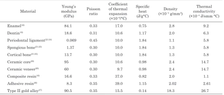
Fig. 3 Temperature distributions under 4ºC thermal condition at 2 s in the restored teeth.
Fig. 4 Compressive stress patterns under 200-N mechanical loading at 4°C for 2 s in enamel.
Fig. 5 Tensile stress patterns under 200-N mechanical loading at 4°C for 2 s in dentin.
separately in enamel, dentin, the inlays, and the adhesive resin.
RESULTS
Figure 3 shows the temperature distributions caused by 4°C cold exposure at 2 s in the restored teeth. The temperature distributions were similar in the restoration models.
Compressive stress yielded the highest values (190–200 MPa) at 4°C in enamel (Fig. 4). Von Mises and
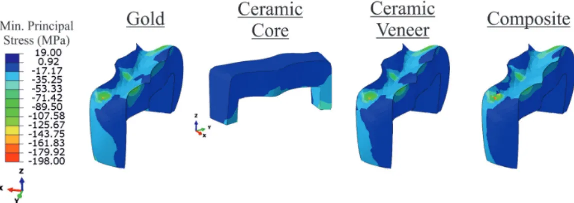
shear stresses had similar values, ranging between 165 MPa and170 MPa. The tensile stress values were lower, ranging between 95 MPa and100 MPa. The functional loading points showed the maximum stress. Secondary stress occurred near the cervical region (80–95 MPa). The stress values were similar in dentin, ranging from 38 MPa to 43 MPa. Except for tensile stress, the maximum stress occurred at the lingual cervical root surface; tensile stress was seen at the buccal cervical root surface (Fig. 5).
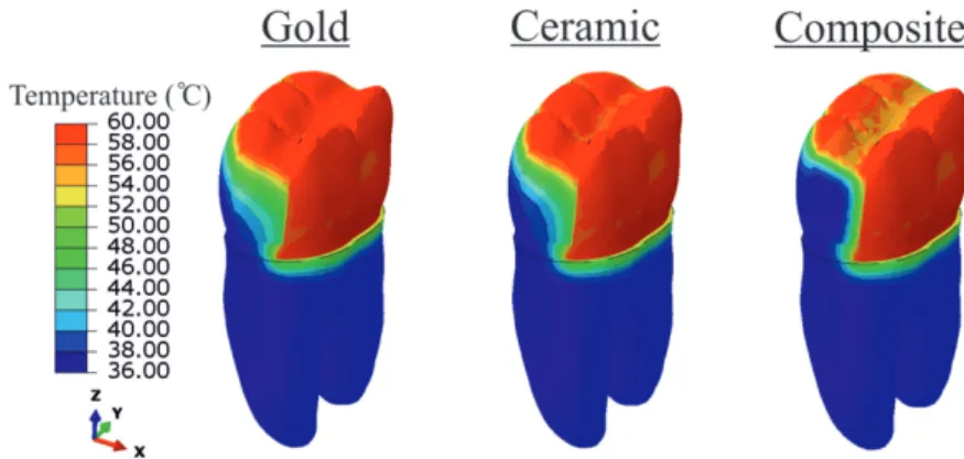
Fig. 6 Compressive stress patterns under 200-N mechanical loading at 4°C for 2 s in inlays.
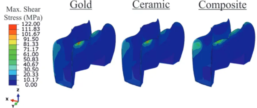
Fig. 7 Shear stress patterns under 200-N mechanical loading at 4°C for 2 s in adhesive resin.
Fig. 8 Temperature distributions under 60ºC thermal condition at 2 s in the restored teeth.
the loading points. Compressive stress had the highest values (170–185 MPa) and showed the following order of magnitude: Type II gold alloy>ceramic>composite resin (Fig. 6). Concerning the adhesive resin, the gold alloy model had the highest values of von Mises, compressive, and shear stresses (100–110 MPa) and lowest tensile stress value (32 MPa). Contrarily, the composite resin model had the highest tensile stress value (70 MPa) and the lowest values of the other stress types (45–55 MPa). The maximum stress in the adhesive resin occurred at
the occlusal margins (Fig. 7).
Figure 8 shows the temperature distributions caused by 60°C hot exposure at 2 s in the restored teeth. The temperature distributions were similar in the restoration models.
In the 60°C with 200-N loading condition, compressive stress yielded the highest value in enamel (195–200 MPa), followed by von Mises and shear stress values (165–175 MPa). Tensile stress ranged between 95 MPa and 100 MPa. Again, the maximum stress values
Fig. 9 Compressive stress patterns under 200-N mechanical loading at 60°C for 2 s in enamel.
Fig. 10 Tensile stress patterns under 200-N mechanical loading at 60°C for 2 s in dentin.
Fig. 11 Compressive stress patterns under 200-N mechanical loading at 60°C for 2 s in inlays.
were recorded at the functional loading points. Secondary stress occurred near the cervical region (80–95 MPa; Fig 9). The values of all the stress types were similar in dentin, ranging from 40 MPa to 50 MPa. Tensile stress occurred at the buccal cervical root surface, whereas the other stresses occurred at the lingual cervical root surface (Fig. 10).
The inlays showed the maximum stress at the loading points (Fig. 11). Compressive stress yielded the highest values (gold alloy>composite resin>ceramic),
ranging between 175 MPa and 185 MPa. The gold alloy model had the highest values of all the stresses (100– 115 MPa) except tensile stress at the occlusal margins of the adhesive resin; the composite resin model had the lowest values of these stresses (65–70 MPa; Fig. 12). Regarding tensile stress, the highest value was noted in the composite resin model (56 MPa), whereas the lowest value was recorded in the gold alloy model (32 MPa).
Fig. 12 Shear stress patterns under 200-N mechanical loading at 60°C for 2 s in adhesive resin.
DISCUSSION
In the present study, the influence of simultaneous thermomechanical loads on stress distribution associated with three types of inlays was investigated by finite element analysis. The different properties of the restorative materials did not affect the stress distribution. However, the stresses resulting from combined thermomechanical loads were considerably higher than those caused by mechanical loading alone2,19,20).
Posterior teeth are subjected to different magnitudes and directions of functional and parafunctional forces19). Intraoral loads vary from 10 N to 431 N3). Oblique loads create higher stress than loads directed along the long axis of a tooth28-30). The oblique loads in this study simulated the force acting on the mandibular molar during the closing phase of mastication. Further, during daily intake of food and drinks, the intraoral temperature varies between 0°C and 67°C31). In this study, 4°C was determined as suitable for cold exposure and 60°C was selected for hot exposure and the thermal exposure time period, simulating the intake of hot and cold food drink, was assumed 2 s, according to the work of Cornacchia
et al.12).
Two properties should be considered when evaluating stress distribution under thermomechanical loads: elastic modulus and coefficient of thermal expansion. Composite resin has low elastic modulus but high coefficient of thermal expansion, whereas gold alloy has high elastic modulus but low coefficient of thermal expansion. These properties explain the similar stress distribution in tooth structures and restorative materials of the inlay models, contradicting previous findings based on mechanical loading alone2,19,20).
Dejak and Mlotkowski32) reported that the restorative material influences adhesive and cohesive failures in adhesive resin. In this study, the stresses in the adhesive resin occurred at the occlusal margins in all the models. However, the stress values in the composite resin model were lower than those in the gold alloy and ceramic models. This result may be attributed to the similar elastic moduli of composite resin, dentin, and
adhesive resin. In spite of the relatively higher thermal expansion coefficiency, composite resin absorbs loads without conducting stresses to adhesive resin.
Shear bond strength is an important factor for success of a restoration. Excessive stress at the adhesive resin-tooth interface can cause adhesive failure and result in microleakage, recurrent caries, and postoperative sensitivity33). The shear bond strength values associated with the adhesive resin used in this study are as follows: enamel, 32.8 MPa; dentin, 15.1 MPa; ceramic, 17.2 MPa; and gold alloy, 5.4 MPa34,35). Notably, the shear stress values at the gingival floor and axial walls in the ceramic and gold alloy models were about 20 MPa. Therefore, adhesive failure can occur in these areas. For cohesive failure, compressive stress did not exceed the compressive strength of the adhesive resin (240 MPa)2).
Three-dimensional finite element analysis does not allow exact reproduction of some clinical situations. First, the adhesive resin had the same thickness around the restorative materials. In reality, the thickness may be non-uniform. Second, the thermal and physical properties of tooth structures and restorative materials vary widely and differ from the linear isotropic properties used in this analysis. In addition, oral temperature changes can influence the mechanical properties of dental restorative materials. Composite resins behave as viscoelastic materials and their mechanical properties are more sensitive to temperature fluctations36,37). Therefore, the data obtained in this study are not completely accurate and must be interpreted cautiously.
CONCLUSIONS
Within the limitations of this study, the following conclusions are drawn:
1. Simultaneous thermomechanical loads caused high stress patterns in inlay-restored teeth. 2. Type II gold alloy, ceramic, and composite resin
inlays showed similar stress distribution in the tooth structures and restorative materials. 3. Simultaneous thermomechanical loads may
floor and axial walls of gold and ceramic inlays. Composite resin inlays may be the better choice to avoid adhesive failure.
REFERENCES
1) Edelhoff D, Sorensen JA. Tooth structure removal associated with various preparation designs for posterior teeth. Int J Periodontics Restorative Dent 2002; 22: 241-249.
2) Yamanel K, Çaglar A, Gülsahi K, Özden UA. Effects of different ceramic and composite materials on stress distribution in inlay and onlay cavities: 3-D finite element analysis. Dent Mater J 2009; 28: 661-670.
3) Roberson TM, Heymann HO, Swift EJ Jr. Sturdevant’s art and science of operative dentistry. 4th ed. St. Louis: Mosby Inc.; 2002. p.569-590.
4) Jacobsen P. Restorative dentistry: an integrated approach, 2nd ed. London: Wiley-Blackwell Publishing; 2008. p.85-98. 5) Jackson RD. Indirect resin inlay and onlay restorations: a
comprehensive clinical overview. Pract Periodont Aesthet Dent 1999; 11: 891-900.
6) Stein PS, Sullivan J, Haubenreich JE, Osborne PB. Composite resin in medicine and dentistry. J Long Term Eff Med Implants 2005; 15: 641-654.
7) Ozen J, Caglar A, Beydemir B, Aydin C, Dalkiz M. Three-dimensional finite element stress analysis of different core materials in maxillary implant-supported fixed partial dentures. Quintessence Int 2007; 38: 355-363.
8) Hopp CD, Land MF. Considerations for ceramic inlays in posterior teeth: a review. Clin Cosmet Investig Dent 2013; 5: 21-32.
9) Jiang W, Bo H, Yongchun G, LongXing N. Stress distribution in molars restored with inlays or onlays with or without endodontic treatment: a three-dimensional finite element analysis. J Prosthet Dent 2010; 103: 6-12.
10) Knosp H, Holliday RJ, Corti CW. Gold in dentistry: alloys, uses and performance. Gold Bull 2003; 36: 93-102.
11) Erpenstein H, Kerschbaum T, Halfin T. Long-term survival of cast-gold inlays in a specialized dental practice. Clin Oral Investig 2001; 5: 162-166.
12) Cornacchia TP, Las Casas EB, Cimini CA Jr, Peixoto RG. 3D finite element analysis on esthetic indirect dental restorations under thermal and mechanical loading. Med Biol Eng Comput 2010; 48: 1107-1113.
13) Farrell CV, Johnson GH, Oswald MT, Tucker RD. Effect of cement selection and finishing technique on marginal opening of cast gold inlays. J Prosthet Dent 2008; 99: 287-292. 14) Summitt JB, Robbins JW, Schwartz RS. Fundamentals
of operative dentistry: a contemporary approach. 2nd ed. Chicago: Quintessence Publishing; 2001.
15) Toparli M, Aykul H, Sasaki S. Temperature and thermal stress analysis of a crowned maxillary second premolar tooth using three-dimensional finite element method. J Oral Rehabil 2003; 30: 99-105.
16) Fenner DN, Robinson PB, Cheung PM-Y. Three-dimensional finite element analysis of thermal shock in a premolar with a composite resin MOD restoration. Med Eng Phys 1998; 20: 269-275.
17) Lin C-L, Chang Y-H, Lin Y-F. Combining structural-thermal coupled field FE analysis and the Taguchi method to evaluate the relative contributions of multi-factors in a premolar adhesive MOD restoration. J Dent 2008; 36: 626-636. 18) Agnihotri H, Bhatnagar N,Rao GV, Jain V, Parkash H, Kar
AK. Evaluation of the onset of failure under mechanical and thermal stresses on luting agent for metal-ceramic and metal crowns by finite element analysis. Contemp Clin Dent 2010; 1: 227-233.
19) Ausiello P, Rengo S, Davidson CL, Watts DC. Stress distributions in adhesively cemented ceramic and resin-composite Class II inlay restorations: a 3D-FEA study. Dent Mater 2004; 20: 862-872.
20) Lin C-L, Chang C-H, Ko C-C. Multifactorial analysis of an MOD-restored human premolar using auto-mesh finite element approach. J Oral Rehabil 2001; 28: 576-585. 21) Wheeler RC. An atlas of tooth form. 4th ed. Philadelphia:
W.B. Saunders Company; 1969.
22) Farah JW, Craig RG, Meroueh KA. Finite element analysis of three- and four-unit bridges. J Oral Rehabil 1989; 16: 603-611.
23) Yang HS, Lang LA, Guckes AD, Felton DA. The effect of thermal change on various dowel- and-core restorative materials. J Prosthet Dent 2001; 86: 74-80.
24) Gale MS, Darvell BW. Thermal cycling procedures for laboratory testing of dental restorations. J Dent 1999; 27: 89-99.
25) Benzing UR, Gall H, Weber H. Biomechanical aspects of two different implant-prosthetic concepts for edentulous maxillae. Int J Oral Maxillofac Implant 1995; 10: 188–198.
26) Güngör MA, Küçük M, Dündar M, Karaoğlu C, Artunç C.Effect of temperature and stress distribution on all-ceramic restorations by using a three-dimensional finite element analysis. J Oral Rehabil 2004; 31: 172-178.
27) O’Brien WJ. Dental materials: properties and selection. 2nd ed. Chicago (IL): Quintessence; 1997.
28) Henry PJ, Bower RC. Post core systems in crown and bridgework. Aust Dent J 1977; 22: 46-52.
29) Pierrisnard L, Bohin F, Renault P, Barquins M. Corono-radicular reconstruction of pulpless teeth: a mechanical study using finite element analysis. J Prosthet Dent 2002; 88: 442-448.
30) Hatzikyriakos AH, Reisis GI, Tsingos N. A 3-year postoperative clinical evaluation of posts and cores beneath existing crowns. J Prosthet Dent 1992; 67: 454-458.
31) Palmer DS, Barco MT, Billy EJ. Temperature extremes produced orally by hot and cold liquids. J Prosthet Dent 1992; 67: 325-327.
32) Dejak B, Mlotkowski A. Three-dimensional finite element analysis of strength and adhesion of composite resin versus ceramic inlays in molars. J Prosthet Dent 2008; 99: 131-140. 33) Anusavice KJ, editor. Phillips’ science of dental materials.
10th ed. Philadelphia: W.B. Saunders; 1996
34) Abo-Hamar SE, Hiller KA, Jung H, Federlin M, Friedl KH, Schmalz G. Bond strength of a new universal self-adhesive resin luting cement to dentin and enamel. Clin Oral Investig 2005; 9: 161-167.
35) Osman SA, McCabe JF, Walls AW. Bonding of adhesive resin luting agents to metal and amalgam. Eur J Prosthodont Restor Dent 2008; 16: 171-176.
36) Ayatollahi MR, Yahya MY, Karimzadeh A, Nikkhooyifar M, Ayob A. Effects of temperature change and beverage on mechanical and tribological properties of dental restorative composites. Mater Sci Eng C Mater Biol Appl 2015; 54: 69-75.
37) Mesquita RV, Geis-Gerstorfer J. Influence of temperature on the visco-elastic properties of direct and indirect dental composite resins. Dent Mater 2008; 24: 623-632.