''Iııbrd Ünı\ Vcı Fak Derg. 49, 21:> 21X. 20{)2
Pathological evaiuation of contralateral testis following various
treatment methods for experimental testicular torsion in rats
Levent SA(;NAK
I,Berk BURGU
ı,Recai TUNCA
2•Rıfkı HAZIRO(;LU!
i i sı Umlogy C1lnic. SSK Ank~ın\ Training Hospita!. Ankara: 'Depanment of Paıhology. Eıeulıy ol' Veının;ıry Medıcıne.
Ankara UnıveLsity. Ankara
Summary: In the research. evalııation of the inlluenees. of the unilateral testiClılar torsıon anci dillereıll treatment resıılls ıhal " applıed to the this side. on eontralateral ıestiele IS aimeci. rol' ıhis aim . .:lO adult (2.5-3 months old) Sprague D~ı\\ley rats \\ere separ;ııed randomly (n: 5) into 8 groups. In eonırol group (group i).the sham operatıon has heen performcd on ıhe teslıcles In ıhe gmup 2 and nı her groups. the ıerı teslıele has hcen macie as torsion at the 720 degree countereloekwi.se ~!ml fixed ıo ıhe scr"ııım. 'I'lıen. ıhe "pn~ıııon;ıl ;ıııempts Iıke detorsıon (group 3. after 24 hours) ~ıml orehieeıomy (group.:l. ~ırtn 24 hoıırs .\IId groııp 'i.;ıfIL'r.:lX Iwıırs iand ın some group.s. Inıraperil()ne~ı1 conisone ın addilion to the orehieetomy (groııp (ı. ;ıfıer 24 Iwurs 'lIld group X. ~11'ln "IX hDULS) aml delDI'sion (groııp 7. ;ııkI' 24 hoıırs) have heen made to ıhıs ıert tesıicle. All cnnlr;ılaıeral tesııcles \VerL' I;ıken h) ıhe orciııectomy 4 weeks arter the tıırsıon ;ınci evalııateci hisıopaıhologically. There was degeneration ,ll ch~lIlglng iıııensily iii the ,ılı
cnnlr~ILııeral teslıcles exeept ıhe cDnırol group. The dillerence hetween the diameters of seminiferous ıuhules ILLShLTn aceL'pted as inıpunanı cxcept ıhe gmup 6 and group 7. In these two grOllps. the contrahııeral hyperemıa. edema 'lIld degener~ıııon aiso \\ere evıdeııılv le". 1\1 the accompanımelıt of these findings. when a puhertial ehildren applied to the clinic if maXIIllIlm 24 hoıırs passed ;ıI'ln ıhe Iınıl;ıter~ı1 ıestieular lorsıoıl. it ha.s heen eoneluded that ıhe adding of short ıerm imınıınoıherary ıo rroten ıhe CDnll';ıl;ıler;ıl ıesticle from ıhe anııgenie stımıılı!.s ıs ı!.sefu!' e\en whieh trcatments that delDrsıon or orehieetomy were applıed.
Key words: CDnıraLıteral ıesticle. hıstoraıhology. r,ıt. testıcular torsion
Sıçanlarda
deneysel testis torsiyonunun
çeşitli sağaitım metodları
ile tedavisi sonrası kontralateral
testisin patolojik değerlendirilmesi
Özet: Çalı~mach unıhııeral testis torsiyonunun ve hu tarafıa uygulanan deği~ik sağaitım Soııııçlarımn kdr~ı testıse DLııı etkılerının değerlendırılmesi ~lIlıaçl~IIJ(Iı. Bıı dmaçla eri~kin (2.5.3 aylık) 40 adet Sprague Dawleysıçan rasılaııı!.s,ıl ol;ırak Yerli X grııbd ayrıldı Kontrol gnıhunda (grup i) testislere yalaneı operasyon (sh,ıın) uygulandı Cnıp 2 ve diğer grurlanl;ı ,Di IL'stlslCl
no
derece sa;11 yelkuvaııı tersine ıorsiyone edilip skroıııma tesrit edıldi. Daha sonra bu testıse deıorsıyon ıgrup :>. 24 sa,11 s(\ıırdi.ııL~I('kıonıi (grup 4. 24 s;ıat sonra \e grııp 5 . .:18 saat sonra) ve hdZI grurlarda or~iektuıniye ek ular;ık inır,lperlıone~11 koılıznıı uyguLıınaLırı (grup Cı. 24 saat sonra) \ c detorsiyon gibı operasyoıılar yapıldı Tüm kolltraLııer,ı1 ıestısler lursıvond;ıı! 4 iıdfı,ı ")I1I;ı orşic:ktomı ile ,ılındlak iıistopatolojik olarak değerlendirildi. Kontrol gruhıı dı~ındaki tiiın grurlmda koııtr~ılaıeral ıesiısierde degl)L'n ~lddL'ııe deıencr;ısyon ınL'veultu. Semınifer tuhulu, çapları arasıncI.ıki fark. grup
cı
\e grı1r 7 dı~ında. önemlı hulundu. 1311ikı gnıpı~ı ,ıyrıcı. koılır;II,lter,ı1 hırereıni. ııdeın ve dejenera,yon da belirgiıı olarak azdi. Bu bulguların eşliğınde unlLııer~ıI ıestis ıorsıyonli Dlu~nm~ \L' ıi/erınden en Lızi<ı 24 saat geçmi~ puhertal çocuklar kliııığe haşvurdıığııııda deınLsiyolı ya d,ı or~ıcktonııckıı bır!.sı ıle hırlık:e koııır~ılaıeral testisı antiJenik uy;ırıdan korııınak için kısa süreli iınımınoterapinin tedavıye eklcnnıesının v;ILırll "Idcağı ,<)ııueııııa \drıldıAn~.ıhıdr kelımeler Hısıor~ltoloJi. koııtralaıend testıs. sıçan. lestıs ıorsiyımu
Introduetion
The testicıılar ıorsion is an aeute disease that ap-pears especial!y in ıhe male children in the adolesccnt age ;ıııd iı appears İn 1/4000 bı:twecn the malcs, under 25 years old (15). i\lıhough the İdentifying of the testİcular ıorsinn is vcr)' easy, the period that is passing between ıhe hegınning and the treatment of the diseasc is very im-porlant (7.12). The allection of the unilatcral testicular \ı)r.sion on the contralateral ıcstidc and fertility have been searched hy several scİentİsts (Ll2, 15) but thcre is no any agreeıııent on thc Icvcl of these affections and Ircat-menı period ancltype.
There are a controvcrsy beıwcen ıhe auıhı)rs: who hclievcs an afkction to thc cüntralatcral tcsıis; ;ıboııı ıhc allccıion mcchanisms üf contralateral lcslis af tn torsion. Whcn some authors suggest that ıhis aneClion is due lo decrease of hlood tlow as a resulı of syıııpathic aCilVdıion (18), 111051 of Ihe others explain this pdtho]ogy \\ııh im-munological ıncehan isms (9, 14)
In this study, eXilmİnalİon of the afkds. ı)j ıhe uni-latcral testicıılar torsion and dillercnl trealıııenl n:sults ıhat İs applied to this sidc, on the contraLıter:ıl tcsıiclc ıs aimed.
214 Levent Sa~nak - Berk Burgu - Recai Tunca - Rıfkı I-Lızıro~ıu
Matcrial and Methods
For this aim, 40 adulı (2.5-3 months old) Sprague Dawlcy rats were used. The applieation was performed on the rats hy the permission. no./dated 17/28.1 i .2000 thaı \vas issued by the Ankara University, Faculıy of Vele-rınary Medicine Ethie Commission.
The groups were formed as foııows: as the animals wen.: separated ranelom!y inıo 8 groups, eaeh group has 5 animals.
Group) (control): The left testieles of this group's rats \vere applied the sham operation.
Group 2 (torsion): Af ter the lefı testicular liberation aı the 720 degree countercloekwise and was fixed to ıhe serotunı suheutaneously by nonabsorbahle sutures. Then. the serotum was ekısed by the ahsorbahle suture materi-als.
Group
:1
(torsion/detOl'sion): The ıert testicle was ımsioned with the same operational principles of group 2 and ıhe sutures \Vere openeel hy entering from the same incision arter 24 hours and nıaele detorsion and the sno-tum was clnsed by ıhe ahsorhahle suture materia!'Group 4 (IOI'sion/Ol'ehieetomy after 24 hours): The Idt tcsticle \Vas torsioned \Vith the same operational prin-cipks of group 2 and the orehieetomy was done by en-[ering from the same incision af ter 24 hours and the scro-tum was closed.
Group 5 (torsion/orehiectomy arter 48 hours): The lert ıesticle was torsioned with the same operational prin-cipks of group 2 and the orchiectomy was done by en-tering from the same ineision after 48 hours and the sero-lunı \Vas closed.
CJrmıp 6 (torsion/orchieetomy after 24 hoursl cortisone): 2mg/kg/day methyl prednisolone has been given intraperitoneally during 4 weeks to the rats after the sanıe processes in group 4 were applied.
Group 7 (torsion/eletorsion/eorıisone): 2mg/kg/day
nıeıhy! preclnisolone has been given intraperitoneally dur-ıng 4 week s ıo the rats af ter the sanıe processes in group
:1
were applied.Group 8 (torsion/orehieetomy arter 48 hoursl cmıisone): 2mg/kg/day methyl prednisolone has been gi"en intraperitoneaııy during 4 weeks to the rats after the same processes in group 5 were applied.
The operational processes were done by using pen-ıobarhiıal sadium anesthesia. The pentobarbital sodium was given hy 26 G insulin injeetor at the 30 mg/kg dos-age as intraperitoneally. The drug was eITeetive beıween
S
ı
s
minuıes anel its effect was disappeared after 45-90 minuıes. Aikr the loeal cleaning by povidone iodine un-dcr the anesthesia, the left testİeles of the rats wereex-plOl'ed hy the incision. In the control group. afıer scroıal ineision and liberatiOlı of the testis. scmlull1 was closeL! by the absOl'bable sutures (sham). AII rats were puı inlo the separate cages in the first 24 hours after ıht.: opera. tional process and the n they wc re uniteel with theır groups.
The animals were kept in the cages that was Llear. iıı 27x 17x42 cm dimensions. reetangular in shape <11](1 ıts over was suitabk for plaeing the speeial waın cup and feedbox and with wire cover in the speeia! lighıing eoneli tİons and room temperature and they were feel hy the spt.:-cial rat feed. At the emI of the 4 weeks, ıht.: orehieetomy was apphed on the right testİcles of all rats and the cu-thanasia has been apphed by over dose anesthesia.
The right testİcles thaı wert.: taken by orehil.:ctomy were fixed in Bouin's solution. The fixeel tissuc samrıks were hloeked in paraIlin vax by applying tht.: rOlıtint.: methods. The sections in 4.-6 ı.ım thiekness that werc tak-en from these bloeks, were stained with hematoxylin-eosin (HE) and evaluated in lig ht microscope. Eaeh rat's body weight and the group averages of the e1aıa thaı be-longs to the testicle weight were ea1cubl(::e1 Tcn st.:mi-nirerous tubules diameters in eaeh animal were Illeasurt.:d by helping of oeular micrometer in the light microseopc and their averages were taken. These averages \\'tre ae-cepted as data of each rat. Thus, the average hody weight, testicle weight and data of the seminiferous tuhuks di-ameters of 5 rats in eaeh group have heen formcd. Krus-kall- Walhs varianee analysis for the evaluation ol' data. and Whitney-U test for the difkrention among ıhe group or groups were used.
Results
Histopathological findings
In contralateral tesudes, the histopathologieal changes in every ease were presented in Tahle
ı,
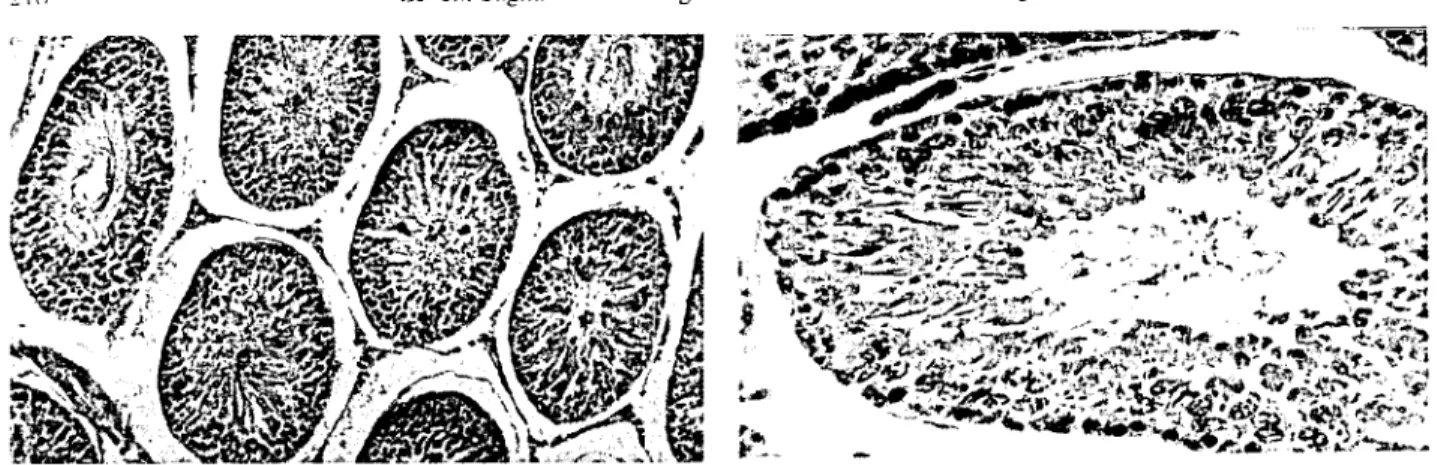
and ıhe average testiele weight, body weight and seminikwus tuhulcs dı-ameters were presented in Tabk 2. The histol, ıgical changes in all groups were evaluated ıogt.:ther. In all groups except the controls, it \Vas fixed thaı seminiknıus tubulcs diameters were getting diminishing and Ihey wc re showing a distrihution randomly as various dimensions.In all testicles that bc10ng to the group 1. it was ob-served that the seminiferous tubules havt.: a regular strue-turc and normal spermatogenesis (Figure 1).
In group 2, in all testicles exeept one ease. ıhe hy-peremia in vessels and interstitiel edema in a lighı in-tensity were ohscrved. There is a vaeuolar and hydropıe degeneration in changing intensity in germinal epithelium that eovers the semİniferous tubules. ]n Silme tubuks. the
Ankara Üniv Vet Fak Derg. 49, 2002 215
Tablc I. 111e cvaiuation of histologieal findings. +: Light. ++: Mediunı, +++: Intensive.
Group Anımal no. Hyperenıia Edenıa Degeneration Spernıaıogeııesıs
i ++ 2 +++ 3 +++ 4 ++ 5 ++ 2 i + + ++ 2 ++ 3 + + ++ 4 + + ++ + 5 + + +++ + 3 i ++ ++ + + 2 + + + + 3 + + + + 4 + + ++ + 5 + + + + 4
ı
++ + + + 2 ++ ++ + 3 ++ +++ ++ 4 ++ + + 5 + + +++ 5ı
+ + + + 2 ++ ++ +++ 3 + ++ + 4. + + + + 5 + + ++ 6 i + + + + 2 + + + + 3 + + 4 + 5 + 7 i + + 2 + + + + 3 + + 4 + + ++ 5 + + + ++s
+ ++ 2 ++ +++ +++ :3 + + +++ 4 + + + + 5 + ++Tahlc 2. 'Ihe average seminiferoııs ıubules diameters. average hody weight and average testiele weight in the groups.
Gmup code Average seminifcrous tubules diameter (micron) Average hody weight (gram) Average tesıicle wcight (gr,ıın)
2 3 4 5 6 7 8 197.0:t 6.670832 '178.8:t 10.76754 *'177,4:t 6.516134 *'ı60.8:t 7.638063 *'160.2 :t 2.8i780
ı
+ 192.2 :t 4.973932 + 193.4:t 9,102747'"ı
73.8 :t 9.340236 229.14:t 18.9196 220.00:t 16.n27 255.60 :t 08.7784 275.60:t 16.0144 263.80:t 08.5288 237,40:t 07.8269 268.20:t 18,9196 268.80:t i4,54iO 2.588 :t 0.1 15 2.4')0 :t 0.1 17 2.882 :t0.210 2.098 :t0.1i i 3.122 :t0.103 2.624 :t 0.084 2.898 :t 0.235 2,4Z0:t 0.216 "'rdJ.05 ımporlallt (whıle compared with control group).LC\ellt Sagnak - Berk Burgu - Recai Tunca - Rıfkı Haııroğlu
FI~llre i !\Imın,tI le~lıcular u~'ue and 'pl'rnıatogeııc~ıs. Group
i "E x ')1)
Fı)'urı' 2. Thc degeııcrauoıı ın ıhe gennınal epitheliunı and ılller-'Jultal edenıa. Group 2. HE x 90.
genninal epiıhelium was completcly removed and base-ment membranes were in a waved appearance (Figure 2). In onlyone case, the spermatogenetic activity was ob-served in a few tubules.
In group 3, there was a vaeuolar and hydropie de-gencration in changing intensity in germinal epiıhelium th::ıt covers the seminiferOlIs tuhules and interstitial ede-m::ı in a light intensity and the vesscls were hyperemie in the testieles. In all case, the spermatogenesis was ob-slT\Td (Figure 3).
The vessels of testieles bclong to the group 4, were hyperemic in an advaneed lcvel and the interstitial tissue was edemaıous in inlensive in one case, and light in-tensive in other cases. The basement membranes were in wa'ed appearance. The gemıinal epithelium was re-nllJ'ed completcly in so mc seminiferous ıubules. In addi-tion ıo some degenerative changes, together with sper-matogenetic aetivitiy were found in a few tubules in one ease.
The vesscls were hyperemic and interstitial tissue was edemaıous in appearance in group 5. The de-gcnerative changes, changing intensity. in the gemlİnal epiıhelium was observed. In some parts, the tubules
di-Fıgure 3. Normal spernıatogcnesıs togeıhcr wiıh degcncrauuıı ın lighı intcn~ity. Group 3. HE x 380
Figure 4. The untidy appcaranee in the semınıferolIS llIbulc~ dı. aıneters. the germinal epithelium that covcrs ~onıe tubule~ arc eompletely reınoved. Group 5. HE x 90.
ameters were quite diminished and the hasement ll1ell1-branes were in waved appearance (Figure 4). Both İn two cases, there was spernıatogenesis in lcss proportion.
The vesscls were hyperemic in a light intensity and interstitial tissue was edematous in two cases in group 6.
In all seminiferous tubules, there were dcgenerative changes in a light intensity. There was spennatogenesis in a lcss proportion in all eases exeept two cases.
The vesscls were hyperemie in a light intensity and interstitial tissue was edematous and ıhe germinal eri thelium had degenerative changes in changing intensity İn three cases in group 7. A few multinueleM gıant eclIs in the degenerated epithelia were noticed in one case. Changing proportions spermatogenesis was ohserved in all eases exeept one.
The vessels were hyperemic in changing intcnsity and interstitial tissue was edematous in three eases 111
group 8. The degenerative ehanging as in changes in-tensity in were ohserved all çases but in two çases these were intensi"e (Figure 5). The germinal epithelium \Vas completely removed in some tubules, and there \Yere large umount of Illultinuclear giant cells in intensi\e cuses
,,,,,,.
-Ankara Üniv Vet Fak Derg. 49.2002 217
Fıgun: 5. The ıntensıvc dcgcncration in thc genninal epithcliuın.
close appcarance. Group 8. HE x 380.
(figure O). The spermatogenesis was in changing pro-portion exeept these two intensiye cases.
Statistical findings
The dillerenee hetween the average hody weight and testiele weights in the gmups were not important in the statistieal evaluation using with the Kruskall- Wallis varianee analysis. However, the din'erenee between semi-nifcrous tubules diameters were important.
Whitney-U test was made for the determination of the dillerences hetween groups. The diminishing in the seminiferous tuhules diameters were found statistieally imponant according to control group exeept the 6ıh and 7'h gmups.
Discussion and Conclusion
In the unilateral testiele torsion, it was showed sta-tisıieally and histopathologieally that the eontralateral tes-tiele was effeeted in changing intensity hy the applied treatment methods. The testiele torsian is an aeute disease espeeially seen in the children who are in the puberty. it is aecepted that the unilateral ıorsion affeets the eontra-lateral testiele and fertility (3,10). but there is no any agreement on the suhject, when this atTection starts the forming af ter the torsion and how it is and whieh treat-menI can be used.
it is recorded that the 4-6 hours torsion period is enoııgh to affeet the contralateral testiele (2). In addition, the torsion degree also is important. In 360 degree tor-sion, there are medium level pathologieal changes in tor-sioned testiele and the full infarct appears aner 4 hours torsion in 720 degree (19).
Nagler and White (14) dcelared that cantralateral tcstinılar atrophy formed hy the torsion is prevented by orchiectomy, and it is unsuccessful with the detorsion. In the testieles were applied detorsion, the immunologie re-aetion starts by the hlood-testiele barrier makes the
Figıırc 6. The dcgcncraıion in the gemıınal cpiıhclıuııı.
ıııııltı-nuclcar giant ecııs and intersititial edeına. Group R.
Hı::
x 90.sperms known were perished as a resulı of ischaemia and it causes a deformation in contralateral testiele and eon-trary to this, it is declared that there is lcss changing on the contralateral testiele, heeause the SOlu'ce is removed in them that the orchiectomy has heen applied. so there \vill not he any immune response (o). it is reponeL! that when the immunosuppressive treatment is given as anti-Iymp'hocyte glohulin (ALP), it decreases the contralateral testiClılar damage hut can not remove eompletely, where-as ALP +splenectomy prevents eompIdely (14). In addi. tion, it is suggested that following detorsion. an adıuv<ınt immunotherapy (hydro.eortisone) in :) day; prnents the antigenie stimulation (I I).
Wallaee ct aL. (20) pointed out that there is an inı-pairment in hIOl)d-testiele harrier of eontralateral testiele af ter 7 days of unilateral testiele torsian and while IgM is determined, the morphologieal changes at this lcvcl is in a smail level and in the 28'h day, IgG took placc insteaL! of IgM and the morphologie impairıııent heeame elem Ko-şar ct aL. (9) in a similm study has determined that. the anti-rat IgG antihodies are contrary to spermatozoa anıi-gen in contralateral testieles tissue i ıııonth afıer of de-torsian.
In addition. Nagler ct aL. (13) has showed that the unilateral testiClılar torsion in immature rats did not l11ake any testieular damage and did not effect the contralateral testiele when theyare reached to the pııberty. The fol-lowing is its comment: Prepubertial tesıinılar tmsıon does not show the same affeet as of postpııbertial torsİon. so why immunologic stimulate that depends on possihly sperm antigens and makes ehanges in the testiele does not exist before puherty. eosentino ct aL. (5) has alsa worked with prepuhertial rats and found that while they reached to the puberty, there is a decreased eontralateral sper-matogenesis as secondary to the torsion.
In the literature, there are als(l same publicatıons that point out the opposite results. In the similar
experi-Lel'enl Sağnak - Berk Burgu - Recai Tunea - Rıfkı Hazıroğlu
218
111l:ntal rescarehes. it is not shown the existenee of eontra-laıeral testiele damage foIlowed by unilateral testieular ıorsion (8.1 7) and it eould not found a deerease in semen parameters (16). In some puhIieations. the immunological damage in the eontralateral testiele af ter the unilateral to 1'-sıon. are also not supported (4.8,i7).
Our results were indieated that while the eontra-lateral testiele was eompared with the control group, ıhe unilateral testiele torsion atIeeted the average semi-nifemus tubuIes diametcrs hy deercasing meaningfuIly. The detorsion arter 24 hours, orehieetomy arter 24 hours, orehieeıom)' af ter 48 hours and orehieetomy+eortisone alkr 41.1hours treatments of Iate period treatments that vvere appIied. aiming to be ahlc to deerease this affeet were not afreetive and also in thest groups. the eontra-laıcral average seminiferous tubules diameters decreased sıatistieal1y. But, in the groups (6 and 7) that detorsion or orehieetomy appIied af ter 24 hours and adding eortisone treatment during i month. the average seminiferous tu-hules diaıııeters were elose to the control group. We sup-pose that eortisone deereases the immunologic afket in the treatment. In addition, eontralateral testieular hyper-emia and edema were elearIy less in these groups white it was compared with other groups and the degeneration als() was less. The spermatogenesis was observed in aIl groups but decreased eleari)' when it was eompared with the control group.
;\s a result, when a puhertial children applied to the elinİe wiıh a unilateral testicle torsion whieh was formed maximum 24 hours ago, wc proposed that a short term il11lmıııotherapy wiIl he uscful hy adding to the treatment el'en \vhieh treatments were applied as detorsion+fixation or orehieetomy to proteet the eontralateral testicle from the antigenie stimulus.
Rcfcrcnces
i. Anderson HA (I 987): Tn[icular salmge jiJilowilıg sper-17w[ic cord [01'SIOII. J Ped Surg. 22. 231 -234.
2. Barkley C, York .IP, Badalament RA, Ncbitt JA, Drago .1, Smith .1.1i.19(3): Tes/lcu/ar torsioıı aııd its eift'ets O/l (.o/lırala[('ral testicle. Urology. 41. i92-i94.
3. Bartsch G. Frank S, Marberger H, Mikuz G (J 9S0): Tesıı('ular Ior.lio/l: lale resull.1 wiıh special rei;ard lo l'er-Illit\' aııd eııdocriııe/wıctioıı. J Crol. 124, 375-378. 4. Cerasaro TS, Nachtsheim DA, Otero F, Parsons CL
(ı 984): The e/(eet oftesıicular ıorsirm {}/i cOlıtralateraltes-ııS aııd ıhe produetirill o{ wıtispt'l'm wıtihodies iıı rabhits. J Urol. 132.577-579.
5. Cosentiono MJ, :'Jishida M, Rebinowitz R, Cockett AT
(I 985): Hisıoloi;ical ch(//li;eS occurring i/ı 1111:'conımlaıeml teSlis or prepuhertal rats suhjec/ed LO 1'(//'llJU'"dumlwns o/ uııilll/eral spennaıic cord torsimı. J Urol. 133. 906-9 iI. 6. Gülmez I, Karacagit M, Sade M, Kandemir B (I ')87)
I]j'eu or tesıicular lors[(}ıı 011 ıhe CllIlıralaleml Inlls aııd prel'eıılioıı or t/w e/reel iJrprednisolmw Etır urul. 13. 340-343.
7. Heindel RM. Pakya RE, Cosentino MJ (i9901 Sl'er-//latic cOl'd tonioıı: Colltralaıeral Tesıieıdar degenemııun ıil various ai;es iıı ıhe ml. J AnoroL. i
ı.
S06-S13.8. Henderson .1B, Williamson RC1\; (1990) Faıilu\ afler torsirııı orthe sperllmlic cord. Br J Urol. 65. :2S-230.
9. Koşar A, Küpeli B, Alçığır G. Ataoğlu H. Sarıca K, Kü-peli S (ı9(9): l//l//ll/lınl"i;ic aSl'ec[ oj leı[/cıt/ur Inn/uıı. Detec[irm oj aıı[isperllı (//uiiJodın in cU17lmlulı'ml Ir'sııc/e. Em Urol. 36. 640-644.
ı
O. Krarup T(ı
978): The testis a/ier ınnio/l. Br.J Un)1. 50. 43-46.ı ı.
Madarihan BA (I987): Tes/lcular .wı/ı'age/ollowing sıwr-matic ('(ml tIJnlrJll. J Ped Surg. 22. 23i-234.12. Madgar I, Lunenfeıd B, Mashiach S. (;oldwasser B. Weissenberg R(I987): FJ/'eel or lesıinda/' Innimı rJll cımı-mlateml [esıis and .I'er/iliır ul //lll/u/,(' /'l1l.1 Arclı AndroL.
19.23724 I.
13. Nagler HM, Dcitch AD, White VR (1984). T(:'S/lıulurl{//'-sioıı: tempoml co/l.ııderatirJlls. Fertil Sıcril. 42. 257-262.
ı
4. Nagler HM, White RD (ı982): 1Iıe el/eel r.j [esllcu/arlu/'-sioıı o/ııhe COlurlı/ateml te.l/ls . .JUrol. 12l!. 1343 1348
ı
S. Rajfer.1 (ı 9(2): COlli;elıi[al Alln//lalies ojıhe Te.lllsIS43-1562. In PC Walsh. MD Rcıık. TA Sıaııc)'. ED V,ıugh,ııı (Eds). Camphell's Urology. Sixıh Eo. \VB Saunders CUlJlr. Philadelphia.
16. Ryan Pc. Whenlan CA, Gafl'ne EF, Fitzpatridı .i1\1
(I(88): The e//'eel oj l/Iıilatt'ml npl"I'IIJ1l"llla/ le.lıicıda/' In/'-.Iimı 011sper//laıngenesis aııd .ler/ilin'. Br .i Uml. 62. 359-366.
17. Saltzman!\, Sadi M, Hoffer A(ı984): Is <lUlOIlIllJ1l/1w.ler-tility a cnll.lequt'llce ol (milaıemi 1t'S[icula/' i.ıche//lııı . .J Urol. 131, 162.
i8. Tanyel FC, Büyükpamukçu N. Hiçsönmez A (1989)
CrJlltmlaleml ıesıicular bınnd jlow d{(l'IIı.~ {(nilolem/
ın-ıicular torsirJll. Br J Urol. 63.522-524
19. Turner TT (I9S8): Acule expemn"Il[al In/lCldar /Oniull
/10eij'eel rm ıhe coııtmlateralle.llls. J Anorol. 6.(ıS-72.
20. Wallacc DMA. Gunter PA, Landon GV, Pugh Re. Hendry Wl' (1982): 51'11ljJ(l/oll'lic orchial'e.\/(/- (/11
('ıpen-melltal aııd clıııiwl studl'. Br.J ürol. 54.765-768 . Rt'ct'll'ed 5 March 2002/ ACCepIl"d 20 Mıııdı 2U02
Correspondence address: A. Levenl SaKııak Ömür Sok. 11l/7 06550 Yukan AyralıCl Aııka/'((. Tl/rkev e-mail: dr.sagıı[email protected]