The effect of physician experience on the
measurement reliability of the Reimers’ hip
migration percentage in children with cerebral
palsy
Pınar Doruk analan, MD1)*, EmınE EcE Yılmaz, MD1), mEhmEt aDam, MD1),
BErrın lEBlEBıcı, MD1)
1) Department of Physical Medicine and Rehabilitation, Faculty of Medicine, Baskent University Adana Research and Education Center: Kazım Karabekir Cad. 4227. Sok. No 37 Yüreğir, Adana, Turkey
Abstract. [Purpose] Reimers’ hip migration percentage (MP) is commonly used to document the extent of hip
displacement in children with cerebral palsy (CP). However, factors such as poor administration of pelvic radio-graphs, a lack of concentration, inexperience, or a busy clinical environment may result in variations in the MP measurements. The aim of this study was to compare the differences in the MP results of two physiatrists with vary-ing levels of experience to determine the role of experience in the measurement’s accuracy. [Subjects and Methods] This retrospective study included 62 hip radiographs of 31 children with spastic CP. Two physiatrists with different experience levels calculated the baseline MP on two occasions six weeks apart. Correlations, intra- and inter-rater reliabilities, and differences in the MPs were compared. [Results] Correlations and inter- and intra-rater reliabilities of the measurements were excellent. There were no statistically significant intra- or inter-rater differences for either of the two measurement points. Inter-rater correlations for each session were 0.94. [Conclusion] Experience does not appear to be a factor in the evaluation of MP, and inter-rater differences do not cause problems regarding patient follow-up. Therefore, repeated pelvic radiographs are not necessary in the evaluation of MP in children with CP unless indicated.
Key words: Reimers’ hip migration index, Cerebral palsy, Experience
(This article was submitted Jun. 11, 2015, and was accepted Jul. 23, 2015) INTRODUCTION
Cerebral palsy (CP) is the most common cause of physical disability in children in developed countries1). CP
is a group of disorders associated with the development of movement and posture that can cause activity limitations attributed to non-progressive disturbances that may occur in the development of the fetal or infant brain1). Hip
displace-ment is a common, severe problem in children with CP, and its development is thought to be related to age, Gross Motor Function Classification System (GMFCS) level, or CP sub-type2–5). Unfortunately, hip displacement in children with
CP is silent in the initial stages and is difficult to detect via physical examination alone. However, detection is possible with repeated radiographic evaluations in conjunction with regular physical examinations.
To optimize the screening procedure, knowledge of the
characteristics of children who are at risk for displacement is important1, 5, 6). In 1980, Reimers developed a measurement
to document the extent of hip subluxation lateral to the ace-tabulum called the migration percentage (MP), which is still used as the key tool to help prevent hip dislocation, monitor its effects, make decisions regarding the most appropriate type of interventions (e.g., a selective dorsal rhizotomy, hip abduction braces, or surgery), and determine the progression of subluxation1, 3, 7). An MP of ≥33% is considered a reaction
threshold, and intensified observation is recommended. In children with an MP of ≥40%, the lateral displacement of the hip increases over time, indicating the need for surgical intervention. For children with an MP between 33% and 40%, it is recommended that treatment be based on other clinical signs as well as the progression of the MP over time as evaluated through repeat examinations performed by the same examiner5). The current study aimed to compare the
differences in the MP calculated by two physiatrists with different levels of experience in order to determine if experi-ence plays a role in the accuracy of the results.
SUBJECTS AND METHODS
The study protocol conforms to the ethical guidelines of the 1975 Declaration of Helsinki and was approved by
J. Phys. Ther. Sci. 27: 3255–3258, 2015
*Corresponding author. Pınar Doruk Analan (E-mail: [email protected]; [email protected]) ©2015 The Society of Physical Therapy Science. Published by IPEC Inc. This is an open-access article distributed under the terms of the Cre-ative Commons Attribution Non-Commercial No DerivCre-atives (by-nc-nd) License <http://creativecommons.org/licenses/by-nc-nd/3.0/>.
Original Article
J. Phys. Ther. Sci. Vol. 27, No. 10, 2015
3256
the local Ethics Committee. Written informed consent was obtained from all participants. This retrospective study included 62 anteroposterior pelvic radiographs of 31 chil-dren (20 boys and 11 girls; mean age 4.2; range 1–14) with spastic CP followed up at our clinic between 2011 and 2013. Patients with CP who had been followed up in our clinic and had radiographs taken were identified using a data search, and those with pelvic radiographs in the hospital’s electronic medical records were included in our study. Radiographs were taken with the patient in the supine position, the pelvis symmetric with respect to rotation, and the femurs in the neutral abduction/adduction position relative to the pelvis.
Radiographs were examined by an independent physiat-rist. Radiographs classified as GMFCS level I–II and those taken after previous reconstructive and salvage surgeries of the hip or pelvis were excluded from the study. MPs were then measured by two raters, a physiatrist with five years of experience and a second-year physiatry resident, blinded to each other. All measurements were recorded by the independent physiatrist. Measurements were repeated six weeks later by the same raters and recorded by the same independent physiatrist. Any MP results recorded previously in the patient’s history by their follow-up physician were not included in our study. Only images stored in the computer system were used for the MP measurements. All radiographs were recorded electronically. Measurements were calculated
using the images on the computer screen and were then recorded confidentially in a location known only to the independent physiatrist.
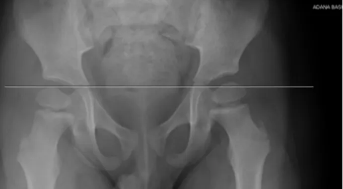
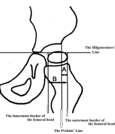
The hospital’s computer software for storing patient radiographic records was used to measure and calculate different angles and distances to minimize measurement errors. To evaluate the MP, the Hilgenreiner line (H) was determined by drawing a line between the Y-cartilages of both sides using the software’s angle measurement option, which allows for precise on-screen measurement of the angle under consideration (Fig. 1a). A second line, known as the Perkins’ line (P), perpendicular to the first one, was drawn through the upper outer side of the acetabulum (Fig. 1b and 1c). Next, the innermost and outermost borders of the femoral head were defined, and two lines were drawn from those inner and outer lines intercepting at an angle of 90° to the H line (Fig. 1d). Finally, using the length measurement option in the software, the length of the acetabular part of the femoral head remaining outside the lines and the total diameter of the femoral head were measured in millimeters. The ratio of these two measurements was then calculated as the ratio of the outer acetabular part of the femoral head to the total diameter of the femoral head. The ratio was then multiplied by 100 to calculate the MP (Fig. 2). Each rater’s own calculations were compared first with each other and then with those of the other rater. The MP results were
clas-Fig. 1a. Hilgenreiner line (H) is a line between the Y-cartilages of both
sides Fig. 1b. The Perkins’ line is perpendicular to Hilgenreiner’s line at the lateral margin of the acetabulum
Fig. 1c. The Perkins’ line is perpendicular to Hilgenreiner’s line at the
3257
sified into three stages: ≤33%, between 33% and 40%, and ≥40%5). Correlations and intra- and inter-rater differences
in the reliability of the recorded MPs were also evaluated according to dislocation severity.
The SPSS Statistics version 17.0 for Windows software package (SPSS Inc., Chicago, IL, USA) was used to perform all statistical analyses in this study. Categorical measurements were given as numbers and percentages and continuous mea-surements as averages and standard deviations, along with the mean and range when needed. The compatibility between raters was tested using a reliability analysis and evaluated using Cronbach’s alpha value. The reliability of the inter-rater concordance correlation was evaluated using the kappa coefficient, and MP values were classified as ≤33%, between 33% and 40%, and ≥40%5). The kappa coefficient strength
of agreement was determined as follows: ≥0.91 very good; 0.90 to 0.71 good; 0.70 to 0.51 moderate; 0.50 and 0.31 fair; and ≤0.30 poor. Distributions were controlled for continuous measurements. As parameters were not normally distributed, the Wilcoxon test was used to compare the measurements, and the inter-rater correlation method was used to compare the two raters’ results. Differences between measurements were evaluated using McNemar’s test. For all tests, a p-value of <0.05 was considered statistically significant.
RESULTS
There were no statistically significant intra- or inter-rater differences for either of the two measurement points (p>0.05). Inter-rater correlations were 0.94 for each session. In addition, there were no significant intra- or inter-rater dif-ferences in the reliability of the MP for different dislocation severity levels. The results are shown in Table 1.
In the reliability analysis test, Cronbach’s alpha correla-tion coefficient values of 0.966 and 0.971 were obtained in
the first and second evaluations, respectively. In the kappa coefficient evaluation, all agreements were classified as moderate. There were no statistically significant differences between the measurements (p>0.05) (Table 2). Similar reli-ability results were found when evaluating the different dislocation severity groups.
DISCUSSION
Hip displacement has a reported prevalence rate of up to 35% and is the second most common deformity in patients with spastic CP1). Increased femoral anteversion and
neck-shaft angle along with the presence of powerful spastic hip adductors and flexors accentuate forces that displace the femoral heads in CP patients1, 7–10). This condition may also
result in secondary acetabular dysplasia and degenerative joint disease, femoral head or windswept hip deformities, scoliosis, severe contractures, pressure ulcers, pain, or a decrease in quality of life as well as difficult nursing and perineal care, problems with positioning while sitting and standing, and ambulation1, 4–11). Early identification and
intervention of hip displacement has been shown to improve treatment outcomes, reduce the number of required recon-structive surgeries, and avoid the need for salvage surgery1).
For children with CP, MP is the most commonly used measurement of lateral displacement due to its predictability and reproducibility. Moreover, it has been accepted as the most valid and reliable technique1, 4–6, 9, 12, 13). However,
mismeasurement can occur as the result of poor patient po-sitioning, a lack of attention to detail when taking the pelvic radiographs, or limited physician experience. In addition, defining the placement of the landmarks may be difficult, particularly when significant acetabular dysplasia is present. Furthermore, a temporary lapse in concentration can lead to an isolated incorrect measurement or the incorrect recording of an accurate measurement, and a busy clinical setting may cause distractions and stress3, 6, 7, 11).
Reimers reported a standard error of ±10% for the MP based on an estimate of the errors involved in the
measure-Table 1. Reimers’ hip migration percentage values MP Values Rater 1 Median (range) Rater 2 Median (range) r 1st evaluation (week 0) 21.2 (0–100) 19.4 (0–100) 0.94 * 2nd evaluation (week 6) 19.3 (0–100) 17.3 (0–100) 0.94 * MP: Migration percentage * p>0.05
Table 2. Correlation between MP measurements
Values Kappa
coefficient Intra-rater (Rater 1) 0.69 * Intra-rater (Rater 2) 0.66 * Inter-rater 1st evaluation (week 0) 0.51 * Inter-rater 2nd evaluation (week 6) 0.58 *
* p>0.05 with McNemar’s test Fig. 2. Reimers’ migration percentage values (MP)
The Hilgenreiner’s line is the horizontal line between the triradi-ate cartilage of both hips. The Perkins’ line is perpendicular to Hilgenreiner’s line at the lateral margin of the acetabulum. A line is without the acetabulum part of the femoral head. B line is the total diameter of the femoral head.
J. Phys. Ther. Sci. Vol. 27, No. 10, 2015
3258
ment of the line to the nearest millimeter3), while Parrot et al.
reported a lower margin of error11). The errors of
measure-ment calculated in our study seem to be acceptable based on Reimers’ estimates.
In a study by Faraj et al.3), two raters with six months
of pediatric orthopedic experience measured the MP at six-week intervals using the same radiographs. They determined that the upper 95% confidence interval for the intra-rater measurements was 13% and for the inter-rater values was 22%. This rather large difference can be interpreted as either a significant improvement or a deterioration, depending on whether the 95th percentile is accepted as the indicator of change3). A subsequent study by Cliffe et al.7) questioned
the reliability of the hip radiographs used in the Faraj et al. study because they believed that the orthopedic trainees did not have sufficient experience and that the results may have been different if other specialists had performed the measurements. Cliffe et al.7) studied 40 hip radiographs
taken in the standard position and read by a trained pediatric radiographer; they reported excellent intra- and inter-rater reliabilities. In the current study, 62 hips were evaluated on standardized pelvic radiographs, and the intra- and inter-rater reliabilities were also determined to be excellent and the measurements to have acceptable margins of error. In addition, all MP measurements in the current study were consistent with each other and yielded no statistically significant differences. However, due to the retrospective nature of the current study, technical problems affecting the MP measurements may possibly have occurred when the radiographs were taken (i.e., an increased incline of the pelvis), particularly when they were not clear.
Parrot et al.11) suggested that in the measurement of
changes in the MP over time, the expected number of errors increases when raters with varied levels of experience are used instead of the same rater. For this reason, inter- and in-tra-rater reliability must be taken into account. Furthermore, other studies have determined that the MP measurements should be performed and compared by the same experienced examiner3, 11, 14). However, our results did not lead to the
same conclusion. Although our raters had different levels of experience in performing MP evaluations, this did not ap-pear to have any bearing on either the intra- and inter-rater reliability or the variations in the measurements.
While Faraj et al.3) suggested that averaging repeated MP
measurements could theoretically reduce the margin of error, our results did not show that repeated measurements were necessary for the routine evaluation of hip displacement. When radiographs are performed under the recommended conditions, the margin of error of a single MP measurement is approximately 10%, and clear decisions can be made when this rate is kept in mind7).
Kim et al.15) analyzed the reliability of different methods
of measuring MP in children with CP via both the classic method (with or without acetabular hip dysplasia), which utilizes the lateral margin of the acetabular roof as the
land-mark for the evaluation of P, and the modified method, in which the lateral margin of the acetabular sourcil is used as the landmark. They found that both methods showed excel-lent intra-rater reliability, but the inter-rater reliability was higher with the classic method. Because the classic method was used for evaluating the MP in our study and as the pres-ence of acetabular hip dysplasia was not taken into account, our intra- and inter-rater reliabilities may have been higher.
In conclusion, our results indicated that the use of physiat-rists with varied experience levels does not affect MP results and that inter-rater variations in the measurements do not cause problems related to patient follow-up. For this reason, repeated pelvic radiographs are not necessary for evaluating the MP in children with CP, particularly when there is no indication that the radiographs were improperly performed.
REFERENCES
1) Shore B, Spence D, Graham H: The role for hip surveillance in children with cerebral palsy. Curr Rev Musculoskelet Med, 2012, 5: 126–134.
[Medline] [CrossRef]
2) Lee CW, Kim SG, Na SS: The effects of hippotherapy and a horse riding simulator on the balance of children with cerebral palsy. J Phys Ther Sci, 2014, 26: 423–425. [Medline] [CrossRef]
3) Faraj S, Atherton WG, Stott NS: Inter- and intra-measurer error in the measurement of Reimers’ hip migration percentage. J Bone Joint Surg Br, 2004, 86: 434–437. [Medline] [CrossRef]
4) Hägglund G, Lauge-Pedersen H, Wagner P: Characteristics of children with hip displacement in cerebral palsy. BMC Musculoskelet Disord, 2007, 8: 101. [Medline] [CrossRef]
5) Hägglund G, Lauge-Pedersen H, Persson M: Radiographic threshold val-ues for hip screening in cerebral palsy. J Child Orthop, 2007, 1: 43–47.
[Medline] [CrossRef]
6) Dobson F, Boyd RN, Parrott J, et al.: Hip surveillance in children with cerebral palsy. Impact on the surgical management of spastic hip disease. J Bone Joint Surg Br, 2002, 84: 720–726. [Medline] [CrossRef]
7) Cliffe L, Sharkey D, Charlesworth G, et al.: Correct positioning for hip radiographs allows reliable measurement of hip displacement in cerebral palsy. Dev Med Child Neurol, 2011, 53: 549–552. [Medline] [CrossRef]
8) Morrell DS, Pearson JM, Sauser DD: Progressive bone and joint abnor-malities of the spine and lower extremities in cerebral palsy. Radiograph-ics, 2002, 22: 257–268. [Medline] [CrossRef]
9) Divecha A, Bhaskar A: Utility of combined hip abduction angle for hip surveillance in children with cerebral palsy. Indian J Orthop, 2011, 45: 548–552. [Medline] [CrossRef]
10) Morton RE, Scott B, McClelland V, et al.: Dislocation of the hips in chil-dren with bilateral spastic cerebral palsy, 1985–2000. Dev Med Child Neu-rol, 2006, 48: 555–558. [Medline] [CrossRef]
11) Parrott J, Boyd RN, Dobson F, et al.: Hip displacement in spastic cerebral palsy: repeatability of radiologic measurement. J Pediatr Orthop, 2002, 22: 660–667. [Medline] [CrossRef]
12) Scrutton D, Baird G: Surveillance measures of the hips of children with bilateral cerebral palsy. Arch Dis Child, 1997, 76: 381–384. [Medline]
[CrossRef]
13) Pons C, Rémy-Néris O, Médée B, et al.: Validity and reliability of radio-logical methods to assess proximal hip geometry in children with cerebral palsy: a systematic review. Dev Med Child Neurol, 2013, 55: 1089–1102.
[Medline] [CrossRef]
14) Hägglund G, Andersson S, Düppe H, et al.: Prevention of dislocation of the hip in children with cerebral palsy. The first ten years of a population-based prevention programme. J Bone Joint Surg Br, 2005, 87: 95–101.
[Medline]
15) Kim SM, Sim EG, Lim SG, et al.: Reliability of hip migration index in chil-dren with cerebral palsy: the classic and modified methods. Ann Rehabil Med, 2012, 36: 33–38. [Medline] [CrossRef]