Address for correspondence: Dr. Kaan Okyay, Başkent Hastanesi Kardiyoloji Anabilim Dalı, Fevzi Çakmak Caddesi, Bahçelievler 06490 Ankara-Türkiye
Phone: +90 312 255 91 99 E-mail: [email protected] Accepted Date: 29.03.2019 Available Online Date: 19.04.2019
©Copyright 2019 by Turkish Society of Cardiology - Available online at www.anatoljcardiol.com DOI:10.14744/AnatolJCardiol.2019.42247
AUTHORS: Kaan Okyay
1, Beste Özben Sadıç
2, Asife Şahinarslan
3,
Murtaza Emre Durakoğlugil
4, Can Yücel Karabay
5, Semiha Emel Eryüksel
6,
Özlem Gülbahar
7, Abdullah Tekin
8REVIEWERS: Aylin Yıldırır
1, Bülent Görenek
9, Oğuz Yavuzgil
10, Ali Serdar Fak
21Department of Cardiology, Faculty of Medicine, Başkent University; Ankara-Turkey 2Department of Cardiology, Faculty of Medicine, Marmara University; İstanbul-Turkey
3Department of Cardiology, Faculty of Medicine, Gazi University; Ankara-Turkey 4Department of Cardiology, Faculty of Medicine, Recep Tayyip Erdoğan University; Rize-Turkey
5Department of Cardiology, Dr. Siyami Ersek Thoracic and Cardiovascular Surgery Training and Research Hospital; İstanbul-Turkey 6Department of Chest Diseases and Critical Care, Faculty of Medicine, Marmara University; İstanbul-Turkey
7Department of Biochemistry, Faculty of Medicine, Gazi University; Ankara-Turkey 8Department of Cardiology, Faculty of Medicine, Başkent University; Adana-Turkey 9Department of Cardiology, Faculty of Medicine, Osmangazi University; Eskişehir-Turkey
10Department of Cardiology, Faculty of Medicine, Ege University; İzmir-Turkey
Turkish Society of Cardiology consensus report on the rational use of
cardiac troponins in daily practice
Background and aim of the document
Cardiac troponins (cTn T and I) are protein molecules that are part of the contractile apparatus of the cardiac muscle. In-crease in these biomarkers represents injury of myocardial cells without giving any evidence for the underlying mechanism (1, 2). In the latest (fourth) Expert Consensus Document of Universal Definition of Myocardial Infarction (2), it has been emphasized that the clinical definition of myocardial infarction (MI, types 1, 2, and 3) refers to the presence of acute myocardial injury de-tected by typical rise and/or fall of cTn in the setting of acute myocardial ischemia evidenced by at least one of the following findings:
• Ischemic symptoms
• New alterations in electrocardiography (ECG) related with ischemia
• Imaging evidence of new loss of viable myocardium or new regional wall motion abnormality
• Demonstration of coronary thrombus
Cardiac troponins are also the mainstay for diagnostic algo-rithms of acute chest pain as explained in the latest European Society of Cardiology (ESC) Guideline to manage acute coronary
syndromes (ACS) in patients presenting without persistent ST-segment elevation (3).
Troponins have typically been used for the diagnosis and prognosis of ACS, but they may be elevated in many stable and unstable cardiac and non-cardiac conditions. The clinicians, mainly the cardiologists, emergency and intensive care physi-cians, and family physiphysi-cians, should be aware of all these enti-ties. The use of new-generation high-sensitivity cTn assays have lowered the diagnostic threshold (specificity) leading to overdi-agnoses of patients with ACS, a vast number of cardiac consul-tations and inappropriate coronary angiograms or unnecessary hospitalizations causing increased complications and cost. The underlying mechanisms and clinical significance of troponin el-evations in some cardiac and non-cardiac conditions have not been completely elucidated. Additionally, many clinicians are not aware of the biochemical assay problems and pre-analytical and analytical factors that may result false-positive troponin measurements (4, 5).
Consequently, there is a need for an up-to-date consensus paper systematically explaining the causes of cTn elevations to make an accurate differential diagnosis. This document does not
aim to evaluate cTn only in acute chest pain. It will first address the identification of pre-analytical and analytical factors affect-ing cTn measurement; then it will discuss cardiac and non-car-diac causes of acute and chronic cTn elevations. The potential underlying mechanisms and clinical and prognostic significance of troponin elevations will be emphasized. We sought to repre-sent messages and recommendations for daily practice with a multidisciplinary approach.
Definition of myocyte injury using cardiac troponins The diagnosis of MI requires assessment of ischemic symp-toms, ECG changes, patient characteristics, and biochemical evidence of myocardial injury. Any molecule should be highly specific and sensitive for myocardial injury to be used as a car-diac biomarker. Since their identification, cTn has become the mainstay for definition of myocardial injury. While both skeletal and cardiac myocytes possess troponin, troponin I (TnI) and troponin T (TnT) have distinct cardiac and skeletal isoforms, whereas troponin C (TnC) is shared in both tissues (6). Thefore, assays for cTnI and cTnT that target the most stable re-gions were developed (7). With the advent of high-sensitive cTn assays, there has been a shift in evaluation of troponin test from binary (negative-positive) results to highly quantitative as-says (8). However, improved sensitivity has identified several non-ischemic cardiac and non-cardiac conditions that have cTn concentrations above the 99th percentile, although not
as high as expected with a major coronary occlusion (9). The definition of MI has been modified over the years because of these advances, and the fourth Universal Definition of MI has been recently published (2). Detection of an elevated cTn value above the 99th percentile upper reference limit is acknowledged
as myocardial injury. The injury is considered acute if there is a rise and/or fall of cTn values, and chronic in the event of per-sistent cTn elevations.
Kinetics of cardiac troponins
In patients with an acute MI, myocyte necrosis ensues after 15 min of ischemia, and troponins become elevated 2–4 h after symptom onset and peak at 24–48 h (10). Early obser-vations revealed an initial peak of troponins followed by sus-tained elevation lasting up to 14 days. This initial peak was evi-dent only in patients with successful reperfusion suggesting a biphasic release (11). The explanation for biphasic profile depends on subcellular localization of cTn. Cardiac troponins are attached to cardiac myofibrils via tropomyosin. Approxi-mately 6%–8% of cTnT and 2%–4% of cTnI is loosely bound that form the cytosolic pool (or the early-release pool), and the remaining cTn is bound as a ternary complex (12). Follow-ing myocardial necrosis, cytosolic pool includFollow-ing free Tn is released first that forms the initial peak; continuous degrada-tion of structurally bound Tn (structural pool) contributes to sustained release of binary and ternary complexes, which is also related to infarct size. Since the cytosolic pool of cTnI is
smaller, biphasic profile of cTnI is not as prominent as cTnT (13). In addition, both binary and ternary forms may undergo post-translational modifications in the cytosol, and are sus-ceptible to proteolysis and oxidation in the circulation. Current assays detect all these different forms, so these changes do not affect sensitivity (7).
Pre-analytical and analytical factors affecting troponin measurement
Alterations of the assays
Because of their myocardial tissue specificity, cTnI and cTnT are accepted as gold standard biomarkers to detect myocardial injury (2). For cTn measurement, numerous contemporary and bedside [point of care (POC)] tests have been approved. In clini-cal practice, standardizing of cTnI assays is not feasible lead-ing to non-identical values, since different antibodies recognize different epitopes of cTnI. In contrast to cTnI, in cTnT assays, same antibodies are used. However, because of use of differ-ent calibrations, the reported values are not the same between fourth and fifth generation (high-sensitive, hs) assays (14, 15). To overcome the equivocacy of assay-to-assay differences, direct comparisons of approved cTn assays are needed (14).
There are some terms used in determination of the analytical view of cTn assays:
• Coefficient of variation (CV) is an estimation of reproducibil-ity of the test (day-to-day imprecision), and it is calculated as the ratio of the standard deviation over the mean value for repeated testing of the same sample over multiple days (14, 16).
• Limit of detection (LOD) is the lowest value measured by pro-gressively dilutions, and used for ruling out MI (14, 17). • The upper reference limit (URL) is the upper limit of the
population of interest, and is defined as the 99th percentile
of the normal distribution. Hence, 1% of otherwise healthy subjects may still have a cTn value higher than the 99th
per-centile URL (16).
High-sensitivity assays should have a CV of <10% at the 99th
percentile value. Additionally, with hs assays, concentrations above the LOD, but below the 99th percentile should be
detect-able for at least >50% of healthy individuals (2, 14).
The use of non-hs-cTn assays without <10% CV at the 99th
percentile URL makes the monitoring of significant changes more difficult, but it does not result in false-positive results. As-says with CVs between 10% and 20% are acceptable for clinical use. However, assays with CVs >20% should not be used. If a cTn assay is not available, the best option is measurement of CK-MB mass activity. As with hs-cTn assays, an increased CK-CK-MB denotes a value above the 99th percentile URL (sex-specific URLs
are recommended for both) (2). Immunoassay interferences
Interferences in immunoassay may cause in confounding re-sults, and they lead the physician to give inappropriate treatments
even to perform unnecessary interventions. Interfering substanc-es may rsubstanc-esult in falsely elevated or falsely low measurements in different assay systems. Laboratories should detect, test, and report suspected interferences. It is of great importance to com-municate with the laboratory for any discordance between the clinical characteristics and the laboratory data (18).
Pre-analytical interferences
Serum, plasma, and anticoagulated whole blood are avail-able specimens for the analyses (14). When hs-cTnI assays are utilized, important differences can be obtained using different samples (serum vs. heparin plasma vs. EDTA plasma) (19).
Several cTn assays are influenced by hemolysis dependent of the amount of cTn and free hemoglobin concentrations. Some assays reported decreased results, and the others are either un-affected by hemolysis or reported falsely elevated results (19). In this point, clinicians should view the properties of the commer-cial kits in coordination with central laboratory.
Fibrin may be present in the blood collection tubes as vis-ible clot or invisvis-ible strands. These fibrin substances may affect cTn assays by interfering with antigen-antibody binding. Fibrin strands can be eliminated if the recommended times subse-quent centrifugations are appointed (18).
Antibody interferences
Both heterophile and anti-animal antibodies may result in im-munoassay interferences. Heterophile antibodies are emerged against many antigens that are not clearly determined. The dif-ferentiation of these antibodies is not always possible (20). Au-toimmune antibodies can cause immunoassay interferences. In many cases, a specific antigen is not well defined. However, in some cases, antibody production occurs because of exposure to intestinal and pulmonary bacteria. Even dietary proteins,
vac-cines, and multiparity may be responsible for this reaction. In-terfering antibodies are detected more in males, and they have been shown to rise in response to blood transfusions and expo-sure to foreign proteins. The presence of RF in blood samples of the patients either with rheumatic or with non-rheumatic diseas-es is rdiseas-esponsible for false-positive troponin assays (20). Human anti-animal antibodies are specific polyclonal antibodies against to specific animal immunogens, most commonly to mouse, but also rat, rabbit, goat, sheep, pig, cattle, and horse antigens (18, 20). These antibodies are also found in subjects having contact with domestic animals such as cat and dog.
Initially, the prevalence of interfering antibodies was report-ed between 10% and 40% depending on the assay and popula-tion of interest. However, the newer assays containing blocking agents added to reagents have lowered the ratio of interference to less than 2% (20). In addition, increased activity of endoge-nous alkaline phosphatase (ALP), or treatment with exogeendoge-nous ALP (asfotase) in patients with phosphatasia may cause inter-ference with some cTnI assays (21, 22).
There are some techniques to deal with antibody interferenc-es. A simple method is the analysis of the sample using an alter-nate assay. Another procedure is measurement before and after applying of a blocking reagent or using heterophile-blocking tubes. Repeated measurement with manufacturer’s diluents is another option. An anti-animal interference can also be eliminated by pre-cipitation with polyethylene glycol prepre-cipitation (PEG 6000) (18).
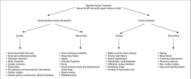
The conditions related with elevation in cardiac troponins There is a wide range of conditions related with elevated cTn. In Figure 1, we have summarized reasons for acute and chronic elevations. From our point of view, classifying of these conditions as cardiac and non-cardiac and as stable and un-stable can be instructive for understanding the underlying
Figure 1. Schematic presentation of the conditions related with elevated cardiac troponins
Elevated Cardiac Troponins (above the 99th percentile upper reference limit)
Acute elevation (and/or fall pattern) Chronic elevation
Noncardiac
• Acute myocardial infarction • Acute pericarditis/myocarditis • Takotsubo syndrome • Aortic dissection • Cardiac contusion • Tachycardia
• Percutaneous coronary/valvular interventions • Cardiac surgery
• Cardiac pacing, cardioversion, ablation therapies
• Acute pulmonary embolism • Respiratory failure • Sepsis • Critically ill patients • Burns
• Acute ischemic stroke and subarachnoid hemorrhage
• Severe anemia • Rhabdomyolysis
• Stable coronary artery disease • Chronic heart failure • Severe hypertension • Hypertrophic cardiomyopathy • Infiltrative cardiac disorders • Cardiotoxic drugs • Turnover of myocardial cells
• Ageing • Renal failure • Pulmonary hypertension • Strenuous exercise • Non-cardiac surgery • Hypo-and hyperthyroidism Noncardiac Cardiac Cardiac
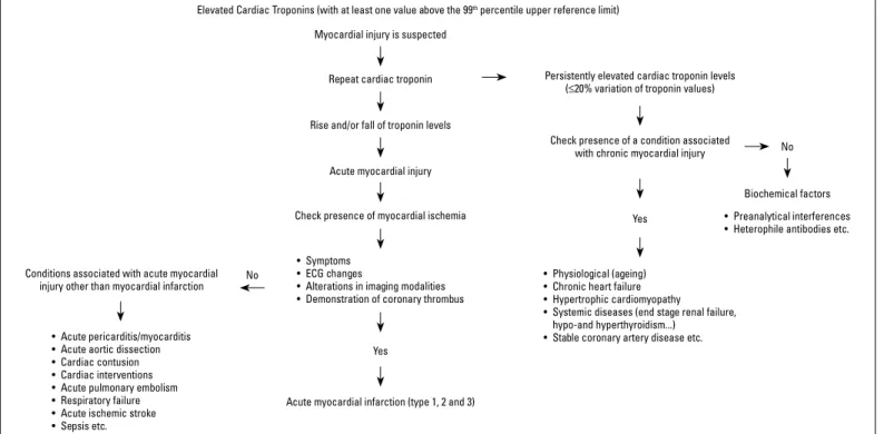
mechanisms and helpful in decision making. In this sense, first, we explain the stable and unstable cardiac and non-cardiac conditions. Later, we propose an algorithm for management of elevated cTn (Fig. 2). We strongly recommend against using the term of “troponinemia”. Any increase in the troponins needs to be taken into account and should be monitored. When per-sistently elevated cTn levels (that means ≤20% variation) are detected, conditions related with chronic cardiac injury should be regarded. When these conditions are excluded, biochemical factors could be the culprit. A typical rise and/or fall of troponin levels (with at least one value above the 99th percentile upper
reference limit) after repeated measure (preferably 3 h later) is called as acute myocardial injury, which is further defined as acute MI when accompanied by myocardial ischemia; other-wise, unstable cardiac and non-cardiac conditions should be considered.
Stable cardiac conditions Chronic heart failure
In patients with advanced heart failure (HF), elevated tropo-nin levels are commonly observed and are indicative of adverse prognosis (23). Increased volume and pressure load results in myocardial wall strain and myocyte death that are accepted to be the underlying mechanisms (23, 24). The relationship between wall strain and myocyte death could be explained by impaired subendocardial perfusion leading to cell death (25). Increased brain natriuretic peptide (BNP) level, which is an indicator for myocardial strain, correlates with increased troponin levels (26). Moreover, in rat myocardium, increased strain resulted in
troponin elevation regardless of ischemia (27). Myocardial cell loss is considered the main underlying mechanism in progres-sion of advanced HF. Sympathetic system and RAS activation, inflammatory mediators, and increased integrin levels as well as oxidative stress may enhance myocardial loss in patients with HF (28, 29). Increased troponin levels were also associated with acute decompensation, progressive disease, and poor prognosis in acute and chronic HF (30, 31).
Hypertrophic cardiomyopathy
In hypertrophic cardiomyopathy (HCMP), troponin rise may be seen because of several factors such as increased myocar-dial volume, increased oxygen need, and decreased flow vol-ume because of remodeling (32). Increased serum troponin level observed in a significant ratio of the patients with HCMP and is an independent predictor of adverse outcome (33). Elevated troponin levels have also been detected in other structural heart diseases associated with left ventricular wall thickening.
Infiltrative cardiac disorders
According to the accumulating substance, some of the infiltra-tive cardiac diseases increase ventricular wall thickness, while others cause chamber enlargement with secondary wall thinning. Cardiac amyloidosis is a primary restrictive cardiomyopathy. Al-though pathophysiology of troponin elevation remains unclear, myocyte compression injury due to extracellular deposition of the amyloid plaque is held to be responsible (34). Dispenzieri et al. (35) showed that in cardiac amyloidosis, troponin levels are more valuable as survival predictors than ECG and symptoms. Likewise,
Figure 2. Algorithm for the management of the cardiac troponin elevation
Elevated Cardiac Troponins (with at least one value above the 99th percentile upper reference limit) Myocardial injury is suspected
Persistently elevated cardiac troponin levels (≤20% variation of troponin values)
Check presence of a condition associated with chronic myocardial injury
Conditions associated with acute myocardial injury other than myocardial infarction
Acute myocardial infarction (type 1, 2 and 3)
Biochemical factors Yes
Yes
No
No
Rise and/or fall of troponin levels
Check presence of myocardial ischemia Acute myocardial injury Repeat cardiac troponin
• Preanalytical interferences • Heterophile antibodies etc. • Physiological (ageing)
• Chronic heart failure • Hypertrophic cardiomyopathy
• Systemic diseases (end stage renal failure, hypo-and hyperthyroidism...)
• Stable coronary artery disease etc. • Acute pericarditis/myocarditis
• Acute aortic dissection • Cardiac contusion • Cardiac interventions • Acute pulmonary embolism • Respiratory failure • Acute ischemic stroke • Sepsis etc.
• Symptoms • ECG changes
• Alterations in imaging modalities • Demonstration of coronary thrombus
hs-TnT value in sarcoidosis is considered an important marker of disease activity and is decreased following steroid therapy (36).
Turnover of myocardial cells, apoptosis
Troponin values increase generally because of myocyte ne-crosis caused by ischemia and MI. In some cases, an increase in troponin levels may be seen without myocyte necrosis. All cells, including cardiomyocytes, have death protocols that are activat-ed when appropriate conditions are met. These protocols can be
activated because of temporary conditions such as apoptosis, preload increase, ischemia, or pulmonary hypertension (37). Our present knowledge is not sufficient to determine by which po-tential effects apoptosis increases troponin levels (38).
Drug toxicity
Many agents can have cardiotoxic effects. Cardiotoxicity is often observed with use of anthracyclines, which are effective drugs for the treatment of solid and hematologic malignancies, Consensus Statements for Elevation of Cardiac Troponins
References
Cardiac troponins (cTn) are the mainstay for definition of myocardial injury. 1-3
Cardiac troponins should be used for the diagnosis and prognosis of acute coronary syndromes. 2, 3 An elevated troponin level should always be interpreted in the context of the clinical presentation
• Typical rise and fall, with at least one value above the 99th percentile URL, in the setting of acute myocardial ischemia (evidenced by ischemic symptoms, and alterations in ECG or imaging modalities) are needed for an accurate diagnosis of myocardial infarction.
• Troponin follow-up along with symptoms and ECG changes are required in case of suspicion
2, 3
A “sole” troponin elevation should not be defined as myocardial infarction. 2, 3
Troponin elevation is not recommended for the purpose of identification of the etiology of myocardial injury. 2, 3 Stable coronary artery disease, chronic heart failure, acute pericarditis, myocarditis, Takatsubo syndrome,
tachycardia, cardiac interventions should be considered among the stable and non-stable cardiac conditions of increased troponins (generally with different enzyme kinetics).
2, 30, 44, 45, 51, 61, 73, 82, 88 Aging, renal failure, sepsis, pulmonary hypertension, acute pulmonary embolism, critically ill patients, acute
cerebrovascular events should be considered among the stable and non-stable non-cardiac conditions of increased troponins (generally with different enzyme kinetics).
92, 96, 100, 113, 117, 123 Troponin elevation is prognostic even when ACS is excluded.
• Troponin levels are recommended to predict prognosis in patients with heart failure, stable chest pain, chronic renal disease, pulmonary diseases, stroke or after cardiac interventions and cardiac surgery. • Troponins may be used as surrogate markers of coronary artery disease mortality for screening and monitoring of healthy subjects.
30, 49-51, 96, 111, 116, 125
Numerous assays are approved for troponin measurement.
• 99th percentile for high-sensitive (hs) cTn assays should be measured with an analytical imprecision
(coefficient of variation -CV-) of 10% or less.
14-16, 18-20 • Non-hs-cTn assays with CV between 10% and 20% may also be used.
• Assays with CVs >20% should not be used.
• Measurement of CK-MB mass activity may be used if a cTn assay is not available.
• Analysis of the sample using an alternate cTn assay, measurement before and after using of heterophile-blocking tubes, and measurement of a series of dilutions may be used to deal with laboratory interferences related with false- positive results.
• Physicians should communicate with the laboratory when a persistent cTn elevation (that means ≤20% variation by repeated measures) cannot be explained by any clinical condition.
and trastuzumab-like drugs, which is an HER-2/neu (Human Epi-dermal Growth Factor Receptor 2) receptor antagonist. While cardiotoxic effects of anthracycline derivatives are dose-depen-dent and irreversible, trastuzumab-like drugs have reversible ef-fects. Cell membrane damage, caused by oxidative stress, reac-tive oxygen species, and lipid peroxidation, is held responsible for anthracycline group cardiotoxicity. Anti-HER2 drug group toxicity has reversible, functional, and structural effects on con-tractive proteins, and mitochondria, therefore rarely causes cell death (39, 40). Chemotherapy-induced troponin rise can predict the forthcoming left ventricular (LV) dysfunction (41, 42).
Cardiac surgery
As in all patient groups, post-operative troponin elevation is as-sociated with poor prognosis. Troponin level increases >10 times of the 99th percentile URL in patients with normal baseline values
on the first 48 h after “on pump” valve or coronary by-pass surgery is an important predictor for the first year survival rate (2, 43).
Percutaneous coronary/valvular interventions
Troponin level increase can be observed in percutaneous coronary interventions in stable settings due to flow discontinua-tion during balloon dilatadiscontinua-tion or ischemia due to distal emboliza-tion, and it is indicative of myocyte necrosis. At least five-fold increase in troponins predicts cardiovascular events at 30 days and one-year follow-up (2). Troponin level increase can also be observed after percutaneous valvular interventions such as transcutaneous aortic valve implantation (TAVI). Pre-interven-tional increased troponin values are observed in most of the pa-tients undergoing TAVI due to critical aortic stenosis. Pre- and post-interventional levels are considered important prognostic factors of one-year survival rate independently from the success of intervention (44).
Cardiac pacing, cardioversion, and ablation therapies Troponin increase can occur following permanent pacemak-er inspacemak-ertion due to minimal myocardial damage caused during endocardial lead implantation (45). A mild but still significant in-crease in troponin levels has been observed following electri-cal cardioversion (CV) in non-valvular atrial fibrillation (AF). This increase is even more significant in patients with increased LV volume and low ejection fraction (EF) (46). Even though tropo-nin increase may show progression of the present HF status, we must also keep in mind that this increase can be due to implant-able cardioverter defibrillator shocks in patients with HF (47).
Radiofrequency ablation, which is performed in various ar-rhythmias such as supraventricular tachycardia (SVT), AF, and ventricular tachycardia, causes cardiac damage via thermal en-ergy, therefore causing troponin increase (48).
Stable coronary artery disease
Several clinical studies have demonstrated that troponin value in otherwise healthy subjects could be a predictor of
sub-sequent adverse cardiac events including mortality (49, 50). The PROMISE (Prospective Multicenter Imaging Study for Evaluation of Chest Pain) study has provided that in patients with stable chest pain and suspected coronary artery disease (CAD), the up-per hsTnI quartiles were independently related with death, acute MI, or hospitalization for unstable chest pain during one-year follow-up period (51). Despite promising results, further data are required using troponins as surrogate markers of CAD mortality to screen and monitor the healthy subjects.
Unstable cardiac conditions Acute coronary syndrome
ACS is a term that includes patients with ST-segment eleva-tion myocardial infarceleva-tion (STEMI), non-ST-segment elevaeleva-tion myocardial infarction (NSTEMI), and unstable angina (UA). Car-diac biomarker elevations are required to distinguish NSTEMI from UA and helpful in patients with chest pain. Many diagnostic algorithms incorporated with serial cTn measurement are pro-posed to rule-in/out in patients with acute chest pain (3, 52, 53). It should be remembered that some diseases like myocarditis, ta-kotsubo cardiomyopathy might produce dynamic changes in cTn levels (53); and late presentation of ACS might not show mean-ingful changes in cTn.
Rapid rule-in and rule-out strategies for patients admitted with chest pain to the emergency department use different time points and cut-off values. The latest ESC Guideline for the man-agement of ACS in patients presenting without persistent ST-seg-ment elevation (3) depicted a comprehensive algorithm. Briefly, in acute chest pain, either 1-h or 3-h strategy should be used. The 1-h strategy is applicable if chest pain onset is >3 h, and there is a high pre-test probability for NSTEMI. Additionally, it can be applied when only high-sensitivity cTn assays are available [hs-cTnT (Elecsys), hs-cTnI (Architect), and hs-cTnI (Dimension Vista) are the validated hs-cTn assays]; and the cut-off levels are assay specific. The guideline recommended using the 3-h strat-egy, in which one delta value is greater than URL during follow-up prompts invasive strategy. We believe that the 3-h strategy is more user friendly and has a high validity, and it should be preferred. These strategies have a negative predictive value (for rule-out) exceeding 98%. On the other hand, the positive predic-tive value (for rule-in) is between 75% and 80% (3).
Apart from its diagnostic usefulness, cTn elevation conveys prognostic value. The patients with elevated cTn have increased mortality rate; and they are likely to have coronary thrombosis, more complex coronary lesions, and diminished ventricular func-tion (54, 55).
Severe hypertension
Increased troponin levels can be seen in hypertensive crisis because of supply-demand mismatch or obstructive CAD. In the study reported by Pattanshetty et al. (56), increased troponin lev-els in patients referring to the hospital because of hypertensive crisis were related with increased adverse cardiac events ratio.
Interestingly, in one-fourth of these patients, obstructive CAD was not seen.
Aortic dissection
Almost 90% of the patients with aortic dissection (AD) have abnormal ECG with 25%–35% having ACS-like ECG features re-sembling NSTEMI and 4%–16% have ECG findings of STEMI (57-60). Troponin positivity was reported from 16% to 33% with stan-dard assays and 54% to 61% with hs assay with no difference between type A and type B dissection. Troponin elevation in the setting of AD may be due to intimal flap obstructing the coro-nary ostia, corocoro-nary ostia dissection, decreased blood pressure, aortic regurgitation, LV pressure and volume overload, increased sympathetic drive leading to microvascular dysfunction, and pre-existing CAD (57-60).
Takotsubo syndrome
Takotsubo syndrome (TTS) is characterized by temporary LV wall motion abnormality that is usually preceded by emotional or physical triggers. The clinical presentation of patients with TTS is very similar to those of ACS subjects. The International Takot-subo Registry (61) found that troponin levels were elevated in 87% of 1750 patients on admission. Recorded troponin values are dis-proportionally low considering the extensive LV involvement (62). The mean troponin levels at admission were found to be similar to those in patients with ACS. Yet, peak values of cTn are lower in TTS than patients with ACS, though comparable values with NSTEMI could be observed (61). Troponin peaks occur earlier, usually at presentation or within 24 h following the onset of symptoms and normalizes faster than STEMI (63). Higher TnI values were ob-served in patients with TTS presenting with cardiogenic shock (64).
Cardiac contusion
Cardiac contusion or currently preferred term blunt cardiac injury (BCI) is usually suspected in blunt thoracic trauma (BTT). There are no definitive diagnostic criteria, but various combina-tions of clinical picture, ECG, troponin, or cardiac imaging were used to define BCI. Therefore, the incidence of BCI in patients with BTT varies from 3% to 56% (65-67). The release of troponin reflect-ing myocardial cell injury is believed to be because of mechanical transmission of force through the chest wall (68). But, troponin ele-vation was also observed in 25%–35% of trauma subjects who had no BTT (67, 69). Among patients with severe traumatic brain injury, around 30% at admission and 41% overall had elevated cTnI (70). Thus, other mechanisms like hypotension due to blood loss, pro-inflammatory cytokines, free radical and oxidative injury, and ad-renergic activation with catecholamine spillover might have a role in troponin elevation (71). Trauma patients with elevated troponins have increased mortality rate even in the absence of BCI (67, 72).
Tachycardias
In a pooled analysis of seven observational studies includ-ing 1155 patients with SVT, 66% of patients were investigated
with troponin (73). Of these, 32% had positive troponin test re-sult. Troponin elevation in the setting of SVT was not predictive of coronary or structural heart disease (74, 75). Myocardial gen demand is increased due to increased heart rate, and oxy-gen delivery is attenuated because of short-diastole in SVT. This may cause ischemia that probably alters the myocyte membrane permeability and might result in the release of cTn from the free cytosolic pool or its loosely attached cytoskeleton. Myocardial stretch was also postulated as another possible mechanism of tachycardia-related troponin elevation (76, 77). The reported pre-dictors of troponin elevation were maximal heart rate, older age, duration of tachycardia, chest pain, and lower diastolic blood pressure (78). The data about the prognostic role of troponin el-evation in SVT is not satisfactory because of limited number of patients. On the other hand, in a recent study performed in 1754 patients with AF admitted with 2754 symptomatic AF episodes to emergency department, elevated hs-TnT levels were indepen-dently related with midterm (median: two years) mortality (79).
Acute pericarditis
Detectable levels of troponin were reported in 32%–71% of patients with acute pericarditis (80, 81). Troponin was beyond the acute MI threshold in 7%–22% of cases (80, 82). Younger age, ST-segment elevation, recent onset of infection, male gender, and pericardial effusion were the properties associated with el-evated troponin levels in patients with pericarditis (80, 83). In one study, troponin elevation was found to be related with mortality in acute pericarditis (84). However, two large studies did not find such a negative prognostic value (80, 85).
Myocarditis
The patients presenting early in the course of the disease were shown to have increased concentrations of cTnT in a small sized biopsy-proven myocarditis study (86). Smith et al. (87) demonstrated that cTnI values were elevated in one of three patients with myo-carditis. Lauer et al. (88) showed that among clinically suspected myocarditis subjects with elevated troponin, myocarditis was evi-dent on 93% of biopsy specimens. However, 44% of subjects with-out troponin elevation had also biopsy-proven myocarditis. Thus, negative troponin does not rule out myocarditis (88). Patients with shorter history of symptoms are more likely to have higher concen-trations of troponins (89). Subjects with fulminant myocarditis have higher troponin elevation than patients with acute myocarditis (90). Peak troponin levels of acute myocarditis are usually lower than that of ACS (91), but release kinetics might mimic ACS (92).
Stable non-cardiac conditions Aging
Elderly people may have elevated hs-cTnT and hs-cTnI, mostly due to increased cardiovascular comorbidities, anemia, decreased renal function with aging, and structural/functional cardiac abnormalities. Gore et al. (93) showed that 10% of men, aged 65–74 years, with no cardiovascular disease had elevated
cTnT above the MI threshold. Reiter et al. (94) reported that mild troponin elevations are common in elderly non-AMI patients. Therefore, they argued that the optimal cut-off levels to separate acute MI from non-cardiac troponin elevations should be higher in elderly as compared with younger patients. Wu et al. (95) found that 44% of the elderly inpatients without ACS had an hs-cTnT level >99th percentile URL, and baseline hs-cTnT level in those
patients was associated with all-cause death after discharge, and the mortality rate increased with increased hs-cTnT level.
Renal failure/end-stage renal disease
Cardiac troponins are frequently elevated in patients with re-nal failure. The prevalence of increased serum cTnT and cTnI increases with severity of renal failure, and cTnT is more fre-quently increased compared with cTnI in asymptomatic patients with end-stage renal disease (ESRD) (96, 97). The mechanisms causing increases in TnT concentrations in patients with renal failure are not clear. Troponin elevations are not solely caused by decreased renal clearance, but also possibly due to direct toxic effects of the uremic state on the myocardium, anemia, hypoten-sion, and accompanying CAD (98).
cTnT is associated with mortality in patients with ESRD (99). The NACB Laboratory Medicine Practice Guidelines (100) recom-mend the use of troponin for diagnosis of MI in all patients with renal failure (regardless of the severity of renal impairment) who have symptoms or electrocardiographic evidence of myocardial ischemia. The guidelines also advise relying on dynamic changes in troponin values of ≥20% in the 6 h after presentation to define acute MI, even in chronically elevated troponin levels.
Pulmonary hypertension
Torbicki et al. (101) reported that cTnT was detected in serum of 14% of patients with chronic precapillary pulmonary hyperten-sion (PH) and was a strong independent marker of mortality. Se-vere PH results in disturbance in the physiological pattern of right ventricular (RV) myocardial perfusion and lower systemic blood pressure, both at rest and during exercise, decreasing coronary perfusion gradient (102). Troponin elevations in these patients may be due to both myocyte death and intracellular degradation of troponin caused by excessive intracellular Ca2+ concentration
in the failing myocardium. Physical Exercise (Strenuous)
The reviews and meta-analysis have shown that cTnT lev-els are frequently elevated after extreme exercise (marathons, triathlons, mountain bicycle races, ultraendurance events) (103, 104). Postulated mechanisms include cellular shifts in cytoplas-mic troponin due to exercise-induced inflammatory cytokines, dehydration, hemoconcentration, and oxidative stressors rather than typical myocardial necrosis. Cardiac magnetic resonance imaging has not shown any functional changes or any detect-able myocardial inflammation or fibrosis after exercise (105). In addition, during strenuous exercise, cTnT can be detected in
less than 2 h and generally return to normal within 24 h, which is different than the course of ACS. One-year clinical follow-up showed no cardiac events or symptoms in the troponin-positive group (106). A recent meta-analysis evaluating elevation in hs-cTn after exercise and pharmacological stress tests revealed that the rising patterns were inconsistent and were not related with inducible myocardial ischemia. So, adding hs-cTn to cardiac stress tests may not improve diagnostic utility (107).
Non-cardiac surgery
In the non-cardiac perioperative surgical setting, troponin elevations may be seen in nearly 8%–11% of the patients with-out apparent ACS in the early post-operative timeframe, and they are associated with increased mortality and longer length of stay. Bleeding, pain, and increased catecholamines may result in tachycardia causing mismatch in oxygen demand or supply, while vasoconstriction or pain may increase blood pressure that in turn increases wall stress; all of which may be responsible from cTnT elevation (108-110).
Hypo-and hyperthyroidism
Thyroid hormones are related with cardiac functions. Cre-atine kinase and troponins may increase in patients with hypo-thyroid without apparent myocardial damage (111, 112). However, despite these rare case reports, studies in consecutive patients with significant hypothyroidism have not reported elevated TnI levels (113) or TnT levels even in patients with increased CK-MB levels (114). Hence, the importance of these findings related to hypothyroidism needs to be determined. Hyperthyroidism may induce tachyarrhythmias and increase oxygen demand resulting in myocyte damage and cTnT release.
Unstable non-cardiac conditions Acute pulmonary embolism
Pulmonary embolism (PE) is one of the most common non-ACS causes of increased troponins (115). Serum troponins are elevated in up to 50% of patients with PE (116). This cTnT re-lease is attributed to the combination of acute pressure overload within the RV, impaired coronary artery flow, and the hypoxic state caused by PE (117). Endothelial damage in the pulmonary vasculature, which has an abundance of angiotensin-converting enzyme, may cause derangements in the renin/angiotensin/al-dosterone system affecting cTnT blood concentrations (118). In contrast to patients with ACS, cTnT peaked after a median of 10 h and remained detectable for a median of only 40 h after admis-sion in patients with PE (119). Troponin elevation is associated with prolonged hypotension and cardiogenic shock, need for ino-tropic support and mechanical ventilation, and increased mortal-ity in patients with PE (95).
Respiratory failure
cTnT may be elevated in advanced/decompensated chronic lung disease. Hypercapnia, hypoxemia and/or respiratory
acido-sis, the worsening of PH resulting in RV hypertrophy, dilation, and subendocardial demand-induced ischemia, the increased work and oxygen cost of breathing, and the increase in LV afterload related to the more negative intrathoracic pressure may all con-tribute to myocardial injury and cTnT released during episodes of exacerbation. Elevated cTnI is a strong and independent pre-dictor of in-hospital death also in patients admitted for acutely exacerbated chronic obstructive pulmonary disease (120).
Sepsis/systemic inflammatory response syndrome
There are modest elevations of troponins in patients with sep-sis, septic shock, and the Systemic inflammatory response syn-drome (SIRS), mostly in the absence of CAD. Causes of troponin elevation in sepsis are multifactorial. It has been suggested that inflammatory cytokines released from neutrophils, particularly tumor necrosis factor-
α
and interleukin-6, are responsible for direct myocardial depression and increased cell membrane per-meability to troponin molecules in sepsis. Decreased myocardial perfusion because of hypotension, increased oxygen consump-tion due to tachycardia, release of noradrenaline and adrenaline with subsequent vasoconstriction, and increased coagulation of capillary bed also may play significant part in myocyte dam-age and subsequent troponin release (95). Although troponin elevation in sepsis is associated with mortality and impaired LV function, routine troponin testing in septic patients is not recom-mended (1).Critically ill patients
Elevations of cTn are common among critically ill patients in the intensive care unit (ICU), and they are associated with in-creased mortality and ICU length of stay regardless of the un-derlying disease state (121). Similar to sepsis, inflammatory cy-tokines are responsible for direct myocardial depression and increased cell membrane permeability to troponin molecules in critically ill patients.
Burns
Cardiac dysfunction associated with severe burns has been suggested by several reports (122, 123). Wang and He (124) re-ported increased cTn levels in patients with severe burns. There was a significant correlation with a positive troponin test and burns greater than 15% total body surface area. The myocardial damage is attributed to adverse effects of inflammatory media-tors on myocardium and severe hypovolemia.
Acute ischemic stroke and subarachnoid hemorrhage An increase in cTn in patients with stroke has been docu-mented in a systematic review of 15 studies using old-genera-tion cTn assays (125). Elevaold-genera-tions that are more significant have been shown in the studies using hs-troponin assays in asso-ciation with both ischemic and hemorrhagic cerebrovascular events (CVE) (126-128). The underlying pathophysiology may be the cardiac damage caused by stunned myocardium with
acute neurological insult resulting in “neurogenic stress car-diomyopathy” that is a relatively new terminology. Neurogenic stress cardiomyopathy indicates acute cardiac damage due to catecholamine excess and unopposed inflammation caused by alterations in autonomic nervous system related to acute CVE (129). On the other hand, similar risk factors such as age, hypertension, diabetes, precipitate stroke, hemorrhagic CVE, and cardiovascular diseases. Thus, the exacerbation of occult cardiac disease in response to stress caused by neurological insult may lead to cTn elevation in these patients (130). Data regarding elevated pro-BNP levels in patients with stroke are compatible with this theory. Moreover, an adverse prognosis in association with elevated cTn values has been suggested in patients with acute CVE (131).
Severe anemia
Although there is no study investigating the rise in cTn lev-els in different hemoglobin levlev-els in large patient populations, severe anemia was shown to be related with increased tro-ponin levels in children with malaria (132, 133). Severe anemia has also been found to relate with mortality in patients with MI and HF (134, 135). Barbarova et al. (136) showed that patients with severe anemia and elevated troponin levels presenting to internal medicine departments due to non-cardiac problems had worse long-term survival if they did not get blood transfu-sion. The main reason of the increase in cTn in case of severe anemia may be resultant tissue hypoxia leading to myocardial injury.
Rhabdomyolysis
Several studies have found increased cTn levels in patients with rhabdomyolysis or neuromuscular diseases (137, 138). Although TnI and TnT are accepted to be expressed by only cardiomyocytes, their messenger RNA can be reexpressed in skeletal muscle disease and may result in misinterpretation of cardiac injury (139, 140). Moreover, TnT is also expressed in fetal skeletal muscle. In case of skeletal muscle injury, it may be reexpressed because of regeneration process included in repair (141). Rhabdomyolysis may cause a rise in cTn level with this mechanism or may include direct cardiac muscle degrada-tion (142, 143).
Conclusion
Increase in cTn has typically been used for the diagnosis and prognosis of ACS. Nevertheless, these biomarkers are also elevated in a variety of stable and unstable cardiac and non-cardiac conditions. The introduction of new-generation hs-cTn assays has allowed detection of these markers even in major-ity of the healthy individuals, also has lowered the specificmajor-ity of the tests in acute chest pain syndromes leading to unnecessary interventions. Consequently, consideration of pre-analytical and
analytical factors affecting troponin measurement and system-atic evaluation of conditions related with troponin elevations are of great importance to obtain accurate diagnosis.
Acknowledgments: The authors thank the members of the Turkish Society of Cardiology Guidelines Committee (Aylin Yıldırır, Bülent Görenek, Kaan Okyay, Gökhan Kahveci, Mustafa Çetin, Regayip Zehir, Ali Baturak, Abdullah Tekin, Beste Özben Sadıç, Asife Şahinarslan, Taner Ulus).
Conflict of interest: None declared.
Peer-review: Externally and internally peer-reviewed.
Authorship contributions: Concept – K.O.; Design – K.O.; Supervision – K.O.; Data collection &/or processing – K.O., B.Ö.S., A.Ş., M.E.D., C.Y.K., S.E.E., Ö.G., A.T.; Analysis &/or interpretation – K.O., B.Ö.S., A.Ş., M.E.D., C.Y.K., S.E.E., Ö.G., A.T.; Literature search – K.O., B.Ö.S., A.Ş., M.E.D., C.Y.K., S.E.E., Ö.G., A.T.; Critical review – A.Y., B.G., O.Y., A.S.F.
References
1. Newby LK, Jesse RL, Babb JD, Christenson RH, De Fer TM, Diamond GA, et al. ACCF 2012 expert consensus document on practical clinical considerations in the interpretation of troponin elevations: a report of the American College of Cardiology Foundation task force on Clinical Expert Consensus Documents. J Am Coll Cardiol 2012; 60: 2427-63. 2. Thygesen K, Alpert JS, Jaffe AS, Chaitman BR, Bax JJ, Morrow DA,
et al.; Executive Group on behalf of the Joint European Society of Cardiology (ESC)/American College of Cardiology (ACC)/American Heart Association (AHA)/World Heart Federation (WHF) Task Force for the Universal Definition of Myocardial Infarction. Fourth Uni-versal Definition of Myocardial Infarction (2018). J Am Coll Cardiol 2018; 72: 2231-64. [CrossRef]
3. Roffi M, Patrono C, Collet JP, Mueller C, Valgimigli M, Andreotti F, et al.; ESC Scientific Document Group. 2015 ESC Guidelines for the management of acute coronary syndromes in patients presenting without persistent ST-segment elevation: Task Force for the Man-agement of Acute Coronary Syndromes in Patients Presenting with-out Persistent ST-Segment Elevation of the European Society of Cardiology (ESC). Eur Heart J 2016; 37: 267-315. [CrossRef]
4. Jaffe AS. Troponin-past, present, and future. Curr Probl Cardiol 2012; 37: 209-28. [CrossRef]
5. Okyay K, Yıldırır A. The preanalytical and analytical factors respon-sible for false-positive cardiac troponins. Anatol J Cardiol 2015; 15: 264-5. [CrossRef]
6. Tiwari RP, Jain A, Khan Z, Kohli V, Bharmal RN, Kartikeyan S, et al. Cardiac troponins I and T: molecular markers for early diagnosis, prognosis, and accurate triaging of patients with acute myocardial infarction. Mol Diagn Ther 2012; 16: 371-81. [CrossRef]
7. Thygesen K, Mair J, Katus H, Plebani M, Venge P, Collinson P, et al.; Study Group on Biomarkers in Cardiology of the ESC Working Group on Acute Cardiac Care. Recommendations for the use of cardiac troponin measurement in acute cardiac care. Eur Heart J 2010; 31: 2197-204. [CrossRef]
8. Galli C, Lippi G. High-sensitivity cardiac troponin testing in routine practice: economic and organizational advantages. Ann Transl Med 2016; 4: 257. [CrossRef]
9. Giannitsis E, Katus HA. Cardiac troponin level elevations not related to acute coronary syndromes. Nat Rev Cardiol 2013; 10: 623-34. 10. Jaffe AS, Babuin L, Apple FS. Biomarkers in acute cardiac
dis-ease: the present and the future. J Am Coll Cardiol 2006; 48: 1-11. 11. Katus HA, Remppis A, Scheffold T, Diederich KW, Kuebler W. Intra-cellular compartmentation of cardiac troponin T and its release kinetics in patients with reperfused and nonreperfused myocar-dial infarction. Am J Cardiol 1991; 67: 1360-7. [CrossRef]
12. Gaze DC, Collinson PO. Multiple molecular forms of circulating cardiac troponin: analytical and clinical significance. Ann Clin Biochem 2008; 45: 349-55. [CrossRef]
13. Park KC, Gaze DC, Collinson PO, Marber MS. Cardiac troponins: from myocardial infarction to chronic disease. Cardiovasc Res 2017; 113: 1708-18. [CrossRef]
14. Apple FS, Sandoval Y, Jaffe AS, Ordonez-Llanos J; IFCC Task Force on Clinical Applications of Cardiac Bio-Markers. Cardiac Troponin Assays: Guide to Under-standing Analytical Characteristics and Their Impact on Clinical Care. Clin Chem 2017; 63: 73-81. [CrossRef]
15. Burtis CA, Ashwood ER, Bruns DE. Tietz Textbook of Clinical Chemistry and Molecular Diagnostics. 5th Ed. Cardiac Function: Chapter 47. Elsevier; 2012. p.1457-522.
16. Jaffe AS, Morrow DA. Troponin testing: analytical considerations. Saperia GM, Ed. UpToDate. Waltham, MA. Accessed on February 13, 2019. Available from: URL: https://www.uptodate.com.
17. Pickering JW, Than MP, Cullen L, Aldous S, Ter Avest E, Body R, et al. Rapid Rule-out of Acute Myocardial Infarction With a Single High-Sensitivity Cardiac Troponin T Measurement Below the Limit of Detection: A Collaborative Meta-analysis. Ann Intern Med 2017; 166: 715-24.
18. Tate J, Ward G. Interferences in immunoassay. Clin Biochem Rev 2004; 25:105-20.
19. Herman DS, Kavsak PA, Greene DN. Variability and Error in Car-diac Troponin Testing: An ACLPS Critical Review. Am J Clin Pathol 2017; 148: 281-95. [CrossRef]
20. Howanitz JH, Bjerner J, Chace NM, Cook BC, Datta P, Doss RC. CLSI. Immunoassay Interference by Endogenous Antibodies; Ap-proved Guideline. CLSI document I/LA30-A. Wayne, PA: Clinical and Laboratory Standards Institute; 2008.
21. Herman DS, Ranjitkar P, Yamaguchi D, Grenache DG, Greene DN. Endogenous alkaline phosphatase interference in cardiac tropo-nin I and other sensitive chemiluminescence immunoassays that use alkaline phosphatase activity for signal amplification. Clin Biochem 2016; 49: 1118-21. [CrossRef]
22. Kishnani PS, Rush ET, Arundel P, Bishop N, Dahir K, Fraser W, et al. Monitoring guidance for patients with hypophosphatasia treated with asfotase alfa. Mol Genet Metab 2017; 122: 4-17. [CrossRef]
23. Horwich TB, Patel J, MacLellan WR, Fonarow GC. Cardiac tro-ponin I is associated with impaired hemodynamics, progressive left ventricular dysfunction, and increased mortality rates in ad-vanced heart failure. Circulation 2003; 108: 833-8. [CrossRef]
24. Nunes JP, Mota Garcia JM, Farinha RM, Carlos Silva J, Magalhães D, Vidal Pinheiro L, et al. Cardiac troponin I in aortic valve disease. Int J Cardiol 2003; 89: 281-5. [CrossRef]
25. Logeart D, Beyne P, Cusson C, Tokmakova M, Leban M, Guiti C, et al. Evidence of cardiac myolysis in severe nonischemic heart failure and the potential role of increased wall strain. Am Heart J 2001; 141: 247-53. [CrossRef]
26. Feng J, Schaus BJ, Fallavollita JA, Lee TC, Canty JM Jr. Preload induces troponin I degradation independently of myocardial isch-emia. Circulation 2001; 103: 2035-7. [CrossRef]
27. Cheng W, Li B, Kajstura J, Li P, Wolin MS, Sonnenblick EH, et al. Stretch-induced programmed myocyte cell death. J Clin Invest 1995; 96: 2247-59. [CrossRef]
28. Fonarow GC, Horwich TB. Combining natriuretic peptides and necrosis markers in determining prognosis in heart failure. Rev Cardiovasc Med 2003; 4 (Suppl 4): S20-8.
29. Peacock WF 4th, De Marco T, Fonarow GC, Diercks D, Wynne J, Apple FS, et al.; ADHERE Investigators. Cardiac Troponin and Out-come in Acute Heart Failure. N Engl J Med 2008; 358: 2117-26. 30. Kociol RD, Pang PS, Gheorghiade M, Fonarow GC, O'Connor CM,
Felker GM. Troponin elevation in heart failure: prevalence, mecha-nisms, and clinical implications. J Am Coll Cardiol 2010; 56: 1071-8. 31. Patil H, Vaidya O, Bogart D. A review of causes and systemic ap-proach to cardiac troponin elevation. Clin Cardiol 2011; 34: 723-8. 32. Krams R, Kofflard MJ, Duncker DJ, Von Birgelen C, Carlier S,
Kliffen M, et al. Decreased coronary flow reserve in hypertrophic cardiomyopathy is related to remodeling of the coronary micro-circulation. Circulation 1998; 97: 230-3. [CrossRef]
33. Kubo T, Kitaoka H, Yamanaka S, Hirota T, Baba Y, Hayashi K, et al. Significance of high-sensitivity cardiac troponin T in hypertrophic cardiomyopathy. J Am Coll Cardiol 2013; 62: 1252-9. [CrossRef]
34. Cantwell RV, Aviles RJ, Bjornsson J, Wright RS, Freeman WK, Oh JK, et al. Cardiac amyloidosis presenting with elevations of car-diac troponin I and angina pectoris. Clin Cardiol 2002; 25: 33-7. 35. Dispenzieri A, Kyle RA, Gertz MA, Therneau TM, Miller WL,
Chan-drasekaran K, et al. Survival in patients with primary systemic amyloidosis and raised serum cardiac troponins. Lancet 2003; 361: 1787-9. [CrossRef]
36. Baba Y, Kubo T, Kitaoka H, Okawa M, Yamanaka S, Kawada Y, et al. Usefulness of high-sensitive cardiac troponin T for evaluating the activity of cardiac sarcoidosis. Int Heart J 2012; 53: 287-92. 37. Weil BR, Young RF, Shen X, Suzuki G, Qu J, Malhotra S, et al. Brief
Myocardial Ischemia Produces Cardiac Troponin I Release and Focal Myocyte Apoptosis in the Absence of Pathological Infarc-tion in Swine. JACC Basic Transl Sci 2017; 2: 105-14. [CrossRef]
38. Hammarsten O, Mair J, Möckel M, Lindahl B, Jaffe AS. Possible mechanisms behind cardiac troponin elevations. Biomarkers 2018; 23: 725-34. [CrossRef]
39. Cote GM, Sawyer DB, Chabner BA. ERBB2 inhibition and heart failure. N Engl J Med 2012; 367: 2150-3. [CrossRef]
40. Ewer MS, Lippman SM. Type II chemotherapy-related cardiac dysfunction: time to recognize a new entity. J Clin Oncol 2005; 23: 2900-2. [CrossRef]
41. Cardinale D, Sandri MT, Martinoni A, Tricca A, Civelli M, Lamantia G, et al. Left ventricular dysfunction predicted by early troponin I release after high-dose chemotherapy. J Am Coll Cardiol 2000; 36: 517-22. [CrossRef]
42. Zamorano JL, Lancellotti P, Rodriguez Muñoz D, Aboyans V, As-teggiano R, Galderisi M, et al.; ESC Scientific Document Group. 2016 ESC Position Paper on cancer treatments and cardiovascu-lar toxicity developed under the auspices of the ESC Committee for Practice Guidelines: The Task Force for cancer treatments and cardiovascular toxicity of the European Society of Cardiology (ESC). Eur Heart J 2016; 37: 2768-801. [CrossRef]
43. Mauermann E, Bolliger D, Fassl J, Grapow M, Seeberger EE, See-berger MD, et al. Association of Troponin Trends and Cardiac Morbidity and Mortality After On-Pump Cardiac Surgery. Ann Thorac Surg 2017; 104: 1289-97. [CrossRef]
44. Chorianopoulos E, Krumsdorf U, Geis N, Pleger ST, Giannitsis E, Katus HA, et al. Preserved prognostic value of preinterventional
troponin T levels despite successful TAVI in patients with severe aortic stenosis. Clin Res Cardiol 2014; 103: 65-72. [CrossRef]
45. Nikolaou NI, Christou AH, Spanodimos SG, Antonatos DG, Korkon-ikitas PI, Patsilinakos SP. Marked troponin elevation after implan-tation of a permanent antibradycardia pacemaker. Hellenic J Car-diol 2011; 52: 489-92.
46. Piechota W, Gielerak G, Ryczek R, Kaźmierczak A, Bejm J, Piecho-ta W. Cardiac troponin I after external electrical cardioversion for atrial fibrillation as a marker of myocardial injury a preliminary report. Kardiol Pol 2007; 65: 664-9.
47. Miranda CH, Schmidt A, Pazin-Filho A. Prognostic evaluation of the troponin I elevation after multiple spontaneous shocks of the implantable cardioverter/defibrillator. Am J Emerg Med 2014; 32: 1085-8. [CrossRef]
48. Reichlin T, Lockwood SJ, Conrad MJ, Nof E, Michaud GF, John RM, et al. Early release of high-sensitive cardiac troponin during com-plex catheter ablation for ventricular tachycardia and atrial fibril-lation. J Interv Card Electrophysiol 2016; 47: 69-74. [CrossRef]
49. de Lamos JA, Drazner MH, Omland T, Ayers CR, Khera A, Rohatgi A, et al. Association of troponin T detected with a highly sensi-tive assay and cardiac structure and mortality risk in the general population. JAMA 2010; 304: 2503-12. [CrossRef]
50. Blankanberg S, Salomaa V, Makarova N, Ojeda F, Wild P, Lackner KJ, et al. Troponin I and cardiovascular risk prediction in the gen-eral population: the BiomarCARE consortium. Eur Heart J 2016; 37: 2428-37. [CrossRef]
51. Januzzi JL Jr, Suchindran S, Hoffmann U, Patel MR, Ferencik M, Coles A, et al.; PROMISE Investigators. Single-Molecule hsTnI and Short-Term Risk in Stable Patients With Chest Pain. J Am Coll Car-diol 2019; 73: 251-60. [CrossRef]
52. Body R. Acute coronary syndromes diagnosis, version 2.0: Tomor-row's approach to diagnosing acute coronary syndromes? Turk J Emerg Med 2018; 18: 94-9. [CrossRef]
53. Cervellin G, Mattiuzzi C, Bovo C, Lippi G. Diagnostic algorithms for acute coronary syndrome-is one better than another? Ann Transl Med 2016; 4: 193. [CrossRef]
54. Heeschen C, Hamm CW, Goldmann B, Deu A, Langenbrink L, White HD. Troponin concentrations for stratification of patients with acute coronary syndromes in relation to therapeutic effi-cacy of tirofiban. PRISM Study Investigators. Platelet Receptor Inhibition in Ischemic Syndrome Management. Lancet 1999; 354: 1757-62. [CrossRef]
55. Lindahl B, Diderholm E, Lagerqvist B, Venge P, Wallentin L; FRISC II (Fast Revascularization during In Stability in CAD) Investigators. Mechanisms behind the prognostic value of troponin T in unstable coronary artery disease: a FRISC II substudy. J Am Coll Cardiol 2001; 38: 979-86. [CrossRef]
56. Pattanshetty DJ, Bhat PK, Aneja A, Pillai DP. Elevated troponin pre-dicts long-term adverse cardiovascular outcomes in hypertensive crisis: a retrospective study. J Hypertens 2012; 30: 2410-5. [CrossRef]
57. Pourafkari L, Tajlil A, Ghaffari S, Chavoshi M, Kolahdouzan K, Par-vizi R, et al. Electrocardiography changes in acute aortic dissec-tion-association with troponin leak, coronary anatomy, and prog-nosis. Am J Emerg Med 2016; 34: 1431-6. [CrossRef]
58. Biagini E, Lofiego C, Ferlito M, Fattori R, Rocchi G, Graziosi M, et al. Frequency, determinants, and clinical relevance of acute coro-nary syndrome-like electrocardiographic findings in patients with acute aortic syndrome. Am J Cardiol 2007; 100: 1013-9. [CrossRef]
59. Vagnarelli F, Corsini A, Bugani G, Lorenzini M, Longhi S, Bacchi Reggiani ML, et al. Troponin T elevation in acute aortic syndromes:
Frequency and impact on diagnostic delay and misdiagnosis. Eur Heart J Acute Cardiovasc Care 2016; 5: 61-71. [CrossRef]
60. Li G, Wu XW, Lu WH, Cheng J, Wu XY, Ai R, et al. High-sensitivity cardiac troponin T: A biomarker for the early risk stratification of type-A acute aortic dissection? Arch Cardiovasc Dis 2016; 109: 163-70. [CrossRef]
61. Templin C, Ghadri JR, Diekmann J, Napp LC, Bataiosu DR, Jag-uszewski M, et al. Clinical Features and Outcomes of Takotsubo (Stress) Cardiomyopathy. N Engl J Med 2015; 373: 929-38. [CrossRef]
62. Ramaraj R, Sorrell VL, Movahed MR. Levels of troponin release can aid in the early exclusion of stress-induced (takotsubo) car-diomyopathy. Exp Clin Cardiol 2009; 14: 6-8.
63. Sharkey SW, Lesser JR, Menon M, Parpart M, Maron MS, Maron BJ. Spectrum and significance of electrocardiographic patterns, troponin levels, and thrombolysis in myocardial infarction frame count in patients with stress (tako-tsubo) cardiomyopathy and comparison to those in patients with ST-elevation anterior wall myocardial infarction. Am J Cardiol 2008; 101: 1723-8. [CrossRef]
64. Song BG, Park SJ, Noh HJ, Jo HC, Choi JO, Lee SC, et al. Clinical characteristics, and laboratory and echocardiographic findings in takotsubo cardiomyopathy presenting as cardiogenic shock. J Crit Care 2010; 25: 329-35. [CrossRef]
65. Gautam PL, Luthra N, Kaur M, Singh J, Wander GS, Tandon R, et al. Evaluation of Myocardial Injury using Standard Diagnostic Tools and Tissue Doppler Imaging in Blunt Trauma Chest. J Clin Diagn Res 2017; 11: OC33-6. [CrossRef]
66. Sybrandy KC, Cramer MJ, Burgersdijk C. Diagnosing cardiac contu-sion: old wisdom and new insights. Heart 2003; 89: 485-9. [CrossRef]
67. Kalbitz M, Pressmar J, Stecher J, Weber B, Weiss M, Schwarz S, et al. The Role of Troponin in Blunt Cardiac Injury After Multiple Trauma in Humans. World J Surg 2017; 41: 162-9. [CrossRef]
68. RuDusky BM. More on myocardial contusion--with additional in-sight on myocardial concussion. Chest 1997; 112: 570-2. [CrossRef]
69. Edouard AR, Benoist JF, Cosson C, Mimoz O, Legrand A, Samii K. Circulating cardiac troponin I in trauma patients without cardiac contusion. Intensive Care Med 1998; 24: 569-73. [CrossRef]
70. Salim A, Hadjizacharia P, Brown C, Inaba K, Teixeira PG, Chan L, et al. Significance of troponin elevation after severe traumatic brain injury. J Trauma 2008; 64: 46-52. [CrossRef]
71. Martin M, Mullenix P, Rhee P, Belzberg H, Demetriades D, Salim A. Troponin increases in the critically injured patient: mechanical trauma or physiologic stress? J Trauma 2005; 59: 1086-91. [CrossRef]
72. Mahmood I, El-Menyar A, Dabdoob W, Abdulrahman Y, Siddiqui T, Atique S, et al. Troponin T in Patients with Traumatic Chest Inju-ries with and without Cardiac Involvement: Insights from an Ob-servational Study. N Am J Med Sci 2016; 8: 17-24. [CrossRef]
73. Fernando H, Adams N, Mitra B. Review article: The utility of tro-ponin and other investigations in patients presenting to the emer-gency department with supraventricular tachycardia. Emerg Med Australas 2019; 31: 35-42. [CrossRef]
74. Schueler M, Vafaie M, Becker R, Biener M, Thomas D, Mueller M, et al. Prevalence, kinetic changes and possible reasons of elevated cardiac troponin T in patients with AV nodal re-entrant tachycardia. Acute Card Care 2012; 14: 131-7. [CrossRef]
75. Bukkapatnam RN, Robinson M, Turnipseed S, Tancredi D, Amster-dam E, Srivatsa UN. Relationship of myocardial ischemia and in-jury to coronary artery disease in patients with supraventricular tachycardia. Am J Cardiol 2010; 106: 374-7. [CrossRef]
76. Higgins JP, Higgins JA. Elevation of cardiac troponin I indicates more than myocardial ischemia. Clin Invest Med 2003; 26: 133-47.
77. Qi W, Kjekshus H, Klinge R, Kjekshus JK, Hall C. Cardiac natriuret-ic peptides and continuously monitored atrial pressures during chronic rapid pacing in pigs. Acta Physiol Scand 2000; 169: 95-102. 78. Sayadnik M, Shafiee A, Jenab Y, Jalali A, Sadeghian S. Predictors of High-Sensitivity Cardiac Troponin T Elevation in Patients with Acute Paroxysmal Supraventricular Tachycardia and Ischemic Heart Disease. Tex Heart Inst J 2017; 44: 306-11. [CrossRef]
79. Niederdöckl J, Simon A, Schnaubelt S, Schuetz N, Laggner R, Sul-zgruber P, et al. Cardiac biomarkers predict mortality in emergency patients presenting with atrial fibrillation. Heart 2019; 105: 482-8. 80. Imazio M, Demichelis B, Cecchi E, Belli R, Ghisio A, Bobbio M, et
al. Cardiac troponin I in acute pericarditis. J Am Coll Cardiol 2003; 42: 2144-8. [CrossRef]
81. Brandt RR, Filzmaier K, Hanrath P. Circulating cardiac troponin I in acute pericarditis. Am J Cardiol 2001; 87: 1326-8. [CrossRef]
82. Bonnefoy E, Godon P, Kirkorian G, Fatemi M, Chevalier P, Touboul P. Serum cardiac troponin I and ST-segment elevation in patients with acute pericarditis. Eur Heart J 2000; 21: 832-6. [CrossRef]
83. Gamaza-Chulián S, León-Jiménez J, Recuerda-Núñez M, Cama-cho-Freire S, Gutiérrez-Barrios A, Vargas-Machuca JC. Cardiac troponin-T in acute pericarditis. J Cardiovasc Med (Hagerstown) 2014; 15: 68-72. [CrossRef]
84. Machado S, Roubille F, Gahide G, Vernhet-Kovacsik H, Cornillet L, Cung TT, et al. Can troponin elevation predict worse prognosis in patients with acute pericarditis? Ann Cardiol Angeiol (Paris) 2010; 59: 1-7. [CrossRef]
85. Imazio M, Brucato A, Barbieri A, Ferroni F, Maestroni S, Ligabue G, et al. Good prognosis for pericarditis with and without myocardial involvement: results from a multicenter, prospective cohort study. Circulation 2013; 128: 42-9. [CrossRef]
86. Franz WM, Remppis A, Kandolf R, Kübler W, Katus HA. Serum tro-ponin T: diagnostic marker for acute myocarditis. Clin Chem 1996; 42: 340-1.
87. Smith SC, Ladenson JH, Mason JW, Jaffe AS. Elevations of car-diac troponin I associated with myocardits. Experimental and clinical correlates. Circulation 1997; 95: 163-8. [CrossRef]
88. Lauer B, Niederau C, Kühl U, Schannwell M, Pauschinger M, Strauer BE, et al. Cardiac troponin T in patients with clinically suspected myocarditis. J Am Coll Cardiol 1997; 30: 1354-9. [CrossRef]
89. Ukena C, Kindermann M, Mahfoud F, Geisel J, Lepper PM, Kandolf R, et al. Diagnostic and prognostic validity of different biomarkers in patients with suspected myocarditis. Clin Res Cardiol 2014; 103: 743-51. [CrossRef]
90. Al-Biltagi M, Issa M, Hagar HA, Abdel-Hafez M, Aziz NA. Circulat-ing cardiac troponins levels and cardiac dysfunction in children with acute and fulminant viral myocarditis. Acta Paediatr 2010; 99: 1510-6. [CrossRef]
91. Eckart RE, Shry EA, Jones SO 4th, Atwood JE, Grabenstein JD. Comparison of clinical presentation of acute myocarditis follow-ing smallpox vaccination to acute coronary syndromes in patients <40 years of age. Am J Cardiol 2005; 95: 1252-5. [CrossRef]
92. Fan Y, Chen M, Liu M, Yang X. Myocarditis with chest pain, normal heart function and extreme increased troponin. Int J Cardiol 2016; 209: 307-9. [CrossRef]
93. Gore MO, Seliger SL, Defilippi CR, Nambi V, Christenson RH, Hashim IA, et al. Age- and-sex-dependent upper reference limits for the high-sensitivity cardiac troponin T assay. J Am Coll Cardiol 2014; 63: 1441-8. [CrossRef]
94. Reiter M, Twerenbold R, Reichlin T, Haaf P, Peter F, Meissner J, et al. Early diagnosis of acute myocardial infarction in the elderly