107
Ankara Üniversitesi Tıp Fakültesi Mecmuası 2010, 63(4) DAHİLİ BİLİMLER / MEDICAL SCIENCES
Olgu Sunumu / Case Reports
Our aim was to present a case of incidentally detected pelvic pancake kidney anomaly with imaging findings in a 65-years-old male patient and to discuss its clinical significance. Pancake kidney, also called pelvic fused kidney, lump or cake kidney, is a rare type of congenital renal fusion anomaly. It is characterized by the presence of a lobulated pelvic renal mass which has a dual parenchymatous system without an intervening septum. Radiologic and scintigraphic findings of the patient hav-ing symptoms of urgency were consistent with a pelvic pancake kidney. Although urinary system anomalies often coexist with malformations of other organs and systems, no associated anomalies could be detected in our case.
Keywords: Kidney,Radionuclide imaging, Tomography, X-Ray Computed, Urography.
Altmışbeş yaşındaki erkek olguda insidental olarak saptanan pankek böbrek anomalisini görüntü-leme bulguları ile sunmayı ve klinik önemini tartışmayı amaçladık. Pelvik füzyon anomalisi gösteren böbrek veya kek böbrek olarak da adlandırılan ‘pankek böbrek’ nadir görülen bir konjenital renal füzyon anomalisidir. Arada septum olmaksızın dual parankim sistemine sahip lobüle konturlu, pel-vik yerleşimli renal kitle olarak karakterizedir. Ani idrar yapma isteği ve idrara sıkışma yakınmaları olan hastanın radyolojik ve sintigrafik bulguları pelvik pankek böbrek ile uyumlu bulundu. Üriner sistem anomalileri sıklıkla diğer organ ve sistem malformasyonlarıyla birliktelik göstermesine rağ-men, olgumuzda eşlik eden başka anomali saptanamadı.
Anahtar Sözcükler: Böbrek, Radyonüklid görüntüleme, Tomografi, X-ışını bilgisayarlı, Ürografi.
1 S.B. Dışkapı Yıldırım Beyazıt Eğitim ve Araştırma Hastanesi Radyoloji
Kliniği
2 Mersin Kadın Doğum ve Çocuk Hastalıkları Hastanesi Radyoloji
Bölümü
3 S.B. Dışkapı Yıldırım Beyazıt Eğitim ve Araştırma Hastanesi Üroloji
Kliniği
4 Gaziosmanpaşa Üniversitesi Tıp Fakültesi Üroloji Anabilim Dalı 5 S.B. Mersin Devlet Hastanesi Nükleer Tıp Bölümü
Pancake Kidney In A Geriatric Patient: Radiologic And
Scintigraphic Findings
Geriatrik Olguda Pankek Böbrek: Radyolojik ve Sintigrafik Bulgular
Alper Dilli
1, Ümit Yaşar Ayaz
2, İdil Güneş Tatar
1, Osman Raif Karabacak
3, Nihat Uluocak
4,
Sevin Ayaz
5Pancake kidney is a rare type of congeni-tal fusion anomaly of the kidney. It is characterized by the presence of a dis-placed, lobulated pelvic renal mass of dual parenchymatous system without an intervening septum. In this case report, we describe ultrasonography (US), excretory urography (EU), com-puted tomography (CT) and renal scintigraphy findings of an inciden-tally detected pancake kidney in a ge-riatric male patient.
Case Report
A 65 year-old male patient was referred to radiology department with symp-toms of urinary urgency. The patient was informed about the procedures that would be performed and oral informed consent was obtained from the patient. His physical examination and laboratory tests were normal. US examination revealed that the kidneys
were not located in their normal loca-tions. In the pelvic region, an anteri-orly located, lobulated mass, relevant with kidney was detected. EU revealed a renal pelvis and calyceal structures which were located on the left side of bony pelvis connecting to the urinary bladder with a short ureter. The caly-ceal structures on the right side were not observed, but the right ureter was seen located on the right side of bony pelvis (Figure 1). In contrast enhanced axial CT images, right and left kidneys could not be visualised in their normal locations. In the pelvic region, a single kidney with lobulated contours and two ureters were detected. (Figure 2 a, b). These findings were consistent with pelvic fused kidney (pancake kid-ney). Nuclear medicine studies were performed afterwards. In static renal scintigraphic examination with Tc-99m DMSA, irregular Tc-Tc-99m uptake was detected in a single mass of
kid-Başvuru tarihi: 02.08.2010 • Kabul tarihi: 28.10.2010 İletişim
Uz.Dr.Alper DİLLİ
S.B. Dışkapı Yıldırım Beyazıt Eğitim ve Araştırma Hastanesi Radyoloji Kliniği
Tel : 0 312 326 00 10 / 1147
E-Posta Adresi : [email protected]
108 Pancake Kidney In A Geriatric Patient: Radiologic And Scintigraphic Findings Ankara Üniversitesi Tıp Fakültesi Mecmuası 2010, 63(4)
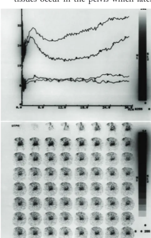
ney which was located in pelvis. Some areas of the kidney demonstrated less activity uptake which correlated with loss of parenchyme function. (Figure 3). Dynamic renal scintigraphic exam-ination with Tc-99m DTPA indicated normal perfusion, concentration and excretion pattern in the pelvic kidney. A reaccumulation pattern was detected after 10th minute which revealed the possibility of vesicoureteral reflux (Fig-ure 4).
Radiological examinations did not reveal any other anomalies related to other organs and systems.
Discussion
In autopsy cases renal ectopia is found in
one of every 400 cases and 85% occur as fused kidney (1). The most com-mon type of the renal ectopia is horse-shoe kidney with an incidence of one in every 700 autopsies (2).
Congenital renal fusion anomalies can be categorized as horseshoe kidney or one of the subtypes of crossed ectopia with or without fusion (3). When a kidney is located on the side opposite from its ure-teric insertion to the urinary bladder, the condition is called as a cross ectopia (4,5).
Looney and Dodd were the first to de-scribe pelvic cake kidney (6). Its exact incidence is not defined in the litera-ture and it is characterized by the pres-ence of a lobulated renal mass located in pelvis which has two fused renal
parenchymes without an interven-ing septum. Each fused kidney has its seperate collecting system. The ureters are short but drain to the urinary blad-der in their normal positions.
In embryological life, two metanephric tissues occur in the pelvis which later
Figure 4: Tc-99m DTPA scintigraphy shows
normal perfusion, concentration and excre-tion pattern in the pelvic kidney. A reac-cumulation pattern was detected after 10th minute, revealing the possibility of vesicoure-teral reflux.
Figure 1: EU demonstrated a renal pelvis
and calyceal structures located on the left side of bony pelvis which connects to the urinary bladder with a short ureter. Only the right ureter is seen on the right side of bony pelvis.
Figure 2 (a, b): Oral and IV contrast enhanced axial CT shows that the kidneys are not
lo-cated in their normal positions (a). In the pelvic region a single mass of kidney with lobulated contours and two ureters are detected (b) .
Figure 3: Tc-99m DMSA scintigraphy shows an irregular Tc-99m uptake in a single mass of
kidney located in pelvis. In some areas of the kidney less activity uptake is seen, which indi-cates loss of parenchyme function.
109
Journal Of Ankara University Faculty of Medicine 2010, 63(4)
Alper Dilli, Ümit Yaşar Ayaz, İdil Güneş Tatar, O. Raif Karabacak, Nihat Uluocak, Sevin Ayaz
show cranial ascent, axial deflection, medial rotation and lateral migration. The embryological basis for pancake kidney is fusion of each metanephric mass in the pelvis during early ascent. An abnormally located umbilical ar-tery may force the metanephric masses into opposition and cause fusion (7). After fusion occurs, cranial ascent to the lomber position is impaired by the retroperitoneal structures. The vascular supply of the pancake kidney is consistent with its arrested ascent and derives from the common iliac artery or terminal aorta. Histologi-cally, fused pelvic kidney shows cystic changes, immature glomeruli and di-lated tubules.
The presence of a pancake kidney may predispose to recurrent urinary tract infections and stones. This is due to
the probable rotation anomaly of the collecting system and short ureters which are prone to stasis and obstruc-tion; but most of the reported cases are asymptomatic (8,9).
In the literature there have been cases of fused pelvic kidneys reported to have concomitant anomalies such as Fallot te-tralogy (10), vaginal absence (11), sacral agenesis and caudal regression (12,13). If a pancake kidney has to undergo
sur-gery, division of the parenchyme pres-ents potential problems such as renal vascular damage, postoperative renal failure and eventual renal failure (14). On the other hand, the isthmus con-necting the inferior poles of horseshoe kidney can be safely divided to facili-tate surgical conditions such as under-lying aortic aneurysms (15).
In conclusion, US, EU and CT were effi-cient, not only in detection and evalu-ation of pancake kidney anomaly in our geriatric patient, but also in exclu-sion of concomitant anomalies as well. Nuclear medicine studies provided to evaluate the static and dynamic images of the kidney and helped to assess its functional status. The presence of pan-cake kidney does not necessarily mean that the patient will have progressive renal failure. But long term follow-up of renal function may help early detec-tion of complicadetec-tions such as urinary tract infection, calculi and obstruction. The presence of concomitant anoma-lies should be investigated, as we did in our case. It is also clinically important to detect pelvic renal fusion anomalies and their accompanying anomalous vasculature before pelvic surgeries.
REFERENCES
1- Shoemaker R, Braasch WF. Fused kidneys. J Urol 1939;41:1-7
2- Eisendrath DN, Phifer FM, Cul-ver HB. Horseshoe kidney. Ann Surg 1925;82(5):735-64
3- Abeshouse BS, Bhisitkul I. Crossed renal ectopia with and without fusion. Urol Int 1959;9:63-91
4- Caldamone AA, Rabinowitz R. Crossed fused renal ectopia, orthotopic multicys-tic dysplasia and vaginal agenesis. J Urol 1981;126(1):105-7
5- Caine M. Crossed renal ectopia without fusion. Br J Urol 1956;28(3):257-8 6- Looney WW, Dodd DL. An ectopic
(pel-vic) completely fused (cake) kidney associ-ated with various anomalies of the abdomi-nal viscera. Ann Surg 1926;84(4):522-4
7- Kelalis PP, Malek RS, Segura JW. Observa-tions on renal ectopia and fusion in chil-dren. J Urol 1973;110(5):588-92 8- Berg OC, Kerns WM. Solitary pelvic
kid-ney. A case report. J Urol 1949;62(3):275-7
9- Kron SD, Meranze DR. Completely fused pelvic kidney. J Urol 1949;62(3):278-85 10- Srivastava RN, Singh M, Ghai OP, et al.
Complete renal fusion (“cake”/“lump” kid-ney). Br J Urol 1971;43(4):391-4 11- Lange AP. Fused pelvic kidney
com-bined with absence of the vagina. J Urol 1979;122(1):92-3
12- Brueziere J, Fretin J. A new case of pyelic fusion. Eur Urol 1980;6(4):249-50
13- Brock JW 3rd, Braren V, Philips K, Win-field AC. Caudal regression with cake kid-ney and a single ureter: a case report. J Urol 1983;130(3):535-6
14- Eze AR, White JV, Pathak AS, et al. “Pan-cake kidney”: a renal anomaly complicat-ing aortic reconstruction. Ann Vasc Surg 1998;12(3):278-81
15- Brown OW, Dosick SM, Blakemore WS. Abdominal aortic aneurysm and horseshoe kidney. A different perspective. Arch Surg 1979;114(7):860-1