An investigation of visual-spatial attention in
children with attention deficit hyperactivity
disorder
Dikkat eksikliği hiperaktivite bozukluğu olan çocuklarda görsel-uzaysal dikkatin araştırılması
Emel Güneş
1, Erhan Nalçacı
1, Aynur Şahin
2, Ayla Aysev
21Ankara University School of Medicine, Department of Physiology, Ankara
2Ankara University School of Medicine, Department of Child Psychiatry, Ankara
Aim: Attention deficit hyperactivity disorder (ADHD ) has been associated with a disturbance in
frontal lobe network function, mediated by the underactivity of right hemispheric frontostriatal regions and loss of normal functional asymmetry. The aim of this study was to examine the right hemisphere functions and lateralization of visual-spatial attention in ADHD, using line bisection task.
Materials and Methods: Eighteen children with a DSM IV diagnosis of ADHD participated with
18 healthy controls, individually matched for age, sex, handedness and school years. In the line bisection task, horizontal lines were presented on the computer screen. Half of the lines were presented in the right hemispatial area, and half in the left hemispatial area. Participants were instructed to move the vertical cursor to the judged center of the line by mouse. Subjects per-formed the task separately with each hand. The difference between fine motor performances was examined using finger tapping task.
Results: In the line bisection task, bisection performance of the children with ADHD was
signifi-cantly lower than that of the normal children. For mean bisection error scores, the controls were transecting the lines to the left of the center when the lines were presented in the left hemispace and to the right of the center when the lines were presented in the right hemispace, performing both hands. The subjects with ADHD showed the same but smaller bias than the controls. The apparent deviations from the actual center in the children with ADHD were not significantly dif-ferent from zero in all conditions. In the finger tapping task, there were no significant differences between two groups for both hands.
Conclusion: These results supported the evidence for deficit in visual-spatial attention consistent
with disability of the right hemisphere in ADHD.
Key words: attention deficit hyperactivity disorder, line bisection task, spatial attention, right
cerebral hemisphere
Amaç: Dikkat Eksikliği Hiperaktivite Bozukluğu (DEHB)’nun sağ fronto-striatal devreyi
ilgilendi-ren bir bozukluğun sonucu olduğu iddia edilmektedir. Bu çalışmada çizgi bölme testi kullanılarak DEHB’de sağ hemisfer işlevlerinin ve görsel uzaysal dikkatin asimetrisinin incelenmesi amaçlan-mıştır.
Gereç ve Yöntem: DSM-IV ölçütlerine göre DEHB tanısı almış, ilaç kullanmayan ve eşlik eden
başka nörolojik veya psikiyatrik bozukluğu olmayan 7-14 yaşları arasında sağ elini kullanan 18 çocuk ile onlarla yaş, cinsiyet, el tercihi ve okul yaşı açısından bire bir eşleştirilmiş 18 sağlıklı çocuk araştırmaya dahil edildi. Bütün çocuklar çizgi bölme testini sağ ve sol elleri ile bilgisayar faresi kullanarak yaptılar. Bilgisayar ekranının yatay olarak sağından ve solundan sunulan çizgilerin tam orta noktalarını işaretlemeleri istendi. Ayrıca her iki gruptaki çocukların ince motor yetenekler açısından farkları parmak vuru testi ile değerlendirildi.
Bulgular: DEHB olan çocuklar çizgi bölme testinde anlamlı olarak daha çok mutlak hata yaparak
kontrol grubuna göre düşük bir performans gösterdiler. Öte yandan kontrol grubu, her iki elle, sağ alanda sunulan çizgileri orta noktanın sağına doğru, sol alanda sunulanları orta noktanın soluna doğru bölerken, DEHB grubunda ise benzer bir kalıp görülmekle birlikte orta noktadan sapmalar sıfır hatadan istatistiksel olarak farksız bulundu. Parmak vuru testinde iki grup arasında anlamlı bir fark yoktu.
Sonuç: Çalışmadan elde edilen bulgular, DEHB’de sağ hemisferin işlev bozukluğu ile uyumlu
ola-rak görsel uzaysal dikkatin bozulduğuna ilişkin kanıtları desteklemektedir.
Anahtar kelimeler: dikkat eksikliği Hiperaktivite Bozukluğu, çizgi bölme testi, uzaysal dikkat, sağ
beyin hemisferi
Received: 07.15.2005 • Accepted: 09.13.2005
Corresponding author
Emel Güneş
Ankara Üniversitesi Tıp Fakültesi Fizyoloji Anabilim Dalı, 06100, Ankara
Phone : (312) 310 30 10-410 E-mail : [email protected]
A
ttention Deficit Hyperactivity Disorder (ADHD) is one of the most common psychiatric disorders of childhood and adolescence. Its primary symptoms include impulsivity, inattention, and hyperactivity. These behavioral deficits arise relatively early in the childhood, typically before the age of seven. The disorder persists into adolescence in 50-80% of cases diagnosed in childhood and into adulthood in 30-50% or more of these same cases (1, 2).There are currently no clear biological markers on etiology or underlying brain abnormality. In terms of the nature of this disorder, there are suggestions that the right hemisphere may be playing a role. Lowered right hemisphere activity, particu-larly in the right frontal regions, has been reported (3, 4, 5).
A number of studies have shown an association between the diagnosis of childhood ADHD and a relative reduction in awareness for visual information presented on the left (6, 7, 8, 9). Normally, visual stimuli evoke predominantly right hemisphere activity, so that attention is directed pref-erentially to the left. Patients with left unilateral neglect due to right hemisphere damage demonstrated a pronounced rightward bias in the visual-spatial task such as line bisec-tion. The line bisection task has been used to demonstrate the asymmetric perception of space in patients with brain lesion and in normal subjects who show pseudoneglect, which can be defined as slight right side neglect.
It is possible to argue that ADHD might be associated with a mild increased risk of spatial bias away from the left. Voeller and Heilman (9) administered a clinical test of neglect (a letter cancellation task, typical laterality task) to a group of boys with the ADHD diagnosis. Their perfor-mance resembled that of adults with unilateral neglect fol-lowing acquired damage to the right hemisphere. Sheppard et al. (8) have shown that ADHD children, again similar to left unilateral neglect patients, tend to bisect horizon-tal lines to the right of the center. However, a study by Ben-Artsy et al. (10) found no laterality trend of group differences in a line bisection task administered to ADHD and control children. Similarly, Adelstein (11) reported no significant difference between boys with ADHD and con-trol children in a line bisection task. Those studies that demonstrated anomalies in spatial biases are in line with the view that ADHD reflect predominantly right-sided frontal-striatal system dysfunction (3,12). Importantly, however, this is by no means a universal finding.
In this research, we aimed to investigate the spatial at-tention of children with ADHD, using a line bisection task. The line bisection task is a simple but very sensitive method to demonstrate visual-spatial asymmetry in human subjects (13, 14). Also, it gives an opportunity to
investi-gate the effects of hand use and the hemispace in which the bisection action is performed. In addition to the line bisection task, we also applied to the subjects a fine motor task in order to eliminate a motor component involved by the line bisection task. The hypothesis of this research is that asymmetric perception of space in normal subjects is reduced in children with ADHD due to a deficit of the right prefrontal circuitry.
Material and methods
Participants
Thirty-six right-handed children between 7-14 years of age participated in this study. The sample consisted of 18 children with ADHD and 18 healthy control subjects, individually matched for age, sex, handedness and school years. Written informed consent was obtained from a par-ent of each subject, and the study was approved by the Eth-ics Committee of the Ankara University School of Medi-cine. Table 1 gives the demographic details of the ADHD sample and their matched controls. A Turkish version for the Wechsler Intelligence Scale for Children-Revised (WISC-R) was used to determine the IQ scores of the par-ticipants (14). IQ scores of all subjects were 85 or above. Handedness was assessed using lateral preference items from Revised Neurological Examination for Subtle Signs Form (16). The children with ADHD (14 males and 4 girls) were diagnosed by the child psychiatrist according to the Diagnostic and Statistical Manual of Mental Disorders, Fourth Edition (DSM-IV) criteria (17). All subjects were combined type, which includes signs of attention deficit and hyperactivity. None of them had comorbid disorder or received stimulant medication, or they had been off medi-cation for at least one month.
Line bisection task
A computerized line bisection task that was developed in our laboratory was used. In this task a solid horizon-tal line was presented at the beginning of each trial. Par-ticipants were instructed to move the vertical cursor by mouse to the judged center of the line. A vertical cursor
Table 1. Demographic details of the patients with ADHD and
matched controls
ADHD group (n=18)
Control group (n=18)
Mean age (months) 121.5 ± 21.8 121.6 ± 20 Sex 14 males, 4 females 14 males, 4 females Handedness 18 right handed 18 right handed Mean grade in school 4.8 ± 1.7 4.5 ± 1.5
was located at either the left or the right end of the hori-zontal line. They were also instructed to sit in front of the screen at a distance of around 60 cm and aligned with the midline. The lines were in ten different lengths (80, 90, 100, 110, 120, 130, 140, 150, 160, 170 mm), which were pseudorandomly allocated to total 40 trials. Half of the target lines were presented in the right hemispatial area, and half were presented in the left hemispatial area. Each participant performed the task twice, once with the right and once with the left hand. The order of hand using was counterbalanced between participants.
The computer recorded for each subject two scores to accuracy better than 1 mm: mean bisection errors (MBE) and mean absolute bisection errors (MABE). MBE score is the distance of the cursor from the actual center of the line to left or right; positive values indicate transections to the right of center and the neglect of the left hemispace, nega-tive values indicate transections to the left and the neglect of the right hemispace. MABE score can give the preci-sion of the subjects’ bisection performance. These scores are separately evaluated for each hand in the left and right hemispatial areas. Thus we were able to examine the effects of hand using and the hemispace in which the line is pre-sented under four conditions: Using the Left hand in the left hemispace presentation (Ll), using the Left hand in the right hemispace presentation (Lr), using the Right hand in the left hemispace presentation (Rl) and using the Right hand in the right hemispace presentation (Rr).
Finger tapping task
The finger tapping task is a fine motor performance task (18, 19). The tapping apparatus consisted of a mouse attached to a wooden rectangular plate (10 x 42 cm). The mouse was connected to a computer for scoring. The sub-jects were asked to tap the mouse with the index finger as quickly as possible and to keep the hand and wrist station-ary on the wooden base. Before the procedure, the task was
demonstrated and then the subjects completed the practice trials twice with each hand. The task was performed for 10 seconds three times with each hand, alternating sides with each trial, and the starting hand was counterbalanced across subjects. For each hand, the mean number of taps per second, averaged across the three trials, was taken as the tapping score of the right and the left hand speed, re-spectively.
All subjects were administered the line bisection task and the finger tapping task in a quite room. First the line bisection task and then the finger tapping task was applied. A demonstration of tasks as well as one practice trial was given before the child started to complete each task.
Statistical analysis:
In the statistical analysis, repeated-measures analy-ses of ANOVAs were used. Also, MBE was compared to zero error, using one-sample-t-test for each hand and each hemispace in the control and the ADHD subjects.
Results
Line bisection task
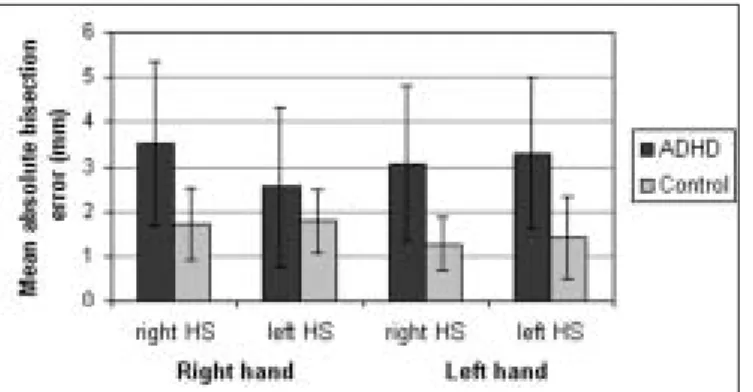
As shown in figure 1, in the line bisection task, MABE scores of the children with ADHD were significantly lower than that of normal children when the line was pre-sented in the left hemispace and the right hemispace, for both hands. MABE scores were analyzed using a 2 x 2 x 2 repeated – measures analysis of ANOVA with groups (ADHD, Control), performing hand (right, left hand) and hemispaces in which lines were presented (left, right hemispace). There was a main effect for the difference
be-Figure 1. Mean absolute bisection errors (mm) for ADHD and control
groups with right and left hand in both hemispace (HS). MABE scores of the children with ADHD were significantly lower than that of normal children in every condition [F(1, 34) = 19.26, p<0.0001].
Figure 2. Mean bisection errors (mm) for ADHD and control groups with
right and left hand in both hemispace (Rl=Right hand, left hemispace; Ll=Left hand, left hemispace; Lr=Left hand, right hemispace; Rr=Right hand, right hemispace). ANOVA yielded a main effect for the hemispaces [F(1,34) = 7.31, p<0.05]. One sample t-test results indicated that the control group was transecting the line significantly to the right in the right hemispace (for right hand t(17) = 3.796, p<0.001; for left hand t(17) = 3.687, p <0.005) and to the left in the left hemispace (for right hand t(17)= -3.165, p <0.01; for left hand t(17)= -2,560, p<0.01). The apparent deviations from the actual center in the children with ADHD were not significantly different from zero in all conditions (p>0.05).
tween the control group and the ADHD group [F(1, 34) = 19.26, p< .0001]. Analyses of ANOVA yielded no another main effect or interaction between the factors.
For MBE scores, figure 2 indicates that the control group was transecting the lines to the left of the center when the lines were presented in the left hemispace and to the right of center when the lines were presented in the right hemispace, performing with both hands. The sub-jects with ADHD showed the same but smaller bias than the control group, except the condition of left hand-right hemispace. In the left hand-right hemispace condition, ADHD group showed leftward bias, when the mean of the control group indicated the rightward error. The MBE scores were submitted to an ANOVA by group, performing hands and hemispaces in which the lines were presented. The main effect for the difference between the control and the ADHD subjects was found to be insignificant [F(1,34) = 0.23]. Also, there was no significant main effect for the performing hand [F(1,34) = 0.50]. However, ANOVA yielded a main effect for the hemispaces [F(1,34) = 7.31, p<0.05]. The interaction between the performing hand and the hemispace was found to be marginally significant [F(1,34) = 3.83, p=0.058]. This interaction indicates that both groups tended to transect the lines in the same direc-tion (to the left in the left hemispace, to the right in the right hemispace), but the subjects with ADHD showed very small amplitude in comparison with the controls.
One sample t-test (comparing mean bisection errors with zero-that is perfect bisection) results indicated that the control group was transecting the line significantly to the right in the right hemispace (for right hand t(17) = 3.796, p< 0.001; for left hand t(17) = 3.687, p <0.005) and to the left in the left hemispace (for right hand t(17)= -3.165, p < 0.01; for left hand t(17)= -2,560, p<0.01). The apparent de-viations from the actual center in the children with ADHD were not significantly different from zero in all conditions (p>0.05).
Finger tapping task
Table 2 indicates the results of the finger tapping task. Even though the control group was slightly faster than the ADHD group, there was no significant difference between the two groups for both hands [F(1,34) = 2.26, p> 0.05]. Analysis of ANOVA indicates that the right hand was sig-nificantly faster than the left hand [F(1,34) = 43.53, p< 0.0001]. There was no interaction between the speed of hands and the groups.
Discussion
The results obtained by the line bisection task in healthy control children indicated that the subjects tended to
tran-sect to the right of the midline when the lines were pre-sented in the right hemispace and to the left of the midline when the lines were presented in the left hemispace. This result is in accordance with the results of Güneş et al. (20) and Çiçek et al. (21) who applied a paper form of line bi-section task to adult subjects. In spite of certain conflicting findings (22, 23), most of the studies either supported or not conflicted with our results (24, 25, 26). This effect of hemispace can be explained by the activation-orientation theory of Kinsbourn (27, 28). According to this theory, presenting a line in a left hemispace activates the right hemisphere, and increased activity in the right hemisphere causes attentional shift to the left and results in a left-sided bisection bias, and vice versa.
In comparison to the control group, significantly lower performance of children with ADHD in line bisection task in-dicated that visual-spatial skills in ADHD was disabled. Neu-rophysiological and neuropsychological studies have suggested that the right hemisphere is dominant for spatial attention (29). This lower performance of children with ADHD in the line bisection task indirectly suggests a disability of the right hemi-sphere functions. On the other hand, a significant transection error was obtained only from the normal subjects, but the chil-dren with ADHD did not significantly tend to the right or to the left. This finding also can be accepted as an indicator of an attentional deficit of the hemispheres, because pseudoneglect is a product of the normal human brain, which shows a func-tional asymmetry. Therefore, disappearance of right hemispatial neglect in the ADHD group was an indirect evidence of the right hemisphere disruption in ADHD. Many previous reports showed that normal people tend to neglect the right hemispace as a result of the superiority of the right cerebral hemisphere in general attention and visual-spatial processes (30, 31). Also, our result is in accordance with Sheppard et al. (8) who previously applied line bisection task to children with ADHD, and found that the children with ADHD off medication bisected signifi-cantly away from the left compared with the controls.
The finger tapping task showed that there is no signifi-cant difference between the ADHD and the control group. Previous studies also could not find a difference between the
Table 2. The comparison of right and left hand speeds between
ADHD and control groups
ADHD group (n=18)
Control group (n=18)
p*
Right hand speed (taps/s) 4.64 ± 0.62 4.96 ± 0.55 ns Left hand speed (taps/s) 4.09 ± 0.49 4.21 ± 0.76 ns
fine motor function in the ADHD group and the control group (1). This finding suggests that the low performance of the ADHD group was not related to a disability of the mo-tor function but the executive function. All findings show us once again that ADHD is a disorder of the prefrontal-striatal circuitry rather than a basic motor disorder.
Results of many investigations suggest that the prob-lems in ADHD may be due to a lateralised disturbance in the frontal lobe network function, mediated by the under activity of predominantly the right hemispheric frontos-triatal regions (9, 32, 33, 34). Functional neuroimaging studies, using single photon emission computed thomog-raphy (SPECT), positron emission thomogthomog-raphy (PET) and functional magnetic resonance imaging (fMRI) have identified anomalies of frontal metabolic activity indexed by diminished cerebral blood flow or glucose metabolism (5, 32, 35). Consistent with such result, structural neuro-imaging studies have identified morphologic differences in frontal and striatal structures of ADHD individuals (3, 34, 36). Recently, activity differences during inhibition in ven-trolateral prefrontal cortical areas, anterior cingulate and frontopolar regions were identified (37). Our results in-directly supported these anatomical findings that indicate the pathology of the right hemisphere in ADHD.
These results have to be replicated with the other stud-ies, regarding to some limitation of our study. Age
distri-bution of subjects (7-14 year) seem to be a large scale for an investigation on ADHD, because of problems of ado-lescent period like depression might be complicated with signs of ADHD. In addition to that, subtypes of ADHD have to be separately investigated. The next study should be designed as divided groups, regarding to narrow age bands and subtypes of ADHD.
In conclusion, the thesis of this study was supported by the findings for the following reasons: Firstly, reduced spatial ability and changed neglect pattern in children with ADHD seems to be related to the disability of the right hemisphere. Secondly, normal fine motor performance in children with ADHD suggests that the disability in these children is more related to the prefrontal executive func-tions than the motor funcfunc-tions.
Acknowledgements
This research was a part of thesis of Emel Güneş and preliminary findings were presented in national and inter-national scientific meetings.
This research was supported by Ankara University Re-search Foundation, Project No. 2003.08.09.095. The au-thors wish to thank Atilla Halil Elhan and Yasemin Genç for statistical analysis.
References
1. Barkley RA. Behavioral inhibition, sustained attention, and executive functions: constructing a unifying theory of ADHD. Psychol Bul 1997; 121:65-94.
2. Tannock R. Attention deficit hyperactivity disorder: advances in cognitive, neurobiological, and genetic research. J Child Psychol Psychiatry 1998; 39:65-99.
3. Castellanos FX, Giedd JN, Marsh WL et al. Quantitative brain magnetic resonance imaging in attention-deficit hyperactivity disorder. Arch Gen Psychiatry 1996; 53:607-616.
4. Lou HC, Henriksen L, Bruhn P et al. Striatal dysfunction in attention deficit and hyperkinetic disorder. Arch Neurol 1989; 46:48-52.
5. Rubia K, Overmeyer S, Taylor E et al. Hypofrontality in attention deficit hyperactivity disorder during higher-order motor control: a study with functional MRI. Am J Psychiatry 1999; 156:891-896. 6. Carter CS, Krener P, Chaderjian M et al. Asymmetrical
visual-spatial attentional performance in ADHD: evidence for a right hemispheric deficit. Biol Psychiatry 1995; 37:789-797.
7. Nigg JT, Swanson JM, Hinshaw SP. Covert visual spatial attention in boys with attention deficit hyperactivity disorder: Lateral effects, metilphenidate response and results for parents. Neuropsychologia 1997; 35:165-176.
8. Sheppard DM, Bradshaw JL, Mattingley JB et al. Effects of stimulant medication on the lateralisation of line bisection judgements of children with attention deficit hyperactivity disorder. J Neurol Neurosurg Psychiatry 1999; 66:57-63.
9. Voeller KKS, Heilman KM. Attention deficit disorder in children: a neglect syndrome. Neurology 1988; 38:806-808.
10. BenArsty A, Glicksohnn J, Soroker N et al. An assessment of hemineglect in children with attention-deficit hyperactivity disorder. Dev Neuropsychol 1996; 12:271-281.
11. Adelstein A. ADHD and attention in the visual-spatial domain. Dissertation Abstracts International: Section B: The sciences and Engineering. 56(2-B), 11098 In: Klimkeit EI, Mattingley JB, Sheppard DM et al. Perceptual asymmetries in normal children and children with attention deficit hyperactivity disorder. Brain Cogn 2003; 52:205-215.
12. Heilman KM, Voeller KKS, Nadeau SE. A possible pathophysiologic substrate of attention deficit hyperactivity disorder. J Child Neurol 1991; 6(suppl): S76-S81.
13. Bradshaw JL, Bradshaw JA, Nathan G et al. Leftwards error in bisecting the gap between two points: stimulus quality and hand effects. Neuropsychologia 1986; 24:849-855
14. Luh KE. Line bisection and perceptual asymmetries in normal individuals:What you see is not what you get. Neuropsychology 1995; 9:435-448.
15. Savaşır I, Şahin N: Wechsler Çocuklar İçin Zeka Ölçeği, Türk Psikologlar Derneği, 1995.
16. Denckla MB. Revised physical and neurological examination for subtle signs. Psychopharmacol Bull 1985; 21:773-800.
17. American Psychiatric Association. Diagnostic and statistical manual of mental disorders (4th ed.) Washington, DC, 1994. 18. Nalçacı E, Kalaycıoğlu C, Çiçek M et al. Relation between
19. Peters M, During B. Handedness measured by finger tapping: A continuous variable. Can J Psychol 1978; 32:257-261.
20. Güneş E, Nalçacı E, Kalaycıoğlu C ve ark. Çizgi bölme testi ve test-yeniden test güvenilirliği. 3P Dergisi 2002; 10:33-39. 21. Çiçek M, Nalçacı E, Kalaycıoğlu C. Line bisection task
performance and resting EEG alpha power. Intern J Neurosci 2003; 113:849-866.
22. Nichelli P, Rinaldi M, Cubelli R. Selective spatial attention and length representation in normal subjects and in patients with unilateral spatial neglect. Brain Cogn 1989; 9:57-70. 23. Mennemeier M, Vezey E, Chatterjee A et al. Contributions
of the left and right cerebral hemispheres to line bisection. Neuropsychologia 1997; 35:703-715.
24. Milner AD, Brechman M, Pagliarini L. To halve and to halve not: an analysis of line bisection judgements in normal subjects. Neuropsychologia 1992; 30:515-526.
25. McCourt ME, Jewell G. Visiospatial attention in line bisection: Stimulus modulation of pseudoneglect. Neuropsychologia 1999; 37:843-855.
26. Post RB, Caufleld KJ, Welch RB. Contributions of object- and space- based mechanisms to line bisection errors. Neuropsychologia 2001; 39:856-864.
27. Kinsbourne M. The cerebral basis of lateral asymmetries in attention. Acta Psychologia 1970; 33:193-201.
28. Kinsbourne M. Hemi-neglect and hemisphere rivalry. Adv Neurol 1977; 18:41-49.
29. Doyon J, Milner B. Right parietal lobe contribution to global visual processing. Neuropsychologia 1991; 29:343-360.
30. Bowers D, Heilman KM. Pseudoneglect: effect of hemispace on tactile line bisection task. Neuropsychologia 1980; 18:491-498. 31. Mesulam MM. Spatial attention and neglect: parietal, frontal,
and cingulate contributions to the mental representation and attentional targeting of salient extrapersonal events. Phil Trans R Soc Lond B 1999,3554:1325-1346.
32. Zametkin AJ, Nordahl TE, Gross J et al. Cerebral glucose metabolism in adults with hyperactivity of childhood onset. New Engl J Med 1990; 323:1361-1366.
33. Filipek PA, Semrud-Clikeman M, Steingard RJ et al. Volumetric MRI analysis comparing subjects having attention-deficit hyperactivity disorder with normal controls. Neurology 1997; 48:589-601.
34. Castellanos FX, Jay MD, Eckburg P et al. Quantitative morphology of the caudate nucleus in attention deficit hyperactivity disorder. Am J Psychiatry 1994; 151:1791-1796. 35. Oner O, Oner P, Aysev A, Kucuk O et al. Regional cerebral blood
flow in children with ADHD: changes with age. Brain Dev 2005; 27:279-85.
36. Aylward EH, Reiss AL, Reader MJ et al. Basal ganglia volumes in children with attention-deficit hyperactivity disorder. Arch Neurol 1996; 11:112-115.
37. Schultz KP, Fan J, Tang CY et al. Response inhibition in
adolescents diagnosed with attention deficit hyperactivity disorder during childhood: An event related fMRI study. Am J Psychiatry 2004;161:1650-1657.
![Table 2 indicates the results of the finger tapping task. Even though the control group was slightly faster than the ADHD group, there was no significant difference between the two groups for both hands [F(1,34) = 2.26, p> 0.05]](https://thumb-eu.123doks.com/thumbv2/9libnet/3917093.46143/4.906.477.838.167.273/table-indicates-results-tapping-control-slightly-significant-difference.webp)
