Serkan Güneyli, Mustafa Gök, Halil Bozkaya, Celal Çınar, Arastu Tizro, Mehmet Korkmaz, Yiğit Akın,
Mustafa Parıldar, İsmail Oran
Diagn Interv Radiol 2015; 21:229–234 © Turkish Society of Radiology 2015
Endovascular management of iatrogenic renal arterial lesions and
clinical outcomes
INTERVENTIONAL RADIOLOGY
ORIGINAL ARTICLEPURPOSE
We aimed to evaluate iatrogenic renal arterial lesions, includ-ing pseudoaneurysm, arteriovenous fistula, and arteriocalice-al fistula, their management by endovascular embolization, and the clinical results.
METHODS
Fifty-five patients (forty males, fifteen females) with a me-dian age of 40 years (range, 8–85 years), who underwent endovascular embolization of iatrogenic renal arterial lesions between March 2003 and December 2013 were included in this retrospective study. Types of iatrogenic lesions and de-tails of embolization procedures were reported. Estimated glomerular filtration rate (eGFR), renal function tests, hemo-globin, and hematocrit levels before and after embolization were recorded and compared.
RESULTS
Median follow-up was 24 months. We identified 53 pseudo-aneurysms, 30 arteriovenous fistulas, and 11 arteriocaliceal fistulas in 55 patients, after percutaneous nephrolithotomy (n=26), renal biopsy (n=21), nephrostomy (n=3), renal sur-gery (n=3), and extracorporeal shock wave lithotripsy (n=2). Median number of pseudoaneurysms was 1 (range, 1–4) with a median size of 7 mm (range, 1.5–35 mm). Fifty-one patients underwent coil embolization. Median number of coils was 5 (range, 2–21) and median renal parenchymal loss was 5% (range, 1%–50%). There were no significant differences between pre- and postoperative eGFR and serum parameters.
CONCLUSION
Iatrogenic renal arterial lesion can be a life threatening con-dition. Superselective coil embolization is a safe, minimally invasive treatment option with minimal renal parenchymal loss and without significant change in renal function.
I
atrogenic renal arterial lesions including pseudoaneurysm (PA),arte-riovenous fistula (AVF), and arteriocaliceal fistula (ACF) are rare, but life-threatening conditions (1). The chief symptom usually includes macroscopic hematuria (2, 3). Catheter angiography is the gold standard for both diagnosis and treatment (1).
Previous studies have evaluated the iatrogenic renal arterial lesions following partial nephrectomy (1, 3–7), but there were only a few stud-ies on iatrogenic renal arterial lesions following any iatrogenic renal interventions (2). In the present study, to the best of our knowledge, we report the largest series of iatrogenic renal arterial lesions following various renal interventions such as biopsy, percutaneous nephrolithot-omy (PCNL), percutaneous nephrostnephrolithot-omy, and partial nephrectnephrolithot-omy. We focused on clinical presentations, imaging findings, management, and outcomes.
Methods
Patients
Data regarding iatrogenic renal arterial lesions treated by endovascu-lar embolization performed at our institution between March 2003 and December 2013 were evaluated retrospectively. A total of 55 patients with iatrogenic renal arterial lesions, who underwent endovascular em-bolization were included in the study. There were 40 males and 15 fe-males with a median age of 40 years (range, 8–85 years). Patients with traumatic renal arterial lesions treated with endovascular embolization were excluded from the study. Informed consent was obtained from all patients.
Data collection
Patients’ demographics, symptoms, lesions, and concomitant diseases were noted. Arterial lesions on angiograms were classified under three groups: PA, AVF, and ACF. We tried to determine whether these lesions were isolated or associated with other lesions. Additionally, the etiology of the iatrogenic lesions was determined. Other variables were as follows: the number and size of PA, the number of AVF, the number of ACF, the location of the lesion, presence of perirenal hematoma, volume of used contrast media, the number and type of embolic material, the rate of pa-renchymal loss after embolization, blood transfusion, 30-day mortality, and length of follow-up. Parenchymal loss was evaluated by comparing angiography images obtained before and after the embolization. Angi-ography images were independently evaluated by two interventional radiologists. Final angiography findings were reached in consensus. The parenchymal loss following the endovascular procedures was catego-From the Department of Radiology (S.G. serkanguneyli@yahoo.
com), Bülent Ecevit University School of Medicine, Zonguldak,
Turkey; the Department of Radiology (M.G.), Kafkas University School of Medicine, Kars, Turkey; the Department of Radiology (H.B., C.Ç., A.T., M.P., İ.O.), Ege University School of Medicine, İzmir, Turkey; the Department of Radiology (M.K.), Dumlupinar University School of Medicine, Kütahya, Turkey; the Department of Urology (Y.A.), Harran University School of Medicine, Şanlıurfa, Turkey.
Received 7 July 2014; revision requested 23 July 2014; final revision received 29 September 2014; accepted 28 October 2014.
Published online 2 April 2015. DOI 10.5152/dir.2014.14286
rized in four groups; <5%, 5%–10%, 10%–25%, and 25%–50%. Early and late complications were determined and computed tomography (CT) angi-ography was performed for follow-up. Preoperative international normalized ratio (INR) and the number of plate-lets were determined. Preoperative and postoperative serum urea, creatinine, hemoglobin, and hematocrit values were noted. These measurements were performed using patient sera and the Modular System (Roche Cobas 8000 System, Roche Diagnostics). Preopera-tive and postoperaPreopera-tive parameters were assessed one day before embolization and two days after embolization, re-spectively. Additionally, to evaluate contrast media-induced nephropathy (CIN), we recorded serum creatinine levels during postoperative day 2 to day 5 and considered the highest levels of serum creatinine in this period. CIN, in which renal function indices begin to deteriorate within 24 h, peaking at ~3–5 days after contrast media admin-istration, is defined as a ≥ 44 µmol/L (0.5 mg/dL) or ≥ 25% increase in serum creatinine levels in the absence of any other cause that might explain deteri-oration of the renal function (8–10). Preoperative and postoperative esti-mated glomerular filtration rates (eG-FRs) were also noted. The eGFR was calculated with the Modification of Diet in Renal Disease equation (11).
Embolization procedure
Transfemoral arteriography was per-formed under intravenous sedation. After obtaining a vascular access by placing a 5 F sheath, selective catheter-ization and angiography of the renal artery (4 F or 5 F Cobra or Simmons 2) were performed. Interlobar arter-ies causing the bleeding were detect-ed and superselective catheterization was performed using microcatheter systems (Progreat Microcatheter; Ter-umo Medical). The feeding arteries were embolized using coils in 51 pa-tients. Coils were placed to occlude flow into the arterial lesion with min-imal parenchymal loss. Embolization was completed when blood flow into the lesion stopped. We used pushable coils (0.018-inch Tornado coils; Cook or VORTX-18 Diamond Shape coils;
detachable coils (GDC coils; Boston Scientific) in two patients. In one pa-tient, coils were used together with glue. Amplatzer vascular plug (AVP) was used as an embolic material in three patients and detachable balloon was used in the remaining patient. These four patients had high-flow fis-tulae and because of migration risk, we used AVP and detachable balloon that permitted endovascular treatment in a more controlled manner than coils. As contrast media, we used Iopamiro 370 (Iopamidol, Bracco), which is a low-os-molar contrast media.
Statistical analysis
Not normally distributed parame-ters were presented as median (min-imum-maximum). Preoperative and postoperative eGFR and serum param-eters were presented as mean±stan-dard deviation. We compared pre- and postoperative values of eGFR, serum urea, creatinine, hemoglobin, and he-matocrit. Statistical analyses were per-formed with paired t test. P < 0.05 was considered to indicate statistical signif-icance.
Results
The most common symptom was macroscopic hematuria (n=37). Four-teen patients had flank pain, and four patients had both hematuria and flank pain. Twenty-seven patients had peri-renal hematoma on radiological exam-inations with a median diameter of 19 mm (range, 5–130 mm). Seventeen tients had hypertension, while no pa-tients had diabetes mellitus. Iatrogenic lesions occurred in the early period fol-lowing the intervention in all but one patient, who developed an iatrogenic lesion two years later. The etiologies of the renal interventions were as follows: PCNL (n=26), renal biopsy (n=21), per-cutaneous nephrostomy (n=3), renal surgery (n=3), and extracorporeal shock wave lithotripsy (n=2). Renal biopsy was performed in native kidneys of 14 patients due to nephritic syndrome (n=6), proteinuria (n=6), systemic lu-pus erythematosus nephropathy (n=1), hypertensive nephropathy (n=1), and in transplanted kidneys of the remain-ing seven patients due to nephritic syndrome (n=6) and renal
insufficien-(n=1). Percutaneous nephrostomy was performed in three patients due to hydronephrosis (n=2) and vesical in-vasion of rectum cancer (n=1). Renal surgeries were partial nephrectomy due to renal tumor (n=1), open nephroli-thotomy due to staghorn kidney cal-culi in ectopic kidney (n=1), and renal transplantation due to hypertensive nephropathy (n=1).
We identified 53 PAs, 30 AVFs, and 11 ACFs in 55 patients. PA was found in 17 patients, while a combination of PA and AVF (Fig. 1), and a combination PA and ACF were diagnosed in 17 and six pa-tients, respectively. AVF was diagnosed in 10 patients, while ACF (Fig. 2) and a combination of AVF and ACF were found in two and three patients, re-spectively. None of the patients had all three types of lesions together. Iatro-genic lesions of eight patients were in transplanted kidneys. Of 47 patients, 19 had iatrogenic lesions in the right kidney and 28 in the left kidney. Of 47 patients, 31 had lesions in the lower pole, 10 in the upper pole and six in the middle segments. All lesions were localized in the distal renal artery.
Median number of PAs per pa-tient was 1 (range, 1–4), with a medi-an size of 7 mm (rmedi-ange, 1.5–35 mm) (Fig. 3). Fifty-one patients underwent coil embolization and a median of 5 coils (range, 2–21) were used in each patient. Rate of parenchymal loss was <5% in 16 patients, 5%–10% in 26 patients, 10%–25% in 11 patients, and 25%–50% in two patients. Medi-an rate of renal parenchymal loss was 5% (range, 1%–50%). A median of 0.5 units (range, 0–6 units) of blood was transfused to patients. Mean INR was 1.0±0.09 and mean platelet count was 289 600±128 770 /µL. Median volume of used contrast media was 55 mL (range, 40–80 mL). Pre- and postoper-ative values of eGFR, serum urea, cre-atinine, hemoglobin, and hematocrit values were not significantly different (Table). After embolization, CIN oc-curred in four patients only.
Median follow-up was 24 months (range, 1–132 months). Six patients had rebleeding in the early follow-up peri-od and underwent reembolization with coils. One of these patients had a mas-sive hematoma in retroperitoneal space
renal artery was embolized with polyvi-nyl alcohol and coils, but the patient ex-pired a few hours after reembolization. One patient had a late complication; she experienced a renal arteriovenous malformation after five years following AVF embolization and the malforma-tion was embolized with Onyx.
Discussion
This study evaluated iatrogenic re-nal arterial lesions, their management
by endovascular embolization, and the clinical results. The initial results were positive, demonstrating high technical and clinical success rates in patients. In the early follow-up period, endovascular embolization did not lead to any deteri-oration of renal function, and rebleed-ing, treated by reembolization, was de-termined in six of 55 patients (10.9%).
Hemorrhage is the most common complication after renal interventions, with a reported incidence of up to 6%
(4–6). It can appear clinically as macro-scopic hematuria or perirenal hemato-ma. PA seems to be the most common lesion after renal interventions (4, 7, 12, 13). However, in a series of 200 patients who underwent laparoscopic partial nephrectomy, PA was not seen in any of 12 patients experiencing hemorrhage (6). Rare complications may include urinary fistula and renal artery thrombosis (5). The time period between renal intervention and
pres-d a
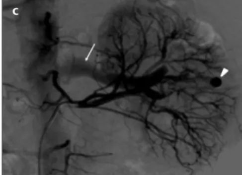
e b
Figure 1. a–e. Axial CT angiography image (a) of a 31-year-old male patient shows
a pseudoaneurysm (arrow) in the left kidney and perirenal hematoma developed following biopsy. Coronal CT angiography reconstruction image (b) shows the pseudoaneurysm (arrow) in the middle segment of the kidney. Selective renal diagnostic angiography image (c) shows the pseudoaneurysm (arrowhead) and filling of renal vein (arrow) indicating an arteriovenous fistula. Superselective renal angiography image (d) shows the pseudoaneurysm (arrow). Control angiography (e) reveals total embolization with coils (arrow).
c
Figure 2. a–c. Axial CT image (a) of a 75-year-old female patient who underwent percutaneous nephrolithotomy shows the right kidney (arrow)
having hydronephrosis and nephrolithiasis. The left kidney (arrowhead) is atrophic. Selective renal diagnostic angiography image (b) shows caliceal filling (arrow) at the upper pole of right kidney indicating an arteriocaliceal fistula. Control angiography (c) reveals total embolization with coils (arrowhead) and parenchymal area devascularized by the embolization (arrow).
a
ence of iatrogenic renal arterial lesions may be varied. Complications usually occur within three weeks following the interventions. However, it should be noted that occasionally complications may occur a long time after the inter-vention.
Richstone et al. (14) reported that 57 out of 4695 patients (1.2%) required selective endovascular embolization following percutaneous renal interven-tions (PCNL was the etiology for 44 of 57 patients). Srivastava et al. (15) re-ported embolization after PCNL in 24 out of 1854 patients and found a com-bination of PA and AVF in four cases. In their study, serum creatinine values were not significantly different after PCNL or embolization. In our series 26 patients underwent embolization after PCNL, and a combination of PA and AVF was present in five of these 26 patients. Moreover, isolated ACF was seen in two patients after PCNL and a combination of ACF and another arte-rial lesion was seen in six of these 26
case with a combination of AVF and caliceovenous fistula after PCNL and pointed out that serum creatinine val-ues increased in this patient. However, caliceovenous fistula was self-limiting unlike arterial bleeding (14, 15, 17).
Today partial nephrectomy, called nephron-sparing surgery, can be per-formed in urology clinics, specifically in the case of solitary kidney, renal insufficiency, or even in patients with a normal contralateral kidney (18). PA and AVF are rare complications of partial nephrectomy. Approximately 40 cases of PA after partial nephrec-tomy have been reported (19–24). In the literature, PA rate was about 0.43% for open partial nephrectomy (4) and 1.7% for laparoscopic partial nephrec-tomy (3). Ghoneim et al. (25) reported their complication rates as 0.6% for open partial nephrectomy and 2.6% for laparoscopic partial nephrectomy. In a multicenter study of 998 patients who underwent minimally invasive partial nephrectomy, Hyams et al. (1)
cular lesions and PA as 2% and 1.7%, respectively. However, the real fre-quency of PA might be higher when considering the asymptomatic PAs. There was only one patient who un-derwent open partial nephrectomy in our study, and a combination of PA and ACF was detected in this patient.
PA is a collection of blood leakage completely out of renal artery, but confined next to the vessel by the surrounding tissue. PA was the most frequent iatrogenic renal arterial le-sion occurring in 40 of 55 patients in our study. In case of a renal arterial injury, hemostatic mechanisms such as decreased blood flow, coagulation, and pressure of surrounding tissues attempt to control bleeding. When these mechanisms fail and blood flow to the injured artery increases, blood leakage may form a PA. A true aneu-rysm is distinguished from a PA by the involvement of all three layers of the arterial wall (intima, media, and ad-ventitia). When the PA becomes larger and blood leakage occurs into the col-lecting system, macroscopic hematuria may occur (26). In the literature, mac-roscopic hematuria and/or flank pain were reported in almost all cases with PA (25, 27). In line with previously re-ported studies, all patients were symp-tomatic in the present series. Albani and Novick (4) reported one asymp-tomatic patient with PA, and Ghoneim et al. (25) reported two asymptomatic patients with PA.
An AVF is an abnormal communica-tion between the arterial and venous systems without an intervening
capil-Figure 3. a–c. Axial CT angiography image (a) of a 43-year-old male patient shows a pseudoaneurysm (arrowhead) in the left kidney, which occurred
following partial nephrectomy and perirenal hematoma (arrow). Selective renal diagnostic angiography image (b) shows the pseudoaneurysm (arrow) at the upper pole of the kidney. Control angiography (c) reveals complete embolization with an Amplatzer vascular plug (arrow).
a b c
Table. Comparison of pre- and postprocedural eGFR and serum parameters
Before embolization After embolization
Parameters (n=55) (n=55) P eGFR (mL/min/1.73 m2) 71.3±32.7 65.5±34.1 0.194 Serum urea (mg/dL) 48.2±30.3 49±30 0.455 Serum creatinine (mg/dL) 1.6±1.4 1.7±1.5 0.202 Serum Hb (g/dL) 10.3±1.6 10.2±1.2 0.903 Serum Htc (%) 30.7±4.8 30.6±3.8 0.893
Data are given as mean±standard deviation. Paired t test was used for statistical analyses. eGFR, estimated glomerular filtration rate; Hb, hemoglobin; Htc, hematocrit.
atic, but may cause macroscopic hema-turia, hypertension or cardiac failure (in rare cases), and renal transplant dysfunction due to “intrarenal steal phenomenon” (27). The frequency of simultaneous PA and AVF is unknown, yet. PA and AVF were present together in 17 of 55 patients, in our study.
An ACF is an abnormal communica-tion between the arterial and caliceal systems. In our study, two patients had isolated ACF while nine patients had ACF with another arterial lesion. Un-like our study, ACF was not mentioned as an iatrogenic lesion following renal interventions in the previous studies.
CT angiography is often used as one of the first suggested radiological examination for diagnosis. In emer-gency conditions, CT angiography is advantageous over the other imaging modalities. Arterial and nephrograph-ic phases of CT angiography should be preferably performed to detect iat-rogenic renal vascular lesions. In our study, all patients were examined by CT angiography (80–100 mL of Iopa-miro 370). Iatrogenic renal arterial le-sions can be best visible on the arterial phase of CT angiography. PA appears as a clear hyperdense lesion on ear-ly phases of CT angiography, while it looks like a cystic lesion on grey-scale ultrasonography. Thus, grey-scale ul-trasonography is not sufficient for differential diagnosis of PA. However, flow within the lesion indicating a PA can be seen on color Doppler ultraso-nography, which can be used in stable patients. Magnetic resonance (MR) an-giography can also be used in patients with iodine contrast allergy (28).
Angiography is required for defini-tive diagnosis of the iatrogenic vascular lesions following renal interventions. Before endovascular embolization, sur-gery was the only treatment choice. Nowadays, endovascular embolization is one of the first minimally invasive treatment options for these lesions (3). Successful renal artery embolization is defined as total and permanent clo-sure of the injured renal artery branch (12). However, it should be considered that closure of proximal vessels or the nearby intact vessels may lead to a re-markable renal parenchymal loss. In our study, coils were used in 51 of 55 patients and we performed successful
embolization in 49 of 55 patients. Six patients underwent reembolization be-cause of early recurrence, and one of these six patients expired a few hours after reembolization. Ghoneim et al. (25) demonstrated that 14 of 15 pa-tients (93%) were successfully treated by embolization and the remaining patient with hemophilia underwent surgery. Sildiroglu et al. (29) stated that thrombin and coils can be used for embolization of large and central PAs with minimal parenchymal loss. Difficulties of the embolization proce-dures include renal insufficiency, tor-tuous vessels, and renal artery steno-sis (12). Carbon dioxide may be used instead of iodinated contrast media to avoid contrast nephropathy in pa-tients with renal insufficiency. In case of a renal artery stenosis, embolization may be performed after the recanaliza-tion treatment of renal artery. Compli-cations of endovascular embolization such as renal artery dissection, post-embolization syndrome (21) and loss of renal function (30) are rare. Renal function can be negatively affected by nephrotoxic iodinated contrast media or devascularization of a part of the renal parenchyma secondary to embo-lization. Superselective embolization performed as distal as possible mini-mizes parenchymal loss of function-al renfunction-al tissue to 0% to 15% (30). No deterioration of renal function after embolization was detected in previous series (12). In accordance with the lit-erature, we did not find a negative ef-fect of embolization on renal function. Only four patients experienced CIN in our study. CIN may be caused by CT angiography examinations performed shortly before embolization or by em-bolization procedures.
Limitations of our study include its retrospective design and variable pa-tient follow-up. Additionally, our eval-uation for renal function impairment is limited to renal function tests and eGFR. Lastly, parenchymal loss can be more correctly evaluated with vol-umetric CT or MR angiography mea-surements, but angiography was not performed in all patients after embo-lization. Because our study was retro-spective, we evaluated parenchymal loss on angiography images. However, according to our best knowledge, this
is the largest reported series of iatro-genic renal arterial lesions following renal interventions in the literature.
In conclusion, renal iatrogenic arterial lesions should be considered in patients with macroscopic hematuria or flank pain following renal interventions. CT and MR angiography are usually per-formed for diagnosis of these lesions. Endovascular embolization can be safe-ly and effectivesafe-ly performed in iatro-genic renal arterial lesions, and it has no negative effect on renal function.
Conflict of interest disclosure
The authors declared no conflicts of interest.
References
1. Hyams ES, Pierorazio P, Proteek O, et al. Iatrogenic vascular lesions after mini-mally invasive partial nephrectomy: a multi-institutional study of clinical and renal functional outcomes. Urology 2011; 78:820–826. [CrossRef]
2. Inci K, Cil B, Yazici S, et al. Renal artery pseudoaneurysm: complication of mini-mally invasive kidney surgery. J Endourol 2010; 24:149–154. [CrossRef]
3. Singh D, Gill IS. Renal artery pseudoan-eurysm following laparoscopic partial ne-phrectomy. J Urol 2005; 174:2256–2259.
[CrossRef]
4. Albani JM, Novick AC. Renal artery pseu-doaneurysm after partial nephrectomy: three case reports and a literature review. Urology 2003; 62:227–231. [CrossRef]
5. Van Poppel H, Bamelis B, Oyen R, Baert L. Partial nephrectomy for renal cell carci-noma can achieve long-term tumor con-trol. J Urol 1998; 160:674–678. [CrossRef]
6. Ramani AP, Desai MM, Steinberg AP, et al. Complications of laparoscopic partial nephrectomy in 200 cases. J Urol 2005; 173:42–47.[CrossRef]
7. Chatziioannou A, Mourikis D, Awad M, Konstantinedes P, Panourgias E, Vlachos L. Embolization of a segmental renal artery pseudoaneurysm after partial ne-phrectomy in a solitary kidney. Urol Int 2000; 64:223–225. [CrossRef]
8. Lindholt JS. Radiocontrast induced ne-phropathy. Eur J Vasc Endovasc Surg 2005; 25:296–304. [CrossRef]
9. Stacul F, van der Molen AJ, Reimer P. Contrast induced nephropathy: updated ESUR Contrast Media Safety Committee guidelines. Eur Radiol 2011; 21:2527– 2541. [CrossRef]
10. Bedolla-Barajas M, Hernández-Colín DD, Morales-Romero J, Serrano-Salinas C. Immediate and nonimmediate reactions induced by contrast media: incidence, severity and risk factors. Asia Pac Allergy 2013; 3:241–248. [CrossRef]
11. Levey AS, Bosch JP, Lewis JB, Greene T, Rogers N, Roth D. A more accurate meth-od to estimate glomerular filtration rate from serum creatinine: a new prediction equation. Modification of Diet in Renal Disease Study Group. Ann Intern Med 1999; 130:461–470. [CrossRef]
12. Heye S, Maleux G, Van Poppel H, Oyen R, Wilms G. Hemorrhagic complications after nephron-sparing surgery: angiographic di-agnosis and management by transcatheter embolization. AJR Am J Roentgenol 2005; 184:1661–1664. [CrossRef]
13. Parsons JK, Schoenberg MP. Renal artery pseudoaneurysm occurring after partial nephrectomy. Urology 2001; 58:105.
[CrossRef]
14. Richstone L, Reggio E, Ost MC, et al. Hemor-rhage following percutaneous renal surgery: characterization of angiographic findings. J Endourol 2008; 22:1129–1135. [CrossRef]
15. Srivastava A, Singh KJ, Suri A, et al. Vascular complications after percutaneous nephroli-thotomy: are there any predictive factors? Urology 2005; 66:38–40. [CrossRef]
16. Rastogi N, Zawacki W, Alencar H. Coex-isting intrarenal arteriovenous and ca-liceovenous fistulae after percutaneous nephrolithotomy: Case report and liter-ature review. Interv Med Appl Sci 2013; 5:81–84. [CrossRef]
17. de la Rosette J, Assimos D, Desai M, et al. The Clinical Research Office of the Endou-rological Society Percutaneous Nephroli-thotomy Global Study: indications, com-plications, and outcomes in 5803 patients. J Endourol 2011; 25:11–17. [CrossRef]
18. Van Poppel H, Dilen K, Baert L. Incidental renal cell carcinoma and nephron sparing surgery. Curr Opin Urol 2001; 11:281–286.
[CrossRef]
19. Cohenpour M, Strauss S, Gottlieb P, et al. Pseudoaneurysm of the renal artery following partial nephrectomy: imaging findings and coil embolization. Clin Ra-diol 2007; 62:1104–1109. [CrossRef]
20. Negoro H, Kawakita M, Koda Y. Renal ar-tery pseudoaneurysm after laparoscopic partial nephrectomy for renal cell carci-noma in a solitary kidney. Int J Urol 2005; 12:683–685. [CrossRef]
21. Schwartz MJ, Smith EB, Trost DW, Vaughan ED Jr. Renal artery emboliza-tion: clinical indications and experi-ence from over 100 cases. BJU Int 2007; 99:881–886. [CrossRef]
22. Bozgeyik Z, Ozdemir H, Orhan I, Cihan-giroglu M, Cetinkaya Z. Pseudoaneurysm and renal arteriovenous fistula after ne-phrectomy: two cases treated by tran-scatheter coil embolization. Emerg Radiol 2008; 15:119–122. [CrossRef]
23. Taneja M, Tan KT. Renal vascular injuries following nephron-sparing surgery and their endovascular management. Singa-pore Med J 2008; 49:63–66.
24. Zelenák K, Sopilko I, Svihra J, Kliment J. Successful embolization of a renal artery pseudoaneurysm with arteriovenous fis-tula and extravasations using Onyx after partial nephrectomy for renal cell carci-noma. Cardiovasc Intervent Radiol 2009; 32:163–165. [CrossRef]
25. Ghoneim TP, Thornton RH, Solomon SB, Adamy A, Favaretto RL, Russo P. Se-lective arterial embolization for pseudo-aneurysms and arteriovenous fistula of renal artery branches following partial ne-phrectomy. J Urol 2011; 185:2061–2065.
[CrossRef]
26. Shapiro EY, Hakimi AA, Hyams ES, Cyna-mon J, Stifelman M, Ghavamian R. Renal artery pseudoaneurysm following lapa-roscopic partial nephrectomy. Urology 2009; 74:819–823. [CrossRef]
27. Kobayashi K, Censullo ML, Rossman LL, Kyriakides PN, Kahan BD, Cohen AM. Inter-ventional radiologic management of renal transplant dysfunction: indications, limita-tions, and technical considerations. Radio-graphics 2007; 27:1109–1130. [CrossRef]
28. Uberoi J, Badwan KH, Wang DS. Renal-ar-tery pseudoaneurysm after laparoscopic partial nephrectomy. J Endourol 2007; 21:330–333. [CrossRef]
29. Sildiroglu O, Saad WE, Hagspiel KD, Mat-sumoto AH, Turba UC. Endovascular man-agement of iatrogenic native renal arterial pseudoaneurysms. Cardiovasc Intervent Radiol 2012; 35:1340–1345. [CrossRef]
30. Poulakis V, Ferakis N, Becht E, Deliveliotis C, Duex M. Treatment of renal-vascular injury by transcatheter embolization: immediate and long-term effects on renal function. J Endourol 2006; 20:405–409. [CrossRef]