https://doi.org/10.1007/s00405-020-05811-4
RHINOLOGY
The role of regulatory T cells in allergic rhinitis and their correlation
with IL‑10, IL‑17 and neopterin levels in serum and nasal lavage fluid
Kadriye Erkan1 · Mete K. Bozkurt2 · Hasibe Artaç3 · Hülya Özdemir3 · Ali Ünlü4 · Emine N. Korucu5 · Çağdaş Elsürer2 Received: 15 October 2019 / Accepted: 18 January 2020 / Published online: 28 January 2020
© Springer-Verlag GmbH Germany, part of Springer Nature 2020
Abstract
Purpose Allergic rhinitis (AR), is an IgE-mediated inflammation of the nose. Regulatory T cells (Tregs) and inflammatory cytokines have been shown to play a critical role in allergic airway inflammation. The aim of the study was to compare the levels of blood T lymphocyte subsets and IL-10, IL-17 and neopterin concentrations in serum and nasal lavage of patients with AR compared to healthy subjects.
Methods The study included 38 subjects with moderate-severe AR and 36 sex- and age-matched controls. Peripheral blood CD3+, CD3+CD4+ and CD4+CD25+Foxp3 percentages were evaluated using flow cytometry. Levels of IL-10, IL-17 and neopterin were measured both in serum and nasal lavage fluid with ELISA and HPLC, respectively.
Results No difference was found in the percentages of T lymphocyte subsets between the two groups (p > 0.05). Serum IL-10 levels were similar (p > 0.05), whereas nasal IL-10 was lower in AR subjects compared to control group (2.22 ± 0.91 and 3.12 ± 1.45 pg/ml, respectively) (p < 0.05). Mean serum and nasal IL-17 were higher in AR (107.7 ± 79.61 and 527.36 ± 738.7 pg/ml) than the control group (76.29 ± 28.94 and 328.9 ± 430.8 pg/ml) (p < 0.05 and p > 0.05). There were no significant differences in serum and nasal neopterin levels (p > 0.05).
Conclusions Although there were no differences in the distribution of lymphocyte subsets between the AR and control groups, the finding of higher levels of serum and nasal IL-17 and lower levels of nasal IL-10 support the cytokine imbalance in the pathogenesis of AR.
Keywords Allergic rhinitis · Regulatory T cells · IL-10 · IL-17 · Neopterin
Introduction
Allergic rhinitis (AR), the most common form of non-infec-tious rhinitis, is characterised by sneezing, nasal congestion, nasal itching and rhinorrhea [1]. It is an immunoglobulin E (IgE)-mediated inflammation of the membranes lining the
nose. Antigen-presenting cells located in the nasal mucosa present allergens to naive T lymphocytes and they initiate a complex series of events, leading to the differentiation of effector T lymphocytes and the production of T helper 2 (Th2) cytokines and antigen-specific IgE [1].
* Mete K. Bozkurt [email protected] Kadriye Erkan [email protected] Hasibe Artaç [email protected] Ali Ünlü [email protected] Emine N. Korucu [email protected] Çağdaş Elsürer [email protected]
1 Otolaryngology Department, Konya Egitim ve Arastirma Hastanesi, Konya, Turkey
2 Otolaryngology Department, Selcuk University School of Medicine, Konya, Turkey
3 Selcuk University School of Medicine, Pediatric Allergy and Immunology Dept, Konya, Turkey
4 Selcuk University School of Medicine, Biochemistry Dept, Konya, Turkey
5 Necmettin Erbakan University, Molecular Biology and Genetics, Konya, Turkey
CD4+CD25+ regulatory T cells (Tregs) represent about 5–10% of human CD4+ T cells, and are commonly identi-fied by the constitutive expression of interleukin-2 (IL-2) receptor α (IL-2Ra; CD25), as well as the transcription tor scurfin, encoded by the forkhead family transcription fac-tor 3 (Foxp3) gene [2–5]. CD4+CD25+ Tregs are essential for healthy immune responses to environmental proteins and the generation of immune tolerance. They suppress the production of IgE and induce IgG4 isotype allergen-specific antibodies that are particularly mediated by interleukin-10 (IL-10) and transforming growth factor (TGF)-β.
The cytokine IL-17 is a proinflammatory Th1 cytokine that was shown to play a role in allergic and autoimmune diseases [6–9]. It activates the expression of a variety of proinflammatory cytokines, chemokines, and cell adhesion molecules by macrophages, dendritic cells, T cells, synovial cells, and endothelial cells.
IL-10 which is mainly produced by CD4+CD25+ Tregs, inhibits the expression of many proinflammatory cytokines and decreases the lifespan of eosinophils, supports the aller-gen tolerance of T cells, mediates alleraller-gen tolerance after allergen-specific immunotherapy and, conversely to these inhibitory effects, prolongs differentiation, proliferation and the lifespan of B cells and increases the production of IgG4 [10–18].
Neopterin is produced by human monocyte-derived mac-rophages and dendritic cells upon stimulation with interfer-ons and due to its stability in biological fluids, it has become a useful marker in cell-mediated Th1-type inflammation, such as infectious and inflammatory diseases [19–21].
The aim of this study was to evaluate the role of Tregs in the pathogenesis of AR and their correlation with inflamma-tory and anti-inflammainflamma-tory cytokines. Therefore, we com-pared the levels of blood T lymphocyte subsets, the ratio of CD4+CD25+ Tregs and IL-10, IL-17 and neopterin concen-trations in serum and nasal lavage of patients with AR with the healthy subjects.
Methods
Patients
The study included AR and healthy subjects aged between 18 and 45 years. After a detailed otolaryngological exami-nation, the subjects were evaluated by an immunology and allergy specialist for family history and for associated sys-temic allergies. The inclusion criteria for AR group were symptoms of rhinitis that should be related to SPT results, positive nasal endoscopic examination and at least one posi-tive SPT and/or serum specific IgE. Subjects were classified as moderate to severe AR according to the ARIA report. They had remarkable symptoms that affect sleep pattern,
school performance and quality of life. Exclusion criteria were as follows: (a) use of intranasal or systemic steroids and/or antihistamine use within the last month; (b) having active rhinosinusitis; and (c) previous history nasal or sinus surgery. The control group included healthy subjects with no history of AR, no allergic symptoms and a normal endo-scopic examination. Blood tests included peripheral blood cell counts, immunoglobulin (Ig) and specific IgE levels. All subjects underwent a skin prick test (SPT) with com-mercial extracts.
Collection of nasal lavage fluid
Nasal lavage was collected while the patient was seated and extended his neck approximately 45° forwards. During this procedure, the patient was not allowed to breathe or swal-low. Nasal lavage fluid was collected by giving 5 cc isotonic saline solution to each nasal cavity. The supplied liquid was collected in a clean kidney cuvette.
Cytokine assay
The levels of cytokines (IL-10 and IL-17) and neopterin in serum and nasal lavage fluids were measured with enzyme-linked immunosorbent assay (ELISA) and high-performance liquid chromatography (HPLC), respectively.
Flow cytometric analysis
Peripheral blood (PB) samples was collected into collection tubes containing sodium heparin. The PBMCs were prepared by Ficoll–Hypaque density centrifugation. Then, the mono-nuclear cells was carefully collected and washed with PBS and cells were stained with Human Treg Kit (Biolegend, San Diego). Monoclonal antibodies included CD3 peridinin chlorophyll protein (PerCp), CD4 allophycocyanin (APC), CD25 phycoerythrin (PE) and FOXP3 fluorescein isothio-cyanate (FITC). Flow cytometry analysis was performed on an FACS Aria III flow cytometer (BD Biosciences, San Jose, USA). The data were further analysed in dot plots and histogram plots using the FACS Diva software package (Becton–Dickinson, CA, USA). Treg cells were defined as CD3+CD4+CD25+FOXP3+ (Fig. 1).
Statistical analysis
All the analyses were performed using a statistical soft-ware package (SPSS for Windows, version 16.0, SPSS Inc., USA). Numerical data were expressed as the mean ± stand-ard deviation (SD). In the analysis of the data, χ2 Chi square
test, T test, NPar test and Mann–Whitney tests were used. Regulatory T cell levels and correlation of IL-10, IL-17 and
neopterin levels were evaluated by Pearson and Spearman correlations. p values less than 0.05 indicate significance.
Results
The study included 38 subjects (20 women, 18 men) and 36 sex- and age-matched controls (19 women, 17 men). The mean age of the AR group was 25.55 ± 5.25 years (range 18–39 years) and 26.64 ± 5.9 years in the control group (range 18–40 years), with no statistically significant difference (p = 0.4). Complete demographic features of the
patients, including medication, smoking status, comorbidi-ties and ARIA classifications are given in Table 1.
Flow cytometry results
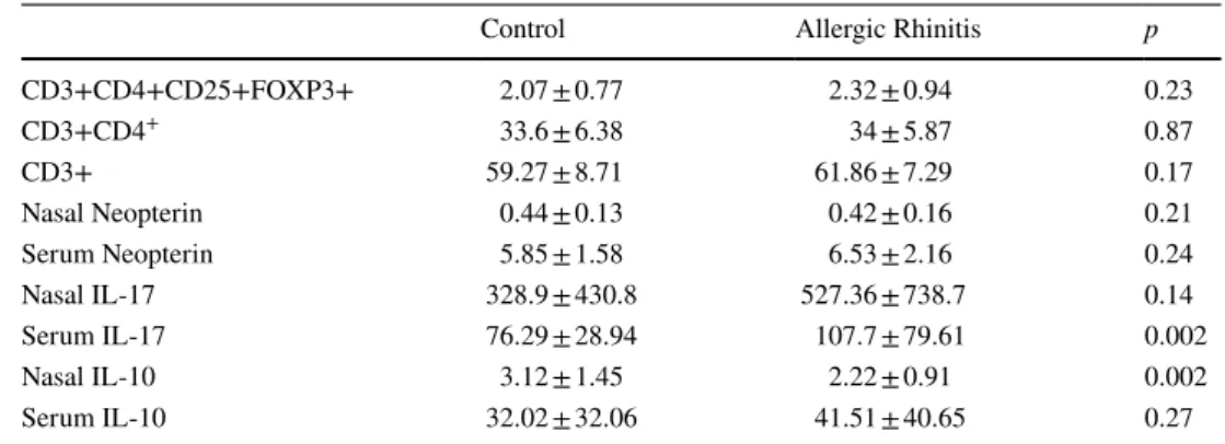
The percentages of CD3 + cells, CD4 + cells and Tregs cells were not different between AR (61.86 ± 7.29, 34 ± 5.87 and 2.32 ± 0.94, respectively) and control groups (59.27 ± 8.71, 33.6 ± 6.38 and 2.07 ± 0.77, respectively) (p > 0.05) (Table 2).
Fig. 1 Flow cytometry dot-plot and histogram surface marker analysis
Table 1 Demographic details of the AR group & controls (AR: Allergic rhinitis, AC: allergic conjunctivitis, CRS: Chronic rhinosinusitis, SPT: skin prick test, HDM: house dust mite)
*Not using any medication within last month
AR group Controls
Gender (n, %)
Female 20 (52.6%) 19 (52.7%)
Male 18 (47.4%) 17 (47.3%)
Age (years) (mean± std) 25.55± 5.25 26.64± 5.89
ARIA classification (n, %) Moderate 14 (36.8%) 0 Severe 24 (63.2%) 0 Smoking (n) 0 0 Comorbidities (n, %) AC 16 (42.1%) 0 CRS 4 (10.5%) 0 Asthma 2 (5.3%) 0 None 16 (42.1%) 36 (100%) Medication* 0 0
IgE (U/ml) Median (min–max) 235 (60–560) 24 (2.80–150)
SPT (n, %) Tree mixture 9 (23.6%) 0 Grass pollen 12 (31.6%) 0 HDM 10 (26.4%) 0 Other 7 (18.4%) 0 Negative 0 36 (100%)
ELISA results
Mean serum IL-10 levels were similar between the two groups, whereas nasal IL-10 was lower in AR subjects com-pared to healthy subjects (2.22 ± 0.91 and 3.12 ± 1.45 pg/ml, respectively) (p < 0.05). Mean serum and nasal IL-17 were higher in the AR (107.7 ± 79.61 and 527.36 ± 738.7 pg/ml) than the control group (76.29 ± 28.94 and 328.9 ± 430.8 pg/ ml; p = 0.002 and p = 0.14). However, there were no signifi-cant differences in serum and nasal neopterin levels between the AR and control groups (p > 0.05, both).
Discussion
In the present study, serum IL-17 level was higher and nasal IL-10 level was lower in patients with AR compared to the healthy control group. Meanwhile, no significant difference was found between the AR and control groups in terms of nasal IL-17 and serum IL-10 levels. In addition, there were no significant differences in the rates of total T cells, CD3+ T cells, CD4+ T cells and Tregs between AR group and the control group. There was no significant difference in neop-terin levels both in serum and in nasal lavage fluid.
Allergic diseases such as asthma and AR are charac-terized by a Th2-skewed immune environment. Th2-type cytokines are upregulated in allergic inflammation, IL-17 is a cytokine released by the Th17 subgroup of CD4+ T cells and play a pathological role in allergic diseases [6–9]. Serum IL-17 level was found to be increased in patients with allergic asthma compared to the non-asthmatic control group [6]. IL-17 level in sputum was found to be correlated with airway hyperresponsiveness [7]. Li et al. showed that Th17 response decreased after specific immunotherapy in patients with house dust mite-induced AR [8]. In another study, the rates of T cells that produce IL-17 and serum IL-17 levels were found to be higher in the allergic group [9].
IL-10 is an inhibitor cytokine that plays an important role in the suppression of allergic reactions [11]. IL-10 decreases IL-4-induced IgE switching [14]. Rapid switch
and expansion of IL-10-producing Tregs and the use of sup-pressive factors represent essential mechanisms in immune tolerance to a high dose of allergens [15]. In the study of Takanaski et al. IL-10 was shown to inhibit survival of, and cytokine production from, eosinophils induced by lipopoly-saccaride [16], In the study of Hussein et al. the IL-10RA gene SNP S138G was shown to contribute to susceptibility to atopic diseases [17]. Grunig et al. found airway hyperac-tivity, eosinophilia and an exaggerated Th2-type response in mice with IL-10 deficiency [18]. In the study of Baumann et al. [22] the nasal lavages of 15 symptomatic allergic and 14 nonallergic subjects were collected during the pollination season and nasal secretion was collected from six allergic subjects after nasal challenge test. After challenge, aller-gic subjects had a significant immediate response of nasal symptoms and a significant increase of IL-4, IL-10, IL-17 and eotaxin-3. During natural pollen exposure, eotaxin-3 and MCP-4 levels were significantly elevated and IL-10 and, unexpectedly, IL-4 were significantly lower in allergic sub-jects compared with nonallergic subsub-jects. In the study by Genç et al., the IL-10 level was decreased in the peripheral blood mononuclear cell culture in the AR group compared to the control group, while no significant difference was found in the nasal fluid [23]. In our study, no significant difference was found in serum IL-10 levels between the control and patient groups, while nasal lavage IL-10 levels were signifi-cantly lower in the patient group. A low nasal IL-10 level was consistent with the studies published in the literature, and it indicated that the cells producing IL-10 should be separately evaluated in the pathogenesis. In our study, the serum IL-17 level was significantly higher in the AR group than the control group, while no significant difference was found in nasal lavage fluid, while nasal IL-10 was lower and serum IL-10 was not significant in patients with AR, sug-gesting that the factors inducing allergic inflammation were systemic and its inhibition occurred locally.
Neopterin, which is a pteridine derivative, increases in many diseases as a specific marker of monocyte/mac-rophage activation [20]. Increased neopterin levels are found in body fluids as a result of stimulation by IL-2 and IFN- γ,
Table 2 Flow cytometry and
ELISA results Control Allergic Rhinitis p
CD3+CD4+CD25+FOXP3+ 2.07 ± 0.77 2.32 ± 0.94 0.23 CD3+CD4+ 33.6 ± 6.38 34 ± 5.87 0.87 CD3+ 59.27 ± 8.71 61.86 ± 7.29 0.17 Nasal Neopterin 0.44 ± 0.13 0.42 ± 0.16 0.21 Serum Neopterin 5.85 ± 1.58 6.53 ± 2.16 0.24 Nasal IL-17 328.9 ± 430.8 527.36 ± 738.7 0.14 Serum IL-17 76.29 ± 28.94 107.7 ± 79.61 0.002 Nasal IL-10 3.12 ± 1.45 2.22 ± 0.91 0.002 Serum IL-10 32.02 ± 32.06 41.51 ± 40.65 0.27
which are the main cytokines of cellular immunity. It has been shown that neopterin is elevated in infections caused by viruses, parasites and intracellular bacteria, malignan-cies, autoimmune diseases, allograft rejection and infections that may develop after transplantation and acute coronary syndrome [21]. Neopterin secretion begins 3 days before T cell proliferation, and it can be detected in serum 2–4 weeks before specific antibody formation [21]. In the study of Unu-var et al. neopterin, tryptophan, and kynurenine levels were found to be higher in patients with asthma and AR than in controls [24]. In this study, we could not find any differences in serum and nasal lavage fluid neopterin levels between AR and control groups.
Treg cells play a basic role in response to allergens by stimulating Th2 cells and the maintenance of immune tol-erance by preventing cytokine secretion [25]. Treg cells have a crucial role, where peripheral tolerance is important, such as in allergic diseases, including asthma, AR, autoim-mune diseases and transplant rejection [26, 27]. Lee et al. [28] and Xu et al. [3] found a decrease in Treg in allergic patients. Lee et al. found that decreased numbers of T(reg) cells in children with allergic airway disease might represent a defect of the T(reg) population while Xu et al. reported that their results indicate that CD4(+) CD25(+) regulatory T cells as well as Foxp3 may play a crucial role in immuno-logical imbalance of AR and increasing Foxp3 and CD4(+) CD25(+) T cells have the potential to be new therapeutic targets for the treatment of AR.
Zhang et al. [29] found a significant decrease in Treg cells in the acute exacerbation period compared to the sta-ble disease stage in asthmatic patients. Akdiş et al. found a decrease in the number of allergen-specific Treg cells in allergic persons [30]. In the study of Stelmaszczyk-Emmel et al., Treg concentration was lower in allergic patients com-pared to the healthy group, and showed a significant correla-tion with serum IgE concentracorrela-tion [31].
In the study of Genc et al. the percentage of FoxP3(+) Tregs were found to be lower in AR patients compared with healthy subjects [23]. In PBMC culture supernatants, IL-10 levels were significantly lower, whereas IL-4, IL-5, and TNF-α levels in nasal lavage fluids were higher in AR patients compared with healthy subjects (p < 0.05).
However, there are some authors reporting no significant difference in Tregs between allergic and nonallergic popula-tions [32–35].
In conclusion, although there were no differences in the distribution of lymphocyte subsets between the AR and con-trol groups, the finding of higher levels of serum and nasal IL-17 and lower levels of nasal IL-10 support the inflamma-tory and antiinflammainflamma-tory cytokine imbalance in the patho-genesis of AR. Systemic and local inflammatory cytokine increase with a relative decrease of anti-inflammatory IL-10
might play a determinant role in the development of AR symptoms.
Compliance with ethical standards
Conflicts of interest The authors declare that they have no conflict of interest.
Research involving human participants and/or animals All procedures performed in studies involving human participants were in accordance with the ethical standards of the institutional research committee of Selcuk University (2015/65) and with the 1964 Helsinki Declaration and its later amendments or comparable ethical standards.
Informed consent Informed consent was obtained from all individual participants included in the study.
References
1. Meng Y, Wang C, Zhang L (2019) Recent developments and highlights in allergic rhinitis. Allergy. https ://doi.org/10.1111/ all.14067
2. Hori S, Nomura T, Sakaguchi S (2003) Control of regulatory T cell development by the transcription factor Foxp3. Science 299:1057–1061
3. Xu G, Mou Z, Jiang H, Cheng L, Shi J, Xu R, Oh Y, Li H (2007) A possible role of CD4+CD25+ T cells as well as transcription fac-tor Foxp3 in the dysregulation of allergic rhinitis. Laryngoscope 117(5):876–880
4. Palomares O, Martin-Fontecha M, Lauener R, Traidl-Hoffmann C, Cavkaytar O, Akdis M, Akdis CA (2014) Regulatory T cells and immune regulation of allergic diseases: roles of IL-10 and TGF-beta. Genes Immun 15(8):511–520. https ://doi.org/10.1038/ gene.2014.45
5. Shevach EM (2001) Certified professionals: cD4(+)CD25(+) sup-pressor T cells. J Exp Med 193(11):F41–F46
6. Wong CK, Ho CY, Ko FW, Chan CH, Ho AS, Hui DS, Lam CW (2001) Proinflammatory cytokines (IL-17, IL-6, IL-18 and IL-12) and Th cytokines (IFN-gamma, IL-4, IL-10 and IL-13) in patients with allergic asthma. Clin Exp Immunol 125(2):177–183 7. Barczyk A, Pierzchala W, Sozanska E (2003) Interleukin-17 in
sputum correlates with airway hyperresponsiveness to methacho-line. Respir Med 97(6):726–733
8. Li CW, Lu HG, Chen DH, Lin ZB, Wang DY, Li TY (2014) In vivo and in vitro studies of Th17 response to specific immuno-therapy in house dust mite-induced allergic rhinitis patients. PLoS One 9(3):e91950. https ://doi.org/10.1371/journ al.pone.00919 50
9. Tsvetkova-Vicheva VM, Gecheva SP, Komsa-Penkova R, Velkova AS, Lukanov TH (2014) IL-17 producing T cells correlate with polysensitization but not with bronchial hyperresponsiveness in patients with allergic rhinitis. Clin Transl Allergy 4(1):3. https :// doi.org/10.1186/2045-7022-4-3
10. Nagalakshmi ML, Murphy E, McClanahan T, de Waal Male-fyt R (2004) Expression patterns of IL-10 ligand and receptor gene families provide leads for biological characterization. Int Immunopharmacol 4(5):577–592. https ://doi.org/10.1016/j.intim p.2004.01.007
11. Akdis CA, Blesken T, Akdis M, Wuthrich B, Blaser K (1998) Role of interleukin 10 in specific immunotherapy. J Clin Invest 102(1):98–106. https ://doi.org/10.1172/JCI22 50
12. Akdis M, Verhagen J, Taylor A, Karamloo F, Karagiannidis C, Crameri R, Thunberg S, Deniz G, Valenta R, Fiebig H, Kegel C, Disch R, Schmidt-Weber CB, Blaser K, Akdis CA (2004) Immune responses in healthy and allergic individuals are characterized by a fine balance between allergen-specific T regulatory 1 and T helper 2 cells. J Exp Med 199(11):1567–1575
13. Enk AH, Angeloni VL, Udey MC, Katz SI (1993) Inhibition of Langerhans cell antigen-presenting function by IL-10. A role for IL-10 in induction of tolerance. J Immunol 151(5):2390–2398 14. Jeannin P, Lecoanet S, Delneste Y, Gauchat JF, Bonnefoy JY
(1998) IgE versus IgG4 production can be differentially regulated by IL-10. J Immunol 160(7):3555–3561
15. Meiler F, Zumkehr J, Klunker S, Ruckert B, Akdis CA, Akdis M (2008) In vivo switch to IL-10-secreting T regulatory cells in high dose allergen exposure. J Exp Med 205(12):2887–2898. https :// doi.org/10.1084/jem.20080 193
16. Takanaski S, Nonaka R, Xing Z, O’Byrne P, Dolovich J, Jordana M (1994) Interleukin 10 inhibits lipopolysaccharide-induced sur-vival and cytokine production by human peripheral blood eosino-phils. J Exp Med 180(2):711–715
17. Hussein PY, Zahran F, Ashour Wahba A, Ahmad AS, Ibrahiem MM, Shalaby SM, El Tarhouny SA, El Sherbiny HM, Bakr N (2010) Interleukin 10 receptor alpha subunit (IL-10RA) gene pol-ymorphism and IL-10 serum levels in Egyptian atopic patients. J Investig Allergol Clin Immunol 20(1):20–26
18. Grunig G, Corry DB, Leach MW, Seymour BW, Kurup VP, Ren-nick DM (1997) Interleukin-10 is a natural suppressor of cytokine production and inflammation in a murine model of allergic bron-chopulmonary aspergillosis. J Exp Med 185(6):1089–1099 19. Hoffmann G, Wirleitner B, Fuchs D (2003) Potential role of
immune system activation-associated production of neopterin derivatives in humans. Inflamm Res 52(8):313–321. https ://doi. org/10.1007/s0001 1-003-1181-9
20. Huber C, Batchelor JR, Fuchs D, Hausen A, Lang A, Nieder-wieser D, Reibnegger G, Swetly P, Troppmair J, Wachter H (1984) Immune response-associated production of neopterin. Release from macrophages primarily under control of interferon-gamma. J Exp Med 160(1):310–316
21. Murr C, Widner B, Wirleitner B, Fuchs D (2002) Neopterin as a marker for immune system activation. Curr Drug Metab 3(2):175–187
22. Baumann R, Rabaszowski M, Stenin I, Tilgner L, Scheckenbach K, Wiltfang J, Schipper J, Chaker A, Wagenmann M (2013) Com-parison of the nasal release of IL-4, IL-10, IL-17, CCL13/MCP-4, and CCL26/eotaxin-3 in allergic rhinitis during season and after allergen challenge. Am J Rhinol Allergy 27(4):266–272. https :// doi.org/10.2500/ajra.2013.27.3913
23. Genc S, Eroglu H, Kucuksezer UC, Aktas-Cetin E, Gelincik A, Ustyol-Aycan E, Buyukozturk S, Deniz G (2012) The decreased CD4+CD25+FoxP3+ T cells in nonstimulated allergic rhinitis patients sensitized to house dust mites. J Asthma 49(6):569–574 24. Ünüvar S, Erge D, Kılıçarslan B, Gözükara Bağ HG, Çatal F,
Girgin G, Baydar T (2019) Neopterin levels and indoleam-ine 2,3-dioxygenase activity as biomarkers of immune sys-tem activation and childhood allergic diseases. Ann Lab Med. 39(3):284–290
25. Pellerin L, Jenks JA, Begin P, Bacchetta R, Nadeau KC (2014) Regulatory T cells and their roles in immune dysregulation and allergy. Immunol Res 58(2–3):358–368
26. Huan J, Culbertson N, Spencer L, Bartholomew R, Burrows GG, Chou YK, Bourdette D, Ziegler SF, Offner H, Vandenbark AA (2005) Decreased FOXP3 levels in multiple sclerosis patients. J Neurosci Res 81(1):45–52. https ://doi.org/10.1002/jnr.20522
27. Shi HZ, Li S, Xie ZF, Qin XJ, Qin X, Zhong XN (2004) Regu-latory CD4+CD25+ T lymphocytes in peripheral blood from patients with atopic asthma. Clin Immunol 113(2):172–178. https ://doi.org/10.1016/j.clim.2004.06.009
28. Lee JH, Yu HH, Wang LC, Yang YH, Lin YT, Chiang BL (2007) The levels of CD4+CD25+ regulatory T cells in pae-diatric patients with allergic rhinitis and bronchial asthma. Clin Exp Immunol 148(1):53–63. https ://doi.org/10.111 1/j.1365-2249.2007.03329 .x
29. Zhang Q, Qian FH, Liu H, Zhou LF, Huang M, Zhang XL, Yin KS (2008) Expression of surface markers on peripheral CD4+CD25high T cells in patients with atopic asthma: role of inhaled corticosteroid. Chin Med J (Engl) 121(3):205–212 30. Akdis M, Burgler S, Crameri R, Eiwegger T, Fujita H, Gomez
E, Klunker S, Meyer N, O’Mahony L, Palomares O, Rhyner C, Ouaked N, Schaffartzik A, Van De Veen W, Zeller S, Zim-mermann M, Akdis CA (2011) Interleukins, from 1 to 37, and interferon-gamma: receptors, functions, and roles in diseases. J Allergy Clin Immunol 127(3):701–721. https ://doi.org/10.1016/j. jaci.2010.11.050
31. Stelmaszczyk-Emmel A, Zawadzka-Krajewska A, Szypowska A, Kulus M, Demkow U (2013) Frequency and activation of CD4+CD25 FoxP3+ regulatory T cells in peripheral blood from children with atopic allergy. Int Arch Allergy Immunol 162(1):16– 24. https ://doi.org/10.1159/00035 0769
32. Geraldes L, Morgado J, Almeida A, Todo-Bom A, Santos P, Paiva A, Cheira C, Pais ML (2010) Expression patterns of HLA-DR+ or HLA-DR− on CD4+/CD25++/CD127low regulatory T cells in patients with allergy. J Investig Allergol Clin Immunol 20(3):201–209
33. Grindebacke H, Wing K, Andersson AC, Suri-Payer E, Rak S, Rudin A (2004) Defective suppression of Th2 cytokines by CD4CD25 regulatory T cells in birch allergics during birch pollen season. Clin Exp Allergy 34(9):1364–1372. https ://doi.org/10.11 11/j.1365-2222.2004.02067 .x
34. Ling EM, Smith T, Nguyen XD, Pridgeon C, Dallman M, Arbery J, Carr VA, Robinson DS (2004) Relation of CD4+CD25+ regulatory T-cell suppression of allergen-driven T-cell acti-vation to atopic status and expression of allergic disease. Lancet 363(9409):608–615. https ://doi.org/10.1016/S0140 -6736(04)15592 -X
35. Bellinghausen I, Klostermann B, Knop J, Saloga J (2003) Human CD4+CD25 +T cells derived from the majority of atopic donors are able to suppress TH1 and TH2 cytokine production. J Allergy Clin Immunol 111(4):862–868
Publisher’s Note Springer Nature remains neutral with regard to jurisdictional claims in published maps and institutional affiliations.