The Biometric Ratios on the Tarsus of the Chinchilla (Chinchilla
lanigera) Based on 3D Reconstructed Images
Sema ÖZKADİF
1,a
Emrullah EKEN
2,bAyşe HALIGÜR
1,c1 Cukurova University, Faculty of Ceyhan Veterinary Medicine, Department of Anatomy, TR-01930 Adana - TURKEY 2 Selcuk University, Faculty of Veterinary Medicine, Department of Anatomy, TR-42003 Konya - TURKEY
a ORCID: 0000-0002-5398-9874; b ORCID: 0000-0001-7426-5325; c ORCID: 0000-0002-3668-4286
Article ID: KVFD-2018-20937 Received: 11.09.2018 Accepted: 04.02.2019 Published Online: 07.02.2019 How to Cite This Article
Özkadif S, Eken E, Halıgür A: The biometric ratios on the tarsus of the Chinchilla (Chinchilla lanigera) based on 3D reconstructed images. Kafkas Univ Vet Fak Derg, 25 (3): 329-333, 2019. DOI: 10.9775/kvfd.2018.20937
Abstract
This study was undertaken to perform a three-dimensional (3D) reconstruction of the tarsal bones of chinchillas using multidetector computed tomography (MDCT) images and reveal biometric ratio of the bones and compare between sexes. For this purpose, a total of 12 adult chinchillas (Chinchilla lanigera) of both sexes (six males and six females) were used. After anesthetizing the animals, MDCT images were obtained in DICOM format, and 3D reconstruction was performed on a computer using the Mimics 13.1 program. The volumes and surface areas of each of the bones that constitute the tarsus of the chinchilla were automatically measured by the program based on the 3D model. After all values of each tarsal bone were expressed as ratios with in tarsus, they were analyzed statistically to reveal differences between sexes. The results showed that there were statistical differences (P<0.05) in calcaneus, talus, central tarsal bone and tarsal bone IV in term of volume ratio and in central tarsal bone, tarsal bone I and tarsal bone IV in term of surface area ratio between sexes. It is considered that 3D tarsus models are useful in revealing anatomic structures and also in assisting clinical diagnosis and treatment.
Keywords: Tarsus, Chinchilla, 3D imaging, Anatomy
Chinchilla (Chinchilla lanigera) Tarsus’unda Üç Boyutlu Rekontrüksiyon
Görüntülerine Dayalı Biyometrik Oranlar
Öz
Bu çalışma şinşillanın tarsal kemiklerinin multidedektör bilgisayarlı tomografi (MDBT) görüntülerini kullanarak üç boyutlu (3B) rekonstrüksiyonunu yapmak ve kemiklerin biyometrik oranlarını ortaya koymak ve cinsiyetler arasında karşılaştırmak amacıyla gerçekleştirildi. Bunun için her iki cinsiyetten (6 erkek, 6 dişi) toplam 12 adet yetişkin şinşilla (Chinchilla lanigera) kullanıldı. Anestezi altında hayvanların MDBT görüntüleri alındıktan sonra DICOM formatında depolandı ve Mimics 13.1 programının olduğu bir bilgisayarda 3B rekonstrüksiyonları gerçekleştirildi. 3B modeli ortaya konulan şinşilla tarsus’unu oluşturan tarsal kemiklerin her birinin hacimleri ve yüzey alanları otomatik olarak program tarafından ölçüldü. Her bir tarsal kemik değerinin tarsus’daki oranları belirtildikten sonra, cinsiyetler arasındaki farklılıkları ortaya çıkarmak için istatistiki analiz yapıldı. Sonuçlar hacim oranına göre calcaneus, talus, os tarsi centrale ve os tarsale IV’de ve yüzey alanı oranına göre os tarsi centrale, os tarsale I ve os tarsale IV’de cinsiyetler arasında istatistiki farkın (P<0.05) olduğunu gösterdi. 3B tarsus modellerinin anatomik yapıları ortaya çıkarmada ve ayrıca klinik tanı ve tedaviye yardımcı olmada yararlı olduğu düşünülmektedir.
Anahtar sözcükler: Tarsus, Şinşilla, 3D görüntüleme, Anatomi
INTRODUCTION
The skeletal dimensions are important when there are no key points that allow the body to be recognized. Sex discrimination is important in the recognition of the body [1].
In forensic medicine anatomically, short bones have some advantages than other bones [2]. Measurements of hand and
tarsal bones have been shown to be sexually dimorphic by previous researchers [3,4].
The tarsal bones are morphologically less recognizable than long bones by non-specialists and can be easily misidentified due to their similarities in animals of similar sizes [5]. Three-dimensional (3D) models of the tarsal bones
İletişim (Correspondence)
+90 322 6133507Measurements obtained from 3D model of bones uses in sexual dimorphism [7]. Computer-based volume calculations
from 3D models and volumetric ratios are significant in determining the gender [8].
A review of the literature reveals studies on the tarsal bones on the leopard (Panthera pardus) [9], the Indian
blackbuck (Antilope cervicapra) [10], rabbit [11], the grasscutter
(Thryonomys swinderianus) [12] and the Indian spotted
deer (Axis axis) [13]; computerized tomography imaging in
dogs [14] and African hedgehogs (Atelerix albiventris) [15];
and the 3D reconstruction of the tarsal joint in mice [16],
laboratory mice, white-footed mice, rats [17], and
red-footed tortoises (Chelonoidis carbonaria) [18]. Furthermore,
research has been undertaken for the 3D reconstruction of human foot bones, and the 3D reconstructed images of the tarsal bones have been utilized in clinical trials, as well as anatomical studies [17,19].
The anatomy of a lot of domestic rodents such as guinea pigs, rats, mice, and hamsters, has been well described. Chinchillas are being popularity as pets [20]. Çevik-Demirkan
et al.[21] investigated the anatomy of the hindlimb of
the chinchilla. In another study, the radiological images of the chinchilla skeleton were analyzed and provided osteological contribution [22]. Also 3D reconstruction of
femur and vertebral column performed and morphometric measurements revealed [23,24]. However, to the best of our
knowledge, no study has been conducted to perform 3D reconstruction of the chinchilla’s tarsal bones, identify their volume and surface area ratios and determine whether there are any differences between the sexes. This current study was carried out to fill this field in the literature.
MATERIAL and METHODS
This study was accepted by the ethics committee of the Veterinary Faculty of Selcuk University on April 27, 2018 (Decision number: 2018/39). In the study, a total of 12 adult chinchillas (Chinchilla lanigera) of both sexes weighing from 500 to 600 g. were used. The 3D models of the tarsal bones were obtained with the Multimodal Immersive Motion rehabilitation with Interactive Cognitive Systems (Mimics) 13.1 software. In order to obtain 3D reconstruction via this program, the MDCT images of the tarsal bones were obtained at high resolution. The animals from which the images were to be taken were anesthetized with a mixture of 60 mg/kg ketamin (Ketalar, Pfizer®) and 6 mg/kg xylazine (Rompun, Bayer®) intravenously. Under anesthesia, the MDCT images were taken of the animals in a prone position. The parameters of the MDCT instrument (Somatom Sensation 64; Siemens Medical Solutions,
range, 0.92 x 0.92. The dosage parameters and scans were performed by utilizing standard protocols and taking the literature [25,26] into consideration. Radiometric resolution
(MONOCHROME2; 16 bits) was obtained at the lowest radiation level and with optimum image quality. The images were stored in DICOM format and transferred to a personal computer installed with Mimics 13.1.
Two of the experts in the field of anatomy obtained similar results by performing reconstructions of tarsal bones at different times. In the automatic segmentation process, the limits of tarsal bones were determined and were assigned different colors (Fig. 1). The limits of the images were determined, and the reconstruction of the tarsal bones was carried out using the 3D transformer component of Mimics 13.1. The volume and surface area of all tarsal bones in the chinchilla both right and left side were measured automatically using the 3D program. After all values of each tarsal bone were expressed as ratios with in tarsus, they were analyzed statistically to reveal differences between sexes. The materiality control of the differences between the average values was undertaken using the SPSS 16.00 software program and an independent t- test.
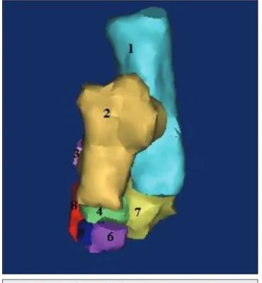
Fig 1. Limitation of tarsal bones on coronal section with different colors
1: Calcaneus, 2: Talus, 3: Medial tibial tarsal bone, 4: Central tarsal bone, 5: Tarsal bone II, 6: Tarsal bone III, 7: Tarsal bone IV, 8: Tarsal bone I
RESULTS
The volume and surface area of the chinchilla tarsal bones were obtained from 3D reconstruction formed using the Mimics 13.1 program to process the MDCT images
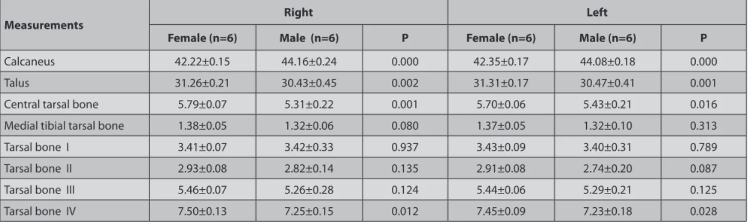
(Fig. 2, Fig. 3). The statistical results the ratio of the mean
values were found significant at the level of P<0.05 (Table
1, Table 2).
The 3D reconstructed images of the tarsal bones of the chinchilla revealed eight bones. The proximal row of the tarsus consisted of the calcaneus articulating with the fibula, the talus articulating with the tibia, and the medial tibial tarsal bone in the medial of the talus. In the distal row were the tarsal bone I to IV. In both proximal and distal rows, the central tarsal bone was observed (Fig. 1,
Fig. 2, Fig. 3). It was determined that the central tarsal bone
did not articulate with the calcaneus and medial tibial tarsal bone.
Both right and left side of the tarsal bones a statistically significant difference was found for calcaneus, talus, central tarsal bone and tarsal bone IV in term of volume ratio between sexes. Also for central tarsal bone, tarsal bone I and tarsal bone IV was seen statistical difference between sexes in term of surface area ratio (Table 1, Table 2). For both male and female chinchillas, the order of the tarsal bones from the greatest to the smallest volume was as follows: the calcaneus, talus, tarsal bone IV, central tarsal bone, tarsal bone III, tarsal bone I, tarsal bone II, and medial tibial tarsal bone. The order of the tarsal bones according to their surface area from the largest to the smallest was; the calcaneus, talus, tarsal bone IV, tarsal bone III, central tarsal bone, tarsal bone I, tarsal bone II, and medial tibial tarsal bone for female chinchillas, and the calcaneus, talus, tarsal bone IV, central tarsal bone, tarsal bone III, tarsal bone I, tarsal bone II, and medial tibial tarsal bone in male chinchillas (Table 1, Table 2).
DISCUSSION
In this study, 3D model obtained from the MDCT images of the tarsal bones in the chinchilla. Three-dimensional reconstructions for bone are clearer and more useful and it is used in tarsal bones [14].
Female chinchillas are larger than male chinchillas. They born larger and grow for a longer time [27]. Depending on
gender, there will be biometric differences between male and female. The most important thing is the difference in the ratio of the measured values.
In this study, the volume and surface area ratio of the tarsal bones differed between the male and female chinchillas. This is consistent with the results of previous study indicating that sexuel dimorphism in chinchilla [27]. Also
sexuel dimorphism were showed in tarsal bones in human [4].
The limitation of this study is the number of the animal. In Fig 2. Dorsal view of 3D model of tarsal bones
1: Calcaneus, 2: Talus 3: Medial tibial tarsal bone, 4: Central tarsal bone, 5: Tarsal bone II, 6: Tarsal bone III, 7: Tarsal bone IV, 8: Tarsal bone I
Fig 3. 3D model of tarsal bones
1: Calcaneus, 2: Talus, 3: Medial tibial tarsal bone, 4: Central tarsal bone, 5: Tarsal bone II, 6: Tarsal bone III, 7: Tarsal bone IV, 8: Tarsal bone I
this study we used 12 chinchillas. If we have more animals we would be able to get strengthen statistical result. Three-dimensional reconstruction method helps user to better understand the anatomical structures that are difficult to understand with other methods by allowing the user to transform 3D image into what they need [28].
Three-dimensional reconsructive models uses in anatomical studies [23,24,29-31] and clinical studies [32-34]. The validity and
reliability of 3D models were proven on comparison of biometric measurement values [35]. Three- dimensional
reconstruction with small bones the section thickness of the MDCT images should be very little.
In conclusion, this was the first study to perform biometric ratios on the tarsus of the chinchilla based on 3D re-constructed images. The 3D volume and surface area ratios of tarsal bones in chinchilla revealed and sexuel dimorphism showed on chinchilla tarsus. Three-dimensional tarsus models can be useful for the investigation of the anatomy and morphology of the tarsal bones with a rather small and complex structure, help clinicians in the diagnosis and treatment processes, assist surgeons in planning operations and in forensic medicine. In further studies the the tarsal joint should be study with its ligaments.
REFERENCES
1. Siddiqi N, Norrish M: Sexual dimorphism from femoral bone dimensions
parameters among African Tribes and South Africans of European descent. Int J Forensic Sci Sexual, 2 (3): 1-11, 2018.
2. Navsa N, Steyn M, Iscan MY: Sex determination from the metacarpals
in a modern South African male and female sample UPS Space University, Pretoria, 2008. www.up.ac.az/dspace/handle.net; Accessed: 23 September 2018.
3. Eshak GA, Ahmed HM, Abdel Gawad EA: Gender determination
from hand bones length and volume using multidetector computed tomography: A study in Egyptian people. J Forensic Leg Med, 18, 246-252, 2011. DOI: 10.1016/j.jflm.2011.04.005
4. Harris SM, Case DT: Sexual dimorphism in the tarsal bones:
Implications for sex determination. J Forensic Sci, 57, 295-305, 2012. DOI: 10.1111/j.1556-4029.2011.02004.x
5. Smart TS: Carpals and tarsals of mule deer, black bear and human: an
osteology guide for the archaeologist. MSc Thesis, Western Washington University, 2009.
6. Jain ML, Dhande SG, Vyas NS: Computer aided diagnosis of human
foot’s bones. IJBES, 1, 17-26, 2014.
7. Brzobohata H, Krajicek V, Horak Z,Veleminska J: Sexual dimorphism
of the human tibia through time: Insights into shape variation using a surface-based approach. PLoS One, 11 (11): e0166461, 2016. DOI:10.1371/ journal.pone.0166461
8. Shearer BM, Sholts SB, Garvin HM, Wärmländer SKTS: Sexual
dimorphism in human browridge volume measured from 3D models of dry crania: A new digital morphometrics approach. Forensic Sci Int, 222 (1-3): 400.e1-400.e5, 2012. DOI: 10.1016/j.forsciint.2012.06.013
9. Podhade DN, Shrivastav AB, Vaish R, Tiwari Y: Morphology and
morphometry of tarsals of the leopard (Panthera pardus). Res J Anim Vet
Fishery Sci, 2, 20-21, 2014.
10. Choudhary OMP, Ishwer S, Bharti SK: Gross and biometrical studies
on the tarsal bones of Indian blackbuck (Antilope cervicapra). IJBAAR, 13,
Calcaneus 42.22±0.15 44.16±0.24 0.000 42.35±0.17 44.08±0.18 0.000
Talus 31.26±0.21 30.43±0.45 0.002 31.31±0.17 30.47±0.41 0.001
Central tarsal bone 5.79±0.07 5.31±0.22 0.001 5.70±0.06 5.43±0.21 0.016
Medial tibial tarsal bone 1.38±0.05 1.32±0.06 0.080 1.37±0.05 1.32±0.10 0.313
Tarsal bone I 3.41±0.07 3.42±0.33 0.937 3.43±0.09 3.40±0.31 0.789
Tarsal bone II 2.93±0.08 2.82±0.14 0.135 2.91±0.08 2.74±0.20 0.087
Tarsal bone III 5.46±0.07 5.26±0.28 0.124 5.44±0.06 5.29±0.21 0.125
Tarsal bone IV 7.50±0.13 7.25±0.15 0.012 7.45±0.09 7.23±0.18 0.028
Table 2. Statistical analysis performed by taking percentage rates of surface area means obtained from 3D images of tarsal bones (mean ± SD)
Measurements Right Left
Female (n=6) Male (n=6) P Female (n=6) Male (n=6) P
Calcaneus 35.56±0.13 35.42±0.30 0.340 35.61±0.15 35.41±0.34 0.211
Talus 25.47±0.11 25.50±0.21 0.771 25.42±0.10 25.48±0.21 0.612
Central tarsal bone 7.60±0.12 7.79±0.08 0.009 7.58±0.13 7.89±0.28 0.034
Medial tibial tarsal bone 2.49±0.08 2.57±0.12 0.264 2.50±0.08 2.54±0.08 0.356
Tarsal bone I 6.46±0.12 6.77±0.18 0.007 6.45±0.13 6.77±0.20 0.011
Tarsal bone II 5.33±0.06 5.33±0.14 0.899 5.35±0.09 5.35±0.16 0.966
Tarsal bone III 7.72±0.11 7.68±0.15 0.681 7.71±0.12 7.63±0.15 0.356
453-456, 2015.
11. Ajayi IE, Shawulu JC, Zachariya TS, Ahmed S, Adah BMJ:
Osteomorphometry of the bones of the thigh, crus and foot in the New Zealand white rabbit (Oryctolagus cuniculus). Ital J Anat Embryol, 117, 125-134, 2012.
12. Onwuama KT, Ojo SA, Hambolu JO, Dzenda T, Zakari FO, Salami SO: Macro-anatomical and morphometric studies of the hindlimb of
grasscutter (Thryonomys swinderianus, Temminck-1827). Anat Histol
Embryol, 47, 21-27, 2018. DOI: 10.1111/ahe.12319
13. Yadav S, Joshi S, Mathur R, Choudhary OP: Morphometry of tarsal
and metatarsal of Indian Spotted Deer (Axis axis). Indian Vet J, 92, 43-46, 2015.
14. Gielen IM, De Rycke LM, Van Bree HJ, Simoens PJ: Computed
tomography of the tarsal joint in clinically normal dogs. Am J Vet Res, 621 (2): 1911-1915, 2001. DOI: 10.2460/ajvr.2001.62.1911
15. Girgiri IA, Yahaya A, Gambo BG, Majama YB, Sule A:
Osteo-morphology of the appendicular skeleton of four-toed african hedgehogs
(Atelerix albiventris) Part (2): Pelvic limb. Glob Vet, 16, 413-418, 2016. 16. Kai Y, Matsumoto K, Kameoka S, Arai S, Matsumoto N, Komiyama K, Shimba S, Honda K: Observation of the tarsus joint in the Mop-3/
Bmal-1 gene knock-out mouse using “In vivo” Micro-CT: Influence of diet and sex on calcification of the tendon of the tarsus joint. J Hard Tissue Biol, 21, 133-140, 2012. DOI: 10.2485/jhtb.21.133
17. Richbourg HA, Martin MJ, Schachner ER, Mcnulty MA: Anatomical
variation of the tarsus in common inbred mouse strains. Anat Rec, 300, 450-459, 2017. DOI 10.1002/ar.23493
18. Bortolini Z, Lehmkuhl RC, Ozeki LM, Tranquilim MV, Sesoko NF, Teixeira CR, Vulcano LC: Association of 3D reconstruction and
conventional radiography for the description of the appendicular skeleton of chelonoidis carbonaria (Spix, 1824). Anat Histol Embryol, 41, 445-452, 2012. DOI: 10.1111/j.1439-0264.2012.01155.x
19. Getman LM, Ross MW, Smith MA: Surgical repair of fractures of the
lateral and medial tibial malleoli in a yearling Arabian filly. Equine Vet
Educ, 24, 496-502, 2012. DOI: 10.1111/j.2042-3292.2011.00328.x
20. Brenner SZG, Hawkins MG, Tell LA, Hornof WJ, Plopper CG, Verstraete FJM: Clinical anatomy, radiography, and computed tomography
of the chinchilla skull. Comp Cont Educ Pract, 27, 933-942, 2005.
21. Çevik-Demirkan A, Özdemir V, Demirkan I: Anatomy of the hind
limb skeleton of the chinchilla (Chinchilla lanigera). Acta Vet Brno, 76, 501-507, 2007. DOI: 10.2754/avb200776040501
22. Gasse CAS: Contribution radiologique et ostéologique à la
connaissance du chinchilla (Chinchilla lanigera). These pour obtenir le grade de Docteur Veterinaire. Ministere de L’agriculture et de la Peche Ecole Nationale Veterinaire de Toulouse. France. 94-97. 2008.
23. Ozkadif S, Varlik A, Kalayci I, Eken E: Morphometric evaluation
of chinchillas (Chinchilla lanigera) femur with different modelling techniques. Kafkas Univ Vet Fak Derg, 22, 945-951, 2016. DOI: 10.9775/ kvfd.2016.15683
24. Ozkadif S, Eken E, Dayan MO, Besoluk K: Determination of
sex-related differences based on 3D reconstruction of the chinchilla
(Chinchilla lanigera) vertebral column from MDCT scans. Vet Med-Czech,
62, 204-210, 2017. DOI: 10.17221/19/2015-VETMED
25. Prokop M: General principles of MDCT. Eur J Radiol, 45, S4-S10, 2003.
DOI: 10.1016/S0720-048X(02)00358-3
26. Kalra MK, Maher MM, Toth TL, Hamberg LM, Blake MA, Shepard J, Saini S: Strategies for CT radiation dose optimization. Radiology, 230,
619-628, 2004. DOI: 10.1148/radiol.2303021726
27. Lammers AR, Dziech HA, German RZ: Ontogeny of sexual
dimorphism in Chinchilla lanigera (Rodentia: Chinchillidae). J Mammal, 82, 79-189, 2001. DOI: 10.1644/1545-1542(2001)082<0179:OOSDIC>2.0.CO;2
28. Yamada K, Taniura T, Tanabe S, Yamaguchi M, Azemoto S, Wisner ER: The use of multi- detector row computed tomography (MDCT) as an
alternative to specimen preparation for anatomical insrtuction. J Vet Med
Educ, 34:143-150, 2007.
29. Jaeger M, Briand D, Borianne P, Bonnel F: Knee anatomy 3D
reconstruction and visualization from CT scans. Surg Radiol Anat, 15 (3): 231-231, 1993.
30. Gezer İnce N, Demircioğlu İ, Yılmaz B, Ağyar A, Dusak A: Martılarda (Laridae spp.) cranium’un üç boyutlu modellemesi. Harran Üniv Vet Fak Derg, 7, 98-101, 2018.
31. Özkadif S, Eken E, Kalaycı I: A three-dimensional reconstructive
study of pelvic cavity in the New Zealand rabbit (Oryctolagus cuniculus).
Sci World J, 2014:489854, 2014. DOI: 10.1155/2014/489854
32. Watanabe Y, Ikegami R, Takasu K, Mori K: Three-dimensional
computed tomographic images of pelvic muscle in anorectal malformations.
J Pediatr Surg, 40, 1931-1934, 2005. DOI: 10.1016/j.jpedsurg.2005.08.010 33. Jun BC, Song SW, Cho JE, Park CS, Lee DH, Chang KH, Yeo SW:
Three-dimensional reconstruction based on images from spiral high-resolution computed tomography of the temporal bone: Anatomy and clinical application. J Laryngol Otol, 119, 693-698, 2005.
34. Miyamoto R, Tadano S, Sano N, Inagawa S, Adachi S, Yamamoto M:
The impact of three-dimensional reconstruction on laparoscopic-assisted surgery for right-sided colon cancer. Wideochir Inne Tech Maloinwazyjne, 12, 251-256, 2017. DOI: 10.5114/wiitm.2017.67996
35. Kim M, Huh KH, Yi WJ, Heo MS, Lee SS, Choi SC: Evaluation of
accuracy of 3D reconstruction images using multi-detector CT and cone-beam CT. Imaging Sci Dent, 42, 25-33, 2012. DOI: 10.5624/isd. 2012.42.1.25