Yazışma Adresi/Address for Correspondence: Dr. Davut Alptekin, Cukurova University Faculty of Medicine, Department of Medical Biology Adana/Turkey Email: [email protected]
Geliş tarihi/Received: 19.09.2018 Kabul tarihi/Accepted: 19.12.2018 Published online: 24.02.2019
ARAŞTIRMA / RESEARCH
Detection of CAG trinucleotide repeat numbers with fragment analysis
in patients diagnosed with Huntington’s disease and in their families
Huntington hastalığı tanısı almış hastalarda ve ailelerinde CAG trinükleotid tekrar
sayılarının fragman analizi ile tespiti
Davut Alptekin1 , Perçin Pazarcı1 , Mehmet Ali Bereketoğlu2 , Mehmet Ali Erkoç1 , Nermin Seda Ilgaz1 , Ümit Lüleyap1
1Cukurova University Faculty of Medicine, Department of Medical Biology, Adana, Turkey 2Su Hospital, Department of Neurology, Mersin, Turkey
Cukurova Medical Journal 2019;44(2):1
Abstract Öz
Purpose: Huntington's Disease (HD) is an autosomal
dominant disorder affecting nervous system. CAG trinucleotide repeat (TNR) increase in Huntingtin gene causes the disease. In normal individuals, 10-35 TNRs are found whereas in HD this number exceeds 36-37. This study aimed to investigate TNR numbers in individuals with HD diagnosed family and to provide genetic counselling for individuals with abnormal alleles.
Material and Methods: Subjects consist of family
members of a male who died at age of 60 due to HD. Randomly selected 57 healthy individuals are also analysed for control. TNR numbers were determined by fragment analysis.
Results: TNR numbers of family members were
determined as 17, 21, 23, 25, 33, 36 and 39. TNR numbers of randomly selected healthy people were found below 26. Individuals with 33 and 36 TNRs were considered as risk groups. Individuals with 39 TNRs were considered as HD patients.
Conclusion: Since some subjects had 39 TNRs, it was
emphasized that these people should be under physician control. Prenatal diagnosis is recommended to those who plan to have children. In addition, subjects with 33 and 36 CAG trinucleotide repeats are advised to inform new generations about HD and that they may be affected in future.
Amaç: Huntington Hastalığı (HD), sinir sistemini
etkileyen otozomal dominant bir hastalıktır. Huntingtin genindeki CAG trinükleotit tekrarı (TNR) artışı hastalığa neden olur. Normal bireylerde 10-35 TNR bulunurken HD'de bu sayı 36-37'yi aşmaktadır. Bu çalışmada, HD tanılı ailede TNR sayılarının araştırılması ve anormal alleli olan bireyler için genetik danışmanlık yapılması amaçlanmıştır.
Gereç ve Yöntem: Örnekler, HD nedeniyle 60 yaşında
ölen bir erkeğin aile üyelerinden oluşmaktadır. Rastgele seçilen 57 sağlıklı birey de kontrol için analiz edilmiştir. TNR sayıları fragman analizi ile belirlenmiştir.
Bulgular: Aile bireylerinin TNR sayıları 17, 21, 23, 25, 33,
36 ve 39 olarak belirlendi. Rastgele seçilmiş sağlıklı kişilerin TNR sayıları 26'nın altında bulundu. 33 ve 36 TNR'li bireyler risk grubu olarak kabul edildi. 39 TNR'li bireyler HD’ye sahip olarak kabul edildi.
Sonuç: Bazı deneklerin 39 TNR'si olduğundan, bu
kişilerin hekim kontrolünde olması gerektiği vurgulanmıştır. Çocuk sahibi olmayı planlayanlara prenatal tanı önerilmektedir. Ek olarak, 33 ve 36 CAG trinükleotid tekrarı olan deneklerin, HD ile ilgili yeni nesilleri bilgilendirmeleri ve gelecekte kendilerinin de etkilenebilecekleri bildirilmektedir.
Keywords: Huntington's disease, Huntingtin gene,
INTRODUCTION
Huntington's disease (HD) is a neurodegenerative disorder which shows autosomal dominant inheritance and is characterized by clinically different phenotypes, such as chorea, dystonia, impaired coordination, cognitive decline, uncontrolled shivering and social dysfunction. With the disease, nerve cell loss and atrophy occur in the caudate and putamen of the brain1-4. HD brain MR images show
significant morphological differences compared to the control group. In particular, it has been reported that basal ganglion and the neck cerebrum are significantly reduced and the cerebral cortex is significantly increased. Early degenerative changes in the striatum and cerebral neck and enlargements in the cerebral cortex were accepted as radiological findings for HD5. In addition, it has been reported
that if one of the parents of an individuals is sick, they are also more likely to think of suicide because of the risk of getting the disease6.
The gene causing HD was detected in 1983 and reported to be present on the fourth chromosome (4p16.3)7. The gene that caused the disease was
named Huntingtin (HTT) and was mostly understood in 1993. It has been reported that CAG (Poly-glutamine) trinucleotide repeats (TNRs) in the first exon of this gene causes the disease8. Normal
individuals have 10-36 CAG TNRs whereas those with HD have more than 37 TNRs9, 10. However, in
some studies, it has been reported that 36-39 TNRs do not cause HD11-13. As the number of TNRs
increases, the severity of the symptoms increases and the disease is seen at an earlier age. The main reason for the occurrence of HD in childhood is that the TNRs in alleles exceed 10014-16.
TNR numbers in alleles show instability in HD. A 1998 study suggested that, TNR numbers lower than 26 are normal, TNR numbers between 27-35 are normal but can increase with time, TNR numbers between 36-39 are abnormal with delayed disease onset, TNR numbers greater than 39 are abnormal and have earlier disease onset17. The alleles with a
TNR number of 27-35 is known as a instable allele, and individuals are at risk because TNR numbers in these alleles increase18, 19. The TNR number is
unstable to remain constant for generations. This
higher number of repetitions. If the number of repetitions in one of the alleles of the father is 34-35, the TNR number in the child may rise to 37 and above, leading to the disease20.
The onset age of HD varies. In some people symptoms may appear within the first 10 years, while others may not show any symptoms until the age of 6021. There are three types of HD which are at
childhood onset HD, youth onset HD and adult onset HD. The childhood HD period starts from birth to the age of 18. In these individuals, mutant alleles cannot pass on to the next generation due to premature deaths14,22. Youth HD usually starts
between the age 18-20. If the TNR count is 60 or more, the disease develops early, starting with symptoms of tremors and contractions. The disease is caused by the fact that the TNR region in the DNA has spontaneously increased by looping, or that it has passed through the mother in some cases but has often passed from the father to the child15, 16, 22-25. As
the number of TNR increases in HD, the age of onset of the disease decreases26. The youngest known case
is a girl with symptoms beginning at 18 months and having 265/14 TNRs14.
The incidence of HD in Turkey is not known precisely. Furthermore, there are not many studies conducted in our country regarding the TNR numbers. In one study, TNR numbers in HTT gene were investigated in 127 HD patients and 122 healthy individuals and repeat numbers reported to be in the range of 38-78 in HD patients and 10-35 in healthy individuals27. In another study, genetic analyses were
performed on 27 HD patients from 19 different families, and the TNR numbers were reported to vary between 40-7628. It has been emphasized that the
resulting symptoms of the TNR increase in HD are similar to diseases such as Fragile X Syndrome, Kennedy Syndrome, Myotonic Dystrophy, so a molecular diagnosis is necessary.
There are many patients diagnosed with HD based on clinical and radiological findings in our region. A sick family has been found in Doruk district (Ceyhan-Adana). Children and close relatives of these patients are living with fear of having HD at a later age and they also think that their children will also suffer from this disease. For this reason, TNR numbers in the HTT gene of patients or individuals under 30 years of
they should definitely have a prenatal diagnosis during pregnancy.
For these reasons, it was aimed to establish a molecular diagnosis in order to avoid confusion with clinically diagnosed Huntington's patients with other neurodegenerative diseases since there was no previous study for the detection of TNR numbers in these individuals. In addition, if the TNR number is above 36-37 in subjects under 30 years of age, these individuals will be recommended to be under the control of a physician. Also, individuals who are married will be recommended to have a prenatal diagnosis during pregnancy to prevent the birth of possible HD patients.
MATERIALS AND METHODS Subjects and DNA isolation
Patient blood samples related to this study were taken from the members of a family who were diagnosed with Huntington's disease after having applied to the neurology clinic of Çukurova University Faculty of Medicine with various complaints. 20 members of this family is found in Ceyhan (Adana) and 3 ml blood samples are collected in tubes containing ethylenediaminetetraacetic acid (EDTA). In addition, 3 ml blood samples are collected from 57 healthy people for control group. DNA is obtained from blood samples using DNA isolation kit according to the manufacturer’s protocol (Invitrogen Inc., California, USA). Each patient and control were informed about the study before blood sampling and was asked to sign an informed consent form approved by the Ethics Committee of the Çukurova University Medical Faculty.
PCR, capillary electrophoresis and TNR number determination
HD related regions with TNRs were amplified using a standard PCR amplification protocol. 11.1 µL distilled water, 4 µL Taq buffer (Thermo Fischer Inc., Massachusetts, USA), 4.8 µL dNTP (240 µmol/L each) (Thermo Fischer Inc., Massachusetts, USA), 1 µL MgCl2 (0.625 mmol) (Thermo Fischer Inc., Massachusetts, USA), 1.5 unit Taq enzyme (Thermo
Fischer Inc., Massachusetts, USA), 10 µL DNA (400-500 ng), 4.8 µL DMSO (120 g/L) (Thermo Fischer Inc., Massachusetts, USA) and 2 µL (20 pmol) of
forward
(5’-6-FAM-ATGAAGGCCTTCGAGTCCCTCAAGTCC-3’) and 2 µL (20 pmol) of reverse primer (5’-CGGTGGCGGCTGTTGCTGCTGCTGCTGCT G-3’) is used for the PCR mixture. PCR conditions are set as 5 min for the primary denaturation at 95 0C,
after that 36 cycles of 1 min at 95 0C for the
denaturation, 1 min at 64 0C for annealing, 1 min at
72 0C for the extension and lastly 10 min at 72 0C for
the final extension.
Statistical analysis
PCR amplified samples using FAM labelled forward primers were then subjected to capillary electrophoresis on an ABI Prism 3130 instrument using POP-7 polymer to measure their length and the length of the region containing the TNR that caused HD was determined. While the number of repeats causing the HD was calculated, the number of non-repeating nucleotides (47 bp) in the region was subtracted from the total number of nucleotides that was determined by capillary electrophoresis, divided by 3, and the number of TNRs of each sample was calculated.
RESULTS
The pedigree was drawn by gathering the information of the family that made up the subject group (Figure 1). According to the information obtained, the first-generation grandfather died from HD, the second-generation individuals were all over the age of 40, and the people numbered 1, 3 and 7 were identified as HD. Individual number 1 was reported to have died from the disease. Individuals 3 and 7 are alive and living as HD patients. All of the people in the second-generation were reported to be married and have children. However, blood samples could be obtained only from individuals 3, 5 and 6 and their children. The others could not be reached because they live abroad (Netherlands).
Figure 1. Pedigree of the family with Huntington’s Disease
Individual 3 (HD patient male) was married and had 2 boys and 3 girls. At the age of 54, however, HD symptoms have begun, he is currently suffering from HD and cannot live without support. His children are between 20-46 years of age. TNR numbers in the HTT gene were determined from the samples we obtained from all of this family. It was determined that there were 23/39 TNRs in the father (individual 3) and 21/23 TNRs in the mother (wife of individual 3). Because one of the father's alleles contained 39 TNRs, the disease was confirmed. When the children of this family are examined, it was determined that the oldest daughter and youngest son were healthy (23/25 TNRs, 23/23 TNRs respectively), and the other three children had potential for future HD due to 23/39 TNRs. These individuals are shown as square patterns in the pedigree because one of their alleles contained 39 TNRs but did not yet show the disease symptoms.
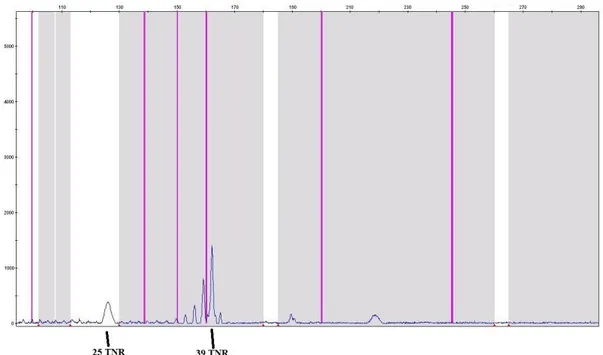
Individual 5 was married and had 2 girls and 2 boys. The mother (individual 5) is 50 years old and looks healthy. When the TNR numbers are examined, it was found that the individual 5 had 25/39 TNRs. Three of her four children were found to have 23 and 25 TNRs in their alleles. However, one daughter, who is 29 years old and healthy for now, was found to have 23/39 TNRs. Figure 2 shows the fragment analysis result of the individual 5 with 25/39 TNRs as an example.
Individual 6 was married and had 4 girls and 1 boy. Blood samples were also taken from this family. The mother (individual 6) is 46 years old and looks healthy. When the TNR numbers are examined, it was found that the individual 6 had 17/33 and her husband had 23/33 TNRs. HD is not expected in this family since the TNRs are not over 36. However, the children of this family were also studied. It was determined that 4 of the children contained 23/33 TNRs, and one contained 23/36 TNRs.
Figure 2. Fragment analysis result of the individual 5 (26/39 TNRs).
Separate from these families where the disease was seen, blood samples were taken from 57 randomly selected healthy subjects and the TNR numbers were determined. Minimum of 9 TNRs and a maximum of 26 TNRs were determined in these individuals. Average number of TNRs in healthy individuals (114 alleles) was found to be 15.15 ± 2.73 (average ± standard deviation).
DISCUSSION
Although the age of onset of HD depends on the number of TNRs, it is very important for children if one of the parents is sick because HD has an autosomal dominant inheritance pattern and the disease symptoms appear mostly after 35 years of age. Until this age, it is very likely that someone will marry and transfer their abnormal allele to their children. Therefore, instead of following the HD diagnosed individuals and conducting a TNR study, a family whose father had 3 HD patient children out of his 8 children and died of HD was selected. Three members of second generation of this family, one patient male and two healthy females, live in the Doruk (Ceyhan-Adana). Other individuals of this generation and their children live in the Netherlands.
Family members living in the Netherlands were not included in study since their blood samples could not be obtained. However, one patient and two healthy individuals from the second generation and their family members were included in the study.
According to our findings, one of the alleles of the individual number 3 (generation II) in the HTT gene contains 39 TNRs, and consequently has HD. In previous studies, it was reported that when the TNR number increases above 36, the disease symptoms begins to appear after the age of 45, and as the number of TNRs increases, onset age of the HD decreases17, 26. As a matter of fact, symptoms in this
patient (individual 3) started at age of 50, and the severity of the symptoms increased with age. This patient is married at the age of 22 and has 3 daughters and 2 sons in which the eldest is 36 years old. It is a very bad situation for children to live in fear because of the risk of HD. The TNR numbers of these children were examined and it was found that three of five children (ages 27-34 years) had 39 TNRs in one of the alleles which they acquired from their father. Due to the fact that their father acquired HD after the age of 50, these children are also expected to show HD symptoms in the coming years and routine doctor control is suggested.
The individual number 5 (generation II, female) is 50 years old and does not have any symptoms of HD yet. However, one of the alleles of this individual was found to have 39 TNRs. This person has had 5 children. Of the 5 children, only one of the alleles of one of her daughter (29 years old) contains 39 TNRs. Although she is 50 years old, she does not have any signs of HD. In some studies, it has been reported that, even in monozygotic twins, the phenotype of the disease may vary due to personal characteristics, environmental and epigenetic factors, and epimutations in the critical DNA region29-31. In this
family, it is suggested that the mother and her daughter should be under doctor's supervision, thinking that they may get HD in the future. With this study, information was provided about the necessity of prenatal diagnosis for persons who had not yet had children and whose TNR number was higher than 36 in one of their alleles. In the generation III, only one of the four people whose TNR number is greater than 36 is single, other three are married and have children. If these individuals are likely to make more children, they would better have a prenatal diagnosis. However, this decision should still be left to couples. In fact, in a study done with 354 couples, in which one of the individuals had increased TNRs, it was emphasized that 75% of women agreed to have prenatal diagnosis on their first and second pregnancies, but all women needed psychological support after prenatal diagnosis32.
Prenatal diagnosis is necessary to prevent the disease from being transmitted to next generations.
The HD status of the subjects was determined precisely and the concerns of those who have healthy alleles were finally ended. In addition, individuals with an increased TNR allele are warned for being at risk of developing disease later in life and are recommended to be under physician control. Moreover, these families have been isolated in rural areas due to HD and marriage with other families has not been possible. With this study, the social pressure on these families was also abolished. In relation to HD, it is reported that if the parents have increased TNRs, they may also have healthy children with Preimplantation Genetic Diagnosis (PGD) method. As a matter of fact, studies on this subject are available and healthy embryos can be obtained by
were analysed and found to be between 9-26 TNRs. These people have no chance of HD. In previous studies, it was emphasized that the probability of emergence of new cases in future generations is higher if TNR number is above 27 in of the alleles8, 9.
It has been reported that in regions where TNRs occur, hairpin loops are formed and these regions are more prone to TNR increase particularly if the second base is Adenine34-36. For this reason, it is
advisable to collect information and data from time to time in society and to follow such people in future generations.
In conclusion, according to many previous studies and the genetic results of this study obtained from these family members who are diagnosed with HD, HD definitely characterized by increase of the CAG TNRs. Since the disease is mostly seen at an old age, new generations marry and transfer abnormal genes to new generations without knowing it. Families with individuals diagnosed with HD in neurology clinic must have genetic analysis and should be informed that they (or their wives) should be given a prenatal diagnosis during pregnancy.
Acknowledgements
The authors are grateful to Cukurova University Research Fund for the financial support to conduct this study (project no: TF2013BAP4).
Yazar Katkıları: Çalışma konsepti/Tasarımı: DA, MAB, ÜL, PP, MAE,
NSI; Veri toplama: PP, MAE, NSI; Veri analizi ve yorumlama: DA, MAB, ÜL, PP, MAE, NSI; Yazı taslağı: DA, MAB, ÜL, PP, MAE, NSI; İçeriğin eleştirel incelenmesi: DA, MAB, ÜL, PP, MAE, NSI; Son onay ve sorumluluk: DA, PP, MAB, MAE, NSI, ÜL; Teknik ve malzeme desteği: DA;MAB, ÜL; Süpervizyon: DA, MAB, ÜL; Fon sağlama (mevcut ise): yok.
Bilgilendirilmiş Onam: Katılımcılardan yazılı onam alınmıştır. Hakem Değerlendirmesi: Dış bağımsız.
Çıkar Çatışması: Yazarlar çıkar çatışması beyan etmemişlerdir. Finansal Destek: Yazarlar finansal destek beyan etmemişlerdir. Author Contributions: Concept/Design :DA, MAB, ÜL, PP, MAE, NSI; Data acquisition: PP, MAE, NSI; Data analysis and interpretation: DA, MAB, ÜL, PP, MAE, NSI; Drafting manuscript: DA, MAB, ÜL, PP, MAE, NSI; Critical revision of manuscript: DA, MAB, ÜL, PP, MAE, NSI; Final approval and accountability: DA, PP, MAB, MAE, NSI, ÜL; Technical or material support: DA, PP, MAB, MAE, NSI, ÜL; Supervision: DA, MAB, ÜL; Securing funding (if available): n/a.
Informed Consent: Written consent was obtained from the
participants.
Peer-review: Externally peer-reviewed.
Conflict of Interest: Authors declared no conflict of interest. Financial Disclosure: Authors declared no financial support
disease carriers: a double blind study. J Med Genet. 1995;32:600-4.
2. Lanska DJ. George Huntington (1850-1916) and hereditary chorea. J Hist Neurosci. 2000;9:76-89. 3. Walker FO. Huntington's disease. Lancet.
2007;369:218-28.
4. Dayalu P, Albin RL. Huntington Disease. Neurol Clin. 2015;33:101-14.
5. Wallace DC, Hall AC. Evidence of genetic heterogeneity in Huntington's chorea. J Neurol Neurosurg Psychiatry. 1972;35:789-800.
6. Nance MA, Myers RH. Juvenile onset Huntington's disease clinical and research perspectives. Mental Retardation and Developmental Disabilities Research Reviews. 2001;7:153-7.
7. Gusella JF, Wexler NS, Conneally PM, Naylor SL, Anderson MA, Tanzi RE et al. A polymorphic DNA marker genetically linked to Huntington's disease. Nature. 1983;306:234-8.
8. Macdonald ME, Ambrose CM, Duyao MP, Myers RH, Lin C, Srinidhi L et al. A novel gene containing a trinucleotide repeat that is expanded and unstable on Huntingtons-Disease chromosomes. Cell. 1993;72:971-83.
9. Duyao M, Ambrose C, Myers R, Novelletto A, Persichetti F, Frontali M et al. Trinucleotide repeat length instability and age of onset in Huntington's disease. Nat Genet. 1993;4:387-92.
10. Snell RG, MacMillan JC, Cheadle JP, Fenton I, Lazarou LP, Davies P et al. Relationship between trinucleotide repeat expansion and phenotypic variation in Huntington's disease. Nat Genet. 1993;4:393-7.
11. Legius E, Cuppens H, Dierick H, Van Zandt K, Dom R, Fryns J-P et al. Limited expansion of the (cag)n repeat of the Huntington gene: A Premutation. Europ J Hum Genet. 1994;2:44-50.
12. Rubinsztein DC, Leggo J, Coles R, Almqvist E, Biancalana V, Cassiman JJ et al. Phenotypic characterization of individuals with 30-40 CAG repeats in the Huntington disease (HD) gene reveals HD cases with 36 repeats and apparently normal elderly individuals with 36-39 repeats. Am J Hum Genet. 1996;59:16-22.
13. McNeil SM, Novelletto A, Srinidhi J, Barnes G, Kornbluth I, Altherr MR et al. Reduced penetrance of the Huntington's disease mutation. Hum Mol Genet. 1997;6:775-9.
14. Milunsky JM, Maher TA, Loose BA, Darras BT, Ito M. XL PCR for the detection of large trinucleotide expansions in juvenile Huntington's disease. Clin Genet. 2003;64:70-3.
15. Nahhas FA, Garbern J, Krajewski KM, Roa BB, Feldman GL. Juvenile onset Huntington disease resulting from a very large maternal expansion. Am J Med Genet A. 2005;137A:328-31.
16. Yoon G, Kramer J, Zanko A, Guzijan M, Lin S, Foster-Barber A et al. Speech and language delay are
early manifestations of juvenile-onset Huntington disease. Neurology. 2006;67:1265-7.
17. Nance MA, Seltzer W, Ashizawa T, Bennett R, McIntosh N, Myers RH et al. Laboratory guidelines for Huntington disease genetic testing. AJHG. 1998;62:1243-7.
18. Potter NT, Spector EB, Prior TW. Technical standards and guidelines for Huntington disease testing. Genet Med. 2004;6:61-5.
19. Semaka A, Creighton S, Warby S, Hayden MR. Predictive testing for Huntington disease: interpretation and significance of intermediate alleles. Clin Genet. 2006;70:283-94.
20. Quarrell OW, Tyler A, Cole G, Harper PS. The problem of isolated cases of Huntington's disease in South Wales 1974-1984. Clin Genet. 1986;30:433-9. 21. Shiwach R. Psychopathology in Huntington's disease
patients. Acta Psychiatr Scand. 1994;90:241-6. 22. Sakazume S, Yoshinari S, Oguma E, Utsuno E, Ishii
T, Narumi Y et al. A patient with early onset Huntington disease and severe cerebellar atrophy. Am J Med Genet A. 2009;149A:598-601.
23. Barbeau A. Parental ascent in the juvenile form of huntington's chorea. The Lancet. 1970;296:937. 24. van Dijk JG, van der Velde EA, Roos RAC, Bruyn
GW. Juvenile Huntington disease. Hum Genet. 1986;73:235-9.
25. Ridley RM, Frith CD, Crow TJ, Conneally PM. Anticipation in Huntington's disease is inherited through the male line but may originate in the female. J Med Genet. 1988;25:589-95.
26. Went LN, Vegter-van der Vlis M, Bruyn GW. Parental transmission in huntington's disease. The Lancet. 1984;323:1100-2.
27. Akbas F, Erginel-Unaltuna N. DNA testing for Huntington disease in the Turkish population. Eur Neurol. 2003;50:20-4.
28. Atac FB, Elibol B, Schaefer F. The genetic analysis of Turkish patients with Huntington's disease. Acta Neurol Scand. 1999;100:195-8.
29. Georgiou N, Bradshaw JL, Chiu E, Tudor A, O'Gorman L, Phillips JG. Differential clinical and motor control function in a pair of monozygotic twins with Huntington's disease. Mov Disord. 1999;14:320-5.
30. Friedman JH, Trieschmann ME, Myers RH, Fernandez HH. Monozygotic twins discordant for Huntington disease after 7 years. Arch Neurol. 2005;62:108-14.
31. Panas M, Karadima G, Markianos M, Kalfakis N, Vassilopoulos D. Phenotypic discordance in a pair of monozygotic twins with Huntington’s disease. Clin Genet. 2008;74:291-2.
32. Bouchghoul H, Clément S-F, Vauthier D, Cazeneuve C, Noel S, Dommergues M et al. Prenatal testing in Huntington disease: after the test, choices recommence. Europ J Hum Genet. 2016;24:1535-40.
33. Blancato JK, Wolfe E, Sacks PC. Preimplantation genetics and other reproductive options in Huntington disease. Huntington Disease: Elsevier; 2017;107-11.
34. Gacy AM, Goellner G, Juranic N, Macura S, McMurray CT. Trinucleotide repeats that expand in human disease form hairpin structures in vitro. Cell. 1995;81:533-40.
35. Khan N, Kolimi N, Rathinavelan T. Twisting right to left: a…a mismatch in a CAG trinucleotide repeat overexpansion provokes left-handed z-DNA conformation. PLoS Comp Biol. 2015;11:e1004162. 36. Lee JK, Conrad A, Epping E, Mathews K, Magnotta
V, Dawson JD et al. Effect of trinucleotide repeats in the Huntington's gene on intelligence. EBioMedicine. 2018;81:212-40.