Annals of Clinical and Analytical Medicine Original Research
Tomris Duymaz1 , Serdar Yuksel2 1Physiotherapy and Rehabilitation, Istanbul Bilgi University, Faculty of Health Sciences 2Department of Orthopedics and Traumatology, Istanbul Bağcılar Training and Research Hospital, Istanbul, Turkey
Ankle ligament injuries: Kinesio tape
Acute treatment of ankle ligament injuries: Is kinesio tape effective?
DOI: 10.4328/ACAM.20119 Received: 2020-01-27 Accepted: 2020-03-23 Published Online: 2020-03-30 Printed: 2020-06-01 Ann Clin Anal Med 2020;11(Suppl 2): S118-122 Corresponding Author: Tomris Duymaz, Istanbul Bilgi University, Faculty of Health Sciences, Physiotherapy and Rehabilitation, Pir Hüsamettin Sk No:20 34440 Beyoğlu, Istanbul, Turkey. E-mail: [email protected] GSM: +90 5446302676
Corresponding Author ORCID ID: https://orcid.org/0000-0003-0917-2098 Abstract
Aim: This study aimed to compare the efficacy of Kinesio taping (KT) and splinting performed on patients with acute ankle ligament injuries on edema, pain, range of motion and disability.
Materials and Methods: This study included 240 patients with an acute ankle sprain. The patients were randomized and divided into two groups each compris-ing 120 patients. Tape and splint were kept on the patients for 5 days. The followcompris-ing parameters were evaluated before and after treatment: circumference measurement for severity of edema (metatarsophalangeal joint, ankle circumference, 5cm above the ankle, 10cm above the ankle), visual analog scale (VAS) pain score, range of motion(ROM) for the ankle and disability level via the Foot Function Index(FFI).
Results: The mean age was 31.10±11.67 years. When the pain and ROM evaluations were compared between the groups, the patients in the KT group had sig-nificantly higher improvement in their pain and ROM compared to the splinting group after treatment (p=0.002, 0.015, 0.523, 0.022, 0.017). The disability levels of the patients treated with KT were significantly improved (p<0.001) at the end of the treatment, but there was no change in patients treated with splinting (p<0.068). According to circumference measurements, the measurement of thickness decreased at all measurement levels in the KT group, whereas the splint-ing group only showed a decrease in the measurement of circumference at 10 cm above the ankle (p=0.001, <0.001, <0.001, 0.001; 0.059, 0.732, 0.238, 0.014). Discussion: KT is considered to be a treatment modality that contributes to fast recovery and healing, which allows patients to mobilise early, doesn’t prevent daily activities such as dressing or taking baths, and increases proprioception and stabilization in the applied area through an increase in sensorial input.
Keywords
Introduction
Ankle injuries are classified among the musculoskeletal system injuries commonly encountered in the general population and athletes [1]. A higher incidence of injuries through the inversion mechanism is due to the fact that the medial ligaments are stronger than the lateral ligaments [2]. The incidence of ankle injury is 1/10,000 per day [2, 3]. Half of these patients recover with medical treatment, and the condition becomes chronic in 40%–60% of the patients. The injury mechanism often involves supination and adduction when the ankle is in plantar flexion. Ankle injuries are classified into three grades: 1) Grade I is mild strain of the ligament without joint instability, 2) Grade II is partial ligament rupture with mild instability of joint and 3) Grade III is total ligament rupture with joint instability. Depending on the severity of pain and swelling, the grade of the injury can be determined 4–5 days after the injury [4,5]. Pain and edema are common symptoms observed during the acute phase of ankle ligament injuries. If successful treatment is not administered in the acute phase, the clinical picture deteriorates and progresses to synovitis, tendinopathy, joint stiffness, muscle weakness, joint instability, persistent pain, and increased swelling. Edema is the most important symptom requiring particular attention during the acute phase of injury. An increase in edema reflects an ongoing inflammatory process and makes the rehabilitation process more difficult. For this reason, the treatment program should primarily be aimed at the rapid elimination of edema [6].
There are three basic modalities in the treatment of ankle injuries: 1) conservative treatment with immobilization by cast or splinting, 2) functional conservative treatment with taping or semi-rigid brace application and 3) surgical treatment [7]. Functional treatment includes taping, bandaging, and bracing instead of surgical treatment or immobilization with cast or splinting [8, 9, 10]. When the efficacy of treatment modalities for reducing edema was compared, intermittent ice applications and functional therapies were found to be more effective in reducing edema and pain [10, 11]. According to previous studies, the action mechanisms of taping and splinting are based on establishing a joint range of motion (ROM), kinaesthesia, neuromuscular response, joint speed, ground reaction forces and postural control [12]. Another modality that has become popular and widespread in recent years is the Kinesio taping (KT) technique. KT is a preferred method in the rehabilitation of various diseases, especially orthopedic injuries, due to its many advantages. KT has been reported as one of the effective methods in eliminating edema [13].
Developed by Dr. Kase in 1970, the KT is an elastic band that has features and elasticity similar to those of human skin. This tape is obtained from a very sensitive and finely woven cotton material [14]. It contains an acrylic adhesive and is porous and elastic. Acrylic adhesive is applied in a wavy pattern similar to a fingerprint and is activated by heat. A total of 20–30 min after application is required for full adhesion of the tape. The most important feature of the KT is that it adapts to the flexing capacity of the skin [15]. The tape minimizes skin irritations and imparts greater elasticity to the skin. Freedom of movement and a feeling of comfort are certain features of the KT that makes it preferable. Its structure is similar to
that of the epidermis layer of the skin due to its thickness and weight. It can adapt to the tension and relaxation of the skin with movement, mimicking its thickness and flexibility. It has an elastic structure that allows it to stretch by 130%–140% of its original length [16]. The structure of the KT does not contain latex or other chemicals that may cause an allergic reaction to the skin. Kinesio tapes provide resistance to water. Due to this feature, it is possible to take a shower or wash the application area while the tape is on the skin. It dries quickly because the air channels on the adhesive surface have a special texture and wide flexibility, and its elastic structure permits the skin to breathe; therefore, it can remain attached to the skin even if it is wet or even if perspiration occurs. This feature solves the problem of tapes being removed due to perspiration [17]. The KT can remain attached to the skin from 24 hours to 7 days without being removed from the application area, depending on the skin structure and environmental conditions. The tape is produced in different colors, which does not indicate anything; every colored tape has the same features, and the selection of different colors during application is left to the choice of the operator or the patient [18]. The KT is stated to have four basic functions in the tissue, including supporting the muscles, reducing the obstruction in the flow of body fluids, stimulating the endogenous analgesic system and correcting joint problems [14,16,19].
This study shows that comparison of the efficacy of KT and splinting performed on patients with acute ankle ligament injuries on edema, pain, range of joint motion and disability.
Material and Methods
This randomized prospective controlled trial was designed, conducted and reported in accordance with the standards of the CONSORT (Consolidated Standards of Reporting Trials) statement. This research has been approved by the IRB of the authors’ affiliated institutions. Our study is a clinical trial. It was registered in Clinical Trials platform number NCT03630757. Written informed consent was obtained from patients who participated in this study.
Participants and trial design
This study included 240 patients with acute ankle sprain admitted to the Emergency Department of Bağcılar Training and Research Hospital. The patients were randomized using randomization software and divided into two groups each comprising 120 patients. KT was applied to the patients in the first group, and splinting was applied to the patients in the second group. Tape and splint were kept on the patients for 5 days. Study inclusion criteria were being older than 18 years, presence of ankle sprain without osseous pathology and injury having occurred within the last 72 h. Exclusion criteria were the presence of fracture, open wound, motor or sensory deficits associated with injury, systemic edema in the lower extremities related to the heart or kidney, venous diseases, and past surgery.
Intervention
KT application: 50-mm wide and 0.5-mm thick KT was applied to the tendinomuscular meridian around the ankle with three I-shaped tapes. Initially, one I-shaped tape was applied along the course of the tibialis anterior muscle, and another I-shaped
tape was then applied to the peroneus longus and brevis muscles. The third I-shaped tape was applied from the abductor digiti minimi muscle and wrapped around the ankle in a figure-of-eight shape to the abductor hallucis muscle, surrounding the ankle over the medial and lateral malleoli (Figure 1). The tape was applied to the skin by applying zero tension, and skin problems were avoided.
In patients in the splint group, 16–18 layers of cotton gauze (15-cm cast gauze) were applied from the tip of the toes to the beginning of the fibula. Following the gauze application, a short leg splint application was performed in a neutral ankle position.
Outcome measurements
The following parameters were evaluated twice once before and once after treatment: circumference measurement for severity of edema (metatarsophalangeal joint, ankle circumference, 5 cm above the ankle, 10 cm above the ankle), VAS pain score, flexion, extension, inversion and eversion range of motion for the ankle with goniometer and disability level via the Foot Function Index (FFI).
The FFI is a widely used, self-administered instrument developed to measure the effects of foot pathologies on pain, disability and activity restriction. Turkish validity and reliability studies of FFI have been previously conducted [20]. The FFI consists of 23 items assessed for three subscales: pain, disability, and activity restriction. The pain subscale, which includes nine items, measures the level of foot pain in various situations, whereas the disability subscale, which includes nine items, identifies the degree of difficulty in performing various functional activities due to foot problems. The activity restriction subscale, which includes five items, is used to assess activity restriction due to foot problems. Patients scored all items using a VAS, taking into account their foot condition 1 week earlier. To calculate subscale and total scale scores, the score of each item was summed, divided by the sum of the maximum scores of the items and multiplied by 100. Higher scores indicate more pain, disability and activity restriction. If the patient is unable to perform activities, such as walking barefoot or using an orthosis, this item may be marked as not valid and removed from the total if possible [20].
Randomization
Patients were randomized using randomization software and divided into two groups. A randomized list was prepared in a computer environment by a statistician to conduct the randomization. In this list, odd numbers were assigned to the control group and the KT group was assigned numbers. The group identification was printed on sequentially numbered cards placed in sealed envelopes.
Blinding
After enrollment, the numbered envelope was opened by the patient and the blinded investigator. Tests before and after treatment were performed by a nurse and a physiotherapist who worked in the field of orthopedics and were completely blinded to the work/study.
Statistical analysis
SPSS version 22.0 statistical software was used to perform statistical analysis of our data. After descriptive statistics were recorded, the Mann–Whitney U test was used for inter-group comparisons of nonparametric data, and the Wilcoxon test was used to compare groups before and after treatment. P < 0.05 was considered statistically significant.
Results
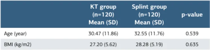
Among the participants, 150 were female and 90 were male. The mean age was 31.10 ± 11.67 years. The demographic data of the patients are shown in Table 1. A homogeneous distribution was observed between the groups in terms of age and BMI (p > 0.05).
When the pain and ROM evaluations were compared between the groups, the patients in the KT group had significantly higher improvement in their pain and ROM compared to the splinting group after treatment (p = 0.002, 0.015, 0.523, 0.022, 0.017) (Table 2).
Table 1. Demographic characteristics of included patients
KT group (n=120) Mean (SD) Splint group (n=120) Mean (SD) p 1 VAS Pre-T 7.52 (1.32) 7.11 (1.45) 0.433 VAS Post-T 2.85 (2.37) 6.00 (1.22) 0.002* p2 <0.001** 0.015* ROM DF Pre-T 10.66 (4.00) 8.55 (3.32) 0.191 ROM DF Post-T 16.09 (6.39) 10.44 (3.39) 0.015* p2 0.002* 0.011* ROM PF Pre-T 30.85 (11.00) 30.77 (12.11) 0.945 ROM PF Post-T 35.95 (8.71) 33.22 (11.67) 0.523 p2 0.062* 0.016*
ROM Inversion Pre-T 12.71 (5.80) 10.77 (3.92) 0.452 ROM Inversion Post-T 20.33 (7.19) 14.00 (3.93) 0.022*
p2 0.002* 0.012*
ROM Eversion Pre-T 9.19 (3.02) 10.00 (3.57) 0.644 ROM Eversion Post-T 15.80 (4.05) 11.00 (4.76) 0.017*
p2 <0.001** 0.236*
*p<0.05, **p<0.001. 1Mann-Whitney U Test, 2Wilcoxon Test. KT: Kinesio taping, VAS: Visual
Analog Scale, ROM: Range of motion. T: Treatment, SD: Standart deviation.
KT group (n=120) Mean (SD) Splint group (n=120) Mean (SD) p-value Age (year) 30.47 (11.86) 32.55 (11.76) 0.539 BMI (kg/m2) 27.20 (5.62) 28.28 (5.19) 0.635 Mann-Whitney U Test. KT: Kinesio taping, BMI: Body mass index, SD: Standart deviation.
Table 2. Comparison of intra and inter groups of pain and
range of motion
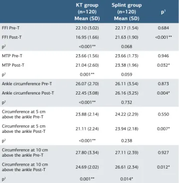
In intra-group evaluations, although pain significantly decreased in both groups and ROM was improved, a higher improvement was observed in the KT group. In addition, the increase in the angle of inversion was higher in the KT group than in the splinting group, and KT application resulted in a greater contribution to the healing process of injuries, leading to increased inversion and decreased eversion (Table 2). The disability levels of the patients treated with KT were significantly improved (p < 0.001) at the end of the treatment, but there was no change in patients treated with splinting (p < 0.068) (Table 3). According to circumference measurements, the measurement of thickness decreased at all measurement levels in the KT group, whereas the splinting group only showed a decrease in the measurement of circumference at 10 cm above the ankle. Oedema in the KT group showed further improvement (p = 0.001, <0.001, <0.001, 0.001; 0.059, 0.732, 0.238, 0.014) (Table 3).
Discussion
Ankle ligament injuries constitute 25% of all musculoskeletal injuries. It is a disease that becomes chronic in later stages and affects the quality of life, leading to an additional cost of healthcare for the affected individual. In recent years, it is argued that functional treatment approaches are the most effective treatment methods [10,11]. In our study, we also found that pain, edema complaints, and disability levels were decreased and ankle range of motion increased when KT was applied, which is one of the functional treatment approaches for patients with acute ankle injuries.
Studies have shown that faster recovery is achieved with early mobilization of patients with acute ankle injuries. Functional treatment modalities comprise healing methods using soft materials such as KT or bandages, which do not cause complications of immobilization and enable patients
to mobilize early. Recent studies have shown that functional treatments such as KT are more beneficial in ankle injuries than rigid treatments such as external ankle support [21,22]. Studies have compared the functional treatment methods and immobilization methods in terms of pain, ROM, edema, patient safety, and complications. Although there is a common view that edema is more effectively reduced by functional treatment methods, no sufficient studies in the literature have reported this view [23]. Previous studies have demonstrated that 3–5 days is the application period for which KT is beneficial and the elastic structure of the tape is maintained during this period. It has been found that the positive effects of KT are lost in applications exceeding 5 days. Based on this information, we also applied KT and splint on our patients for the first 5 days [24,25].
The basic factor in the mechanism of ankle injury is the increase in the supination angle in the subtalar joint together with inversion. Subtalar joint can be taken into pronation with KT, but splinting does not support pronation. For this reason, there is a common view that KT offers more benefits than splinting [12]. In two recent studies, the efficacy of splinting, soft-bracing and taping were compared, and it was reported that splinting provides more stabilization than soft-bracing and taping has the same support as splinting. A study investigating the neuromuscular response in the injured area reported that the EMG response was prolonged and the EMG amplitude in the KT-applied ankle was increased 1.6 times, suggesting positive effects on the neuromuscular response. It was reported that the increased movement speed in the direction of supination in the injured ankle could be reduced by 25%–40% with KT [26]. When we evaluated the range of joint motion in our study, a 60%, 80%, and 70% increase in dorsiflexion, inversion and eversion angles, respectively, was observed in patients in the KT group than in patients in the splinting group.
According to recent studies, KT applications provided positive results in edema drainage when compared to placebo KT, as in the case of manual lymph drainage. KT can reabsorb the interstitial fluid through the lymphatic system [25]. At the same time, KT reduces the pressure on the epidermis, thereby reducing the pressure on the lymphatic cells and accelerating the flow to the lumen of the lymphatic vessels to provide fluid reabsorption. The reduction of pressure in the epidermis in the KT-applied area is due to the effect of microwaves in the tape structure during active motion. As in manual techniques, it can also create a friction effect on the skin. KT reduces edema by improving proprioception due to sensorial input provided by tissue pressure [2]. Aguilar-Ferra’ndiz et al. performed KT in postmenopausal women with lower extremity edema due to chronic venous insufficiency and observed that the amount of extracellular fluid in the lower extremity, pain and disease severity decreased, whereas functionality increased. According to the study of Aguilar-Ferra’ndiz et al., KT promotes edema reabsorption through changes in hydrostatic pressure in the transudate phase of the early stages of edema, when the protein density has not yet increased [24]. In our study, when the circumference measurements of KT-applied patients were compared with those of splinted patients, the circumference decreased by 1.98 cm around the MTP, 3.57 cm around the
KT group (n=120) Mean (SD) Splint group (n=120) Mean (SD) p 1 FFI Pre-T 22.10 (3.02) 22.17 (1.54) 0.684 FFI Post-T 16.95 (1.66) 21.63 (1.90) <0.001** p2 <0.001** 0.068 MTP Pre-T 23.66 (1.56) 23.66 (1.73) 0.946 MTP Post-T 21.04 (2.60) 23.38 (1.96) 0.032* p2 0.001** 0.059
Ankle circumference Pre-T 26.07 (2.70) 26.11 (3.54) 0.873 Ankle circumference Post-T 22.45 (3.08) 26.16 (3.25) 0.004*
p2 <0.001** 0.732
Circumference at 5 cm
above the ankle Pre-T 23.88 (2.14) 24.22 (2.29) 0.550 Circumference at 5 cm
above the ankle Post-T 21.11 (2.24) 23.94 (2.18) 0.007*
p2 <0.001** 0.238
Circumference at 10 cm
above the ankle Pre-T 27.80 (3.34) 27.11 (2.39) 0.927 Circumference at 10 cm
above the ankle Post-T 24.69 (2.02) 26.61 (2.34) 0.012*
p2 0.001** 0.014*
*p<0.05, **p<0.001. 1Mann-Whitney U Test, 2Wilcoxon Test. KT: Kinesiotaping, FFI: Foot function index, MTP: Metatarsophalangeal joint, T: Treatment, SD: Standart deviation.
Table 3. Comparison of intra and inter groups of disability and
ankle, 2.49 cm at 5 cm above the ankle and 2.61 cm at 10 cm above the ankle, and KT application was better than splinting in reducing the edema.
Ankle injuries become chronic and cause disability in 60% of patients [2]. Boyce et al. evaluated pain with VAS and disability with foot ankle outcome score by performing functional treatment on patients with ankle injury, but they did not report any clear conclusion. They attributed this to the small sample size [27]. Bilgic et al. compared two functional treatments, splint treatment (n = 26) and bandaging (n = 25) in patients with ankle injuries and found that elastic bandage application achieved significantly better healing results in ROM and edema evaluations after 1 week, but they found no difference between groups in pain evaluations [28]. When we compared the pain and disability level of patients participating in our study, KT application resulted in 50% and 30% further reductions in pain and disability levels, respectively, in comparison to splinting.
Conclusion
In conclusion, it was found that KT application in patients with acute ankle ligament injuries admitted to emergency services resulted in higher reductions in edema, pain severity, and disability levels compared to splinting and provided a higher increase in ankle ROM. KT is considered to be a treatment modality that contributes to fast recovery and healing, which allows patients to mobilize early; does not interfere with daily activities, such as dressing or taking baths, and increases proprioception and stabilization in the applied area through an increase in sensorial input. We believe that its effectiveness can be further proven by long-term follow-up studies with increased case diversity and a larger sample size.
Scientific Responsibility Statement
The authors declare that they are responsible for the article’s scientific content including study design, data collection, analysis and interpretation, writing, some of the main line, or all of the preparation and scientific review of the contents and approval of the final version of the article.
Animal and human rights statement
All procedures performed in this study were in accordance with the ethical standards of the institutional and/or national research committee and with the 1964 Helsinki declaration and its later amendments or comparable ethical standards. No animal or human studies were carried out by the authors for this article.
Funding: None Conflict of interest
None of the authors received any type of financial support that could be considered potential conflict of interest regarding the manuscript or its submission. References
1. Hootman JM, Dick R, Agel J. Epidemiology of collegiate injuries for 15 sports: summary and recommendations for injury prevention initiatives. J Athlet Train. 2007; 42(2):311–19.
2. Czajka CM, Tran E, Cai AN, DiPreta JA. Ankle sprains and instability. Med Clin North Am. 2014; 98(2):313-29.
3. Waterman BR, Owens BD, Davey S, Zacchilli MA, Belmont Jr PJ. The epidemiology of ankle sprains in the United States. J Bone Jt Surg. 2010; 92(13):2279–84. 4. van den Bekerom MP, van Kimmenade R, Sierevelt IN, Eggink K, Kerkhoffs GM, van Dijk CN, et al. Randomized comparison of tape versus semi-rigid and versus lace-up ankle support in the treatment of acute lateral ankle ligament injury. Knee Surg Sports Traumatol Arthrosc. 2016;24(4):978-84.
5. van RM R, van Os AG, Bernsen RM, Luijsterburg PA, Koes BW, Bierma-Zeinstra SM. What is the clinical course of acute ankle sprains? A systematic literature review. Am J Med. 2008;121(4):324–31. DOI: 10.1016/j.amjmed.2007.11.018. 6. Bleakley CM, O’Connor SR, Tully MA, Rocke LG, Macauley DC, Bradbury I, et al. Effect of accelerated rehabilitation on function after ankle sprain: randomised controlled trial. BMJ. 2010;340:c1964. DOI: 10.1136/bmj.c1964.
7. Kerkhoffs GM, van den Bekerom M, Elders LA, van Beek PA, Hullegie WA, Bloemers GM, et al. Diagnosis, treatment and prevention of ankle sprains: an
evidence-based clinical guideline. Br J Sports Med. 2012;46(12):854-60. 8. Kemler E, van de Port I, Backx F, van Dijk CN. A systematic review on the treatment of acute ankle sprain Braces versus other functional treatment types. Sports Med. 2011; 41(3):185–97.
9. Petersen W, Rembitzki IV, Koppenburg AG, Ellermann A, Liebau C, Brüggemann GP, et al. Treatment of acute ankle ligament injuries: a systematic review. Arch Orthop Trauma Surg. 2013; 133(8):1129–41.
10. Kerkhoffs GM, Rowe BH, Assendelft WJ, Kelly K, Struijs PA, van Dijk CN. Immobilisation and functional treatment for acute lateral ankle ligament injuries in adults. Cochrane Datab Syst Rev. 2002; (3): CD003762.
11. Bleakley CM, McDonough SM, MacAuley DC, Bjordal J. Cryotherapy for acute ankle sprains: a randomised controlled study of two different icing protocols. Br J Sports Med. 2006; 40:700-5.
12. Arnold BL, Docherty CL. Bracing and rehabilitation-what’s new. Clin Sports Med. 2004; 23(1):83-95.
13. Windisch C, Brodt S, Röhner E, Matziolis G. Effects of Kinesio taping compared to arterio-venous Impulse System™ on limb swelling and skin temperature after total knee arthroplasty. Int Orthop. 2017;41(2):301-7.
14. Kase K, Wallis J, Kase T. Clinical Therapeutic Applications of the Kinesio Taping Method, 2nd ed. Tokyo: Kinesio Taping Association; 2003:5-8.
15. Kim JH, Cho MR, Park JH, Shin JC, Cho JH, Park GC, et al. The effects of Kinesiotape on acute lateral ankle sprain: study protocol for a randomized controlled trial. Trials. 2018;19(1):125.
16. Basset KT, Lingman SA, Ellis RF. The use and treatment efficacy of kinaesthetic taping for musculoskeletal conditions: a systematic review. N Z J Physiotherap. 2010; 38(2):56–62.
17. Morris D, Jones D, Ryan H, Ryan CG. The clinical effects of Kinesio® Tex taping: a systematic review. Physiother Theory Pract. 2013; 29(4):259–70. 18. Mostafavifar M, Wertz J, Borchers J. A systematic review of the effectiveness of kinesio taping for musculoskeletal injury. Phys Sportsmed. 2012; 40(4):33–40. 19. Williams S, Whatman C, Hume PA, Sheerin K. Kinesio taping in treatment and prevention of sports injuries: a meta-analysis of the evidence for its effectiveness. Sports Med. 2012; 42(2):153–64.
20. Yalıman A, Şen Eİ, Eskiyurt N, Budiman E. Turkish Translation and Adaptation of Foot Function Index in Patients with Plantar Fasciitis. Turk J Phys Med Rehab. 2014;60:212-22.
21. Lin CW, Hiller CE, de Bie RA. Evidence-based treatment for ankle injuries: a clinical perspective. J Man Manip Ther. 2010;18(1):22-28.
22. Witjes S, Gresnigt F, van den Bekerom MP, Olsman JG, van Dijk NC. The ANKLE TRIAL (ankle treatment after injuries of the ankle ligaments): what is the benefit of external support devices in the functional treatment of acute ankle sprain? A randomised controlled trial. BMC Musculoskelet Disord. 2012;13:21.
23. Günay S, Karaduman A, Oztürk BB. Effects of Aircast brace and elastic bandage on physical performance of athletes after ankle injuries. Acta Orthop Traumatol Turc. 2014;48(1):10-16.
24. Aguilar-Ferra´ndiz ME, Castro-Sa´nchez AM, Matara´n-Pen˜arrocha GA, Guisado-Barrilao R, Garcı´a-Rı´os MC, Moreno-Lorenzo C. A randomized controlled trial of a mixed Kinesio taping-compression technique on venous symptoms, pain, peripheral venous flow, clinical severity and overall health status in postmenopausal women with chronic venous insufficiency. Clin Rehabil. 2014 ;28(1):69–81.
25. Białoszewski D, Woz´niak W, Zarek S. Clinical efficacy of kinesiology taping in reducing edema of the lower limbs in patients treated with the Ilizarov methodpreliminary report. Ortopedia Traumatologia Rehabilitacja. 2009;11(1):46–54.
26. Eils E, Demming C, Kollmeier G, Thorwesten L, Volker K, Rosenbaum D. Comprehensive testing of 10 different ankle braces Evaluation of passwive and rapidly induced stability in subjects with chronic ankle instability. Clin Biomech (Bristol, Avon). 2002;17:526– 35.
27. Boyce SH, Quigley MA, Campbell S. Management of ankle sprains: a randomised controlled trial of the treatment of inversion injuries using an elastic support bandage or an Aircast ankle brace. Br J Sports Med. 2005;39(2):91-96. 28. Bilgic S, Durusu M, Aliyev B, Akpancar S, Ersen O, Yasar SM, et al. Comparison of two main treatment modalities for acute ankle sprain. Pak J Med Sci 2015;31(6): 1496-9.
How to cite this article:
Tomris Duymaz, Serdar Yuksel. Acute treatment of ankle ligament injuries: Is kinesio tape effective? Ann Clin Anal Med 2020;11(Suppl 2): S118-122