Access this article online Quick Response Code:
Website: www.njcponline.com
DOI: 10.4103/1119-3077.181362
How to cite this article: Kurt A, Erkose-Genc G, Uzun M, Emrence Z, Ustek D, Isik-Ozkol G. The antifungal activity and cytotoxicity of silver containing denture base material. Niger J Clin Pract 2017;20:290-5.
This is an open access article distributed under the terms of the Creative Commons Attribution-Non Commercial-Share Alike 3.0 License, which allows others to remix, tweak, and build upon the work non-commercially, as long as the author is credited and the new creations are licensed under the identical terms.
For reprints contact: [email protected] Acceptance Date: 30-10-2015
Objective: Denture base materials are susceptible to fungal adhesion, which is an important etiological issue in the pathogenesis of denture stomatitis. The purpose of this in vitro study was to evaluate the antifungal activity and cytotoxicity of denture base material containing silver microparticles. Materials
and Methods: The polymethyl methacrylate (PMMA) denture base material was
used, and silver microparticles were added to the polymer powder in different concentrations by volume (0%, 0.25%, 0.5%, and 1%). Their antifungal activity against Candida albicans was assessed in terms of colony-forming units. PMMA disc specimens containing silver microparticles were eluted with culture medium for 1, 2, and 5 days. The cytotoxicity of the eluates to cultured L929 mouse fibroblast cells was evaluated using a real-time cell analysis (RTCA) system and the 3-[4,5-dimethylthiazol-2-yl]-2,5 diphenyl tetrazolium bromide (MTT) assay. Results: The antifungal effect against C. albicans increased with the percentage of silver microparticles (P < 0.05). For both tests, both RTCA and the MTT assay, no time- or silver-dependent cytotoxicity of PMMA denture base material containing silver microparticles was observed. Conclusions: PMMA denture base material containing silver microparticles have antifungal activity and no cytotoxic effect.
The Antifungal Activity and Cytotoxicity of Silver Containing Denture
Base Material
Address for correspondence: Dr. A Kurt, Department of Prosthodontics, Faculty of Dentistry, Bezmialem Vakıf University, Adnan Menderes Bulvarı Vatan Caddesi Fatih, Istanbul, Turkey. E-mail: [email protected] dental materials are due to their release of small amounts of various incorporated substances into the physiological environment.[13] Their antifungal effects may also be related to a wide spectrum of cytotoxic effects; however, any potential cytotoxic effect must be minimized in dental materials.[14] Silver ions are biologically active,[15,16] so silver may have adverse effects on human cells.[17] Although the literature reports various studies related to silver nanoparticles with antifungal applications in PMMA denture base materials,[10-13] silver nanoparticles have higher toxicity than silver microparticles.[16,18,19] In addition, the incorporation of silver micro-particles in PMMA denture base material may be easier, and producing silver microparticles may be more efficient
I
ntroductIonPolymethyl methacrylate (PMMA) heat-polymerized resin has continued to be the most frequently used material for denture bases since its introduction in 1937.[1] However, this material is susceptible to fungal adhesion,[2] which is an important etiological issue in the pathogenesis of denture stomatitis.[3] Although Candida species other than Candida albicans (C. albicans) have also been isolated from denture stomatitis lesions, C.
albicans is their primary pathogen.[4] Because the first
step in the development of infection is fungal adherence to PMMA denture base materials,[5] preventing C.
albicans adherence may help to treat denture stomatitis.
Silver particles may have strong antifungal effects in various biomedical applications.[6-8] Production of silver nanoparticles involves a series of chemical processes that are different than those for silver microparticles,[9] and various studies have examined the antifungal effects of PMMA denture base materials containing silver nanoparticles.[10-13] The antifungal properties of these
A Kurt, G Erkose-Genc1, M Uzun1, Z Emrence2, D Ustek3, G Isik-Ozkol4
A
bstr
A
ct
Keywords: Candida albicans, cytotoxicity, denture base, silver microparticles,
Department of
Prosthodontics, Faculty of Dentistry, Bezmialem Vakif University, 1Department
of Medical Microbiology, Faculty of Medicine, Istanbul University, 2Department
of Genetics, Institute for Experimental Medicine, Istanbul University,
3Department of Medical
Genetics and REMER, School of Medicine, Medipol University, 4Department of
Prosthodontics, Faculty of Dentistry, Istanbul University, Istanbul, Turkey
than producing silver nanoparticles in daily clinical practice. Therefore, new studies are necessary to evaluate PMMA denture base material containing silver micro-particles under in vitro conditions.
According to The International Organization for Standardization (ISO), dental materials must be evaluated for biocompatibility before being applied to patients.[14] The ISO recommends that this biocompatibility first be assessed by in vitro cytotoxicity assays using isolated cells and has recommended several techniques (e.g., 3-[4,5-dimethylthiazol-2-yl]-2,5 diphenyl tetrazolium bromide [MTT] and 2, 3-bis-(2-methoxy-4-nitro-5-sulfophenyl)-2 H-tetrazolium-5-carboxanilide assays and agar and filter diffusion tests).[20] These techniques provided information about the time points to be investigated but ignored the kinetic effects. For this reason, to better assess the cytotoxicity of materials, these techniques can support by the real-time cell analysis (RTCA). RTCA enables constant and noninvasive monitoring of the proliferation of living cells because the micro-sensor perceives changes in the viable cell numbers in the wells.[21] Using the different cytotoxicity tests, that measure the viability of cells may have more reliable results.
The purpose of this in vitro study was to evaluate the antifungal activity and cytotoxicity of a denture base material containing silver micro-particles with different percentages. The first research hypothesis was that PMMA denture base material containing different percentages of silver microparticles would not show antifungal activity against C. albicans. The second research hypothesis was that silver microparticles do not have high cytotoxicity in combination with PMMA denture base material when assessed using two different cytotoxicity assays.
M
AterIAls AndM
ethodsTable 1 list the heat-polymerized PMMA denture base material (Meliodent, Heraeus Kulzer GmbH and Co., Hanau, Germany) and silver microparticles (Ferro Corporation Cleveland, Ohio, USA) selected for this experiment. C. albicans ATCC 2091 was purchased as a stock culture (KUKENS study group, Department of Microbiology, University of Istanbul, Turkey) for antifungal activity assay. The L929 mouse fibroblast cell line (NCTC clone 929 [L-cell, L929, derivative of strain L; ATCC CCL-1]) was used to determine the cytotoxic effects of the samples.
Sample preparation
Four groups of samples were prepared with different percentages of silver microparticles by volume: 0% (control), 0.25%, 0.5%, and 1%. The silver microparticles were incorporated into the polymer powder of the
PMMA denture base material; the powder had a consistent liquid volume ratio of 3:1. When the mixed material reached the dough stage, it was put into molds prepared using Teflon discs to create samples. Ten disc-shaped samples (10 mm in diameter and 2 mm thick) per group were fabricated for the antifungal activity assay, and 36 disc-shaped samples (10 mm in diameter and 1 mm thick) per group were fabricated for the cytotoxicity assays from PMMA denture base material. All discs were polymerized in water for 7 h at 70°C, stored for 3 h at 100°C in an automated polymerization unit (Kavo EWL 5501; Kavo Electrotechnisches Werk GmbH, Leutkirch, Germany), and left at 37°C in distilled water for 24 h.[22] Prior to the assay, the discs were cleansed ultrasonically in distilled water for 20 min and exposed to ultraviolet light for another 20 min to kill any microorganisms that may have caused contamination during fabrication.[23] Antifungal activity assay
The C. albicans strain was placed on Sabouraud Dextrose Agar (SDA) (Becton Dickinson, Sparks, MD, USA) and incubated at 37°C for 48 h. After this period, a suspension containing 106 of C. albicans/mL was prepared in sterile saline solution with the aid of a spectrophotometer (Shimadzu Corp., Kyoto, Japan) to achieve a wavelength of 530 nm and optical density of 0.284. Each disc specimen was placed in a sterile tube containing 2 mL of fungal suspension and incubated at 37°C for 90 min. Then, each disc specimen was transferred to tubes containing 10 mL of sterile saline solution, and the adhered cells were dispersed by vortexing. Dilutions of this solution were prepared, and aliquots of 0.1 mL were plated on SDA. After incubation, the numbers of colony-forming units of the microorganism were determined; the reduction in viable, adherent cells was calculated by comparison with control specimens.[24]
Cytotoxicity assays
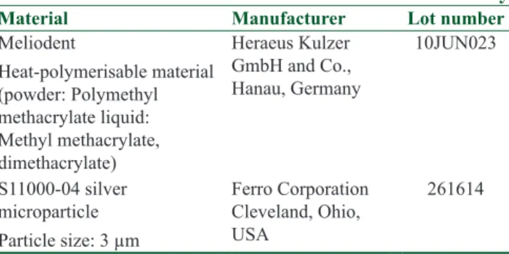
Complete cell culture medium without serum was used for elution. Eluates of the samples were prepared by placing three PMMA denture base material discs into their own sterile tubes with 9 mL of elution medium.[25] Table 1: Denture base material and silver used in this study
Material Manufacturer Lot number
Meliodent Heat-polymerisable material (powder: Polymethyl methacrylate liquid: Methyl methacrylate, dimethacrylate) Heraeus Kulzer GmbH and Co., Hanau, Germany 10JUN023 S11000-04 silver microparticle Particle size: 3 µm Ferro Corporation Cleveland, Ohio, USA 261614
expressed as a percentage of the untreated control cells. Eluates were analyzed in quadruplicate.
Statistics
Data were analyzed using GraphPad Prism 5.0 (GraphPad Software, La Jolla, CA, USA) and are presented as means ± standard deviations (SDs). Statistically significant differences were assessed using the Kruskal-Wallis test, followed by the Mann-Whitney U-test. Pooled data were subjected to analyses of variance with post hoc Tukey’s tests. Differences were considered statistically significant at P < 0.05.
r
esultsAntifungal activity assay
Table 2 provides the mean values and SDs for the antifungal activity against C. albicans of denture base material containing silver microparticles. The addition of silver microparticles to the denture base material significantly reduced the adherence of C. albicans to the surface (P < 0.05).
Cytotoxicity assays Real-time cell analysis assay
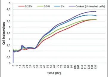
The CI values of 1-day eluates with the RTCA assay were measured over 138 h [Figure 1]. Table 3 provides the mean values and SDs of the CI values and the viability (%) at different time points. At the acute and They were then incubated at 37°C for 1, 2, and 5 days in
a humidified atmosphere of 5% CO2 and 95% air. Elution times were identified to extend intervals used in previous reports[23,26-28] to identify any trends in cytotoxicity over time. The eluates were sterilized by filtration through a 0.22 µm filter (Millex-GP; EMD Millipore Corp., Billerica, MA, USA), and samples were transferred to new tubes containing fresh elution medium after each elution period. Elution medium with no specimen was also incubated as a negative control. Next, 10% fetal bovine serum (Biochrom, Berlin, Germany) was added to the eluates, and they were stored at -20°C until the cytotoxicity assay.
Two in vitro cytotoxicity assay techniques were used: RTCA xCELLigence System and the MTT Cell Proliferation Kit (Roche Diagnostics GmbH, Mannheim, Germany).
Real-time cell analysis assay
The RTCA assay was used to evaluate cell viability according to the manufacturer’s protocol. After seeding 150 µL cell suspensions into the wells (3 × 10⁴ cells/ well) of the E-plate 96, cells were treated with the 1-day eluates in 50 µL volumes at 24 h and monitored every 10 min for 138 h. Untreated cells were used as controls, and eluates were analyzed in quadruplicate. To quantify the number of viable cells, the data were expressed in a cell index (CI) and exported to Excel software (Microsoft Corporation, Seattle, WA, USA). Cell viability at specific time points was expressed as a percentage of the untreated control cells. Statistical analyses of the data were performed at 24, 56, and 138 h to observe immediate, acute, and chronic cell responses, respectively.
3-[4,5-dimethylthiazol-2-yl]-2,5 diphenyl tetrazolium bromide assay
The MTT test was performed to indicate the acute cell response in 1-, 2-, and 5-day eluates. The MTT Cell Proliferation Kit (Roche Diagnostics GmbH, Mannheim, Germany) contained a labeling reagent and a solubilization solution. First, 3 × 10⁴ cells/100 µL medium were plated per well in 96-well plates and incubated at 37°C for 24 h in a humidified atmosphere of 5% CO2 and 95% air. All eluates were added in 50 µL volumes, and cells were treated for 28 h. After treatment, 10 µL of MTT labeling reagent was added to each well and incubated for another 4 h. Then, 100 µL of the solubilization solution was added to each well and incubated overnight. After incubation, colorimetric absorbance was measured at 580 nm (reference wavelength at 650 nm) using a microtiter plate reader (Universal Microplate Reader ELX 800; Bio-Tek Instruments Inc., Winooski, VT, USA) in accordance with the manufacturer’s protocol. Cell viability was
Table 2: Mean (±SD) numbers of colony forming units of Candida albicans
Material Percentage of silver
0 (control) 0.25 0.5 1
Meliodent 11.11 (±10.93) 3.20 (±2.29)*2.50 (±1.89)*1.70 (±1.02)*
*P<0.05 versus 0% control group. SD=Standard deviation
Figure 1: Cell index value of 1-day elutes was measured throughout 138
wearers.[3] While denture hygiene, trauma, systemic diseases, and immune system deficiency also play roles in denture stomatitis, the invasion of Candida species is associated with its progression;[2] in fact, the adherence of
C. albicans to PMMA denture base materials is the first
step in the development of infection.[5] Significantly, the current study showed that PMMA denture base material containing silver microparticles showed antifungal activity by reducing the adhesion of C. albicans. In addition, the results demonstrated a pattern of antifungal activity that increased with the percentage of silver. The findings are consistent with those of other studies that used silver to prevent C. albicans adhesion to treat denture stomatitis.[10-13]
According to ISO standards, a reduction of cell viability by more than 30% is considered to indicate cytotoxicity.[20] By this standard, and corroborated by two separate and unrelated cytotoxicity assays, the PMMA denture base material containing different percentages of silver microparticles were not cytotoxic. The MTT test illustrated the acute cell response in 1-, 2-, and 5-day eluates. In RTCA, the CI values of 1-day eluates were measured throughout 138 h with the RTCA assay [Figure 1] and expressed as the value of cell viability at immediate, acute, and chronic cell responses [Table 3]. Despite the same cell numbers, the MTT assay showed that all silver groups increased cell viability, but RTCA showed that the 0.25% group significantly decreased cell viability when compared to control groups for acute cell response and 1-day eluates. This could be due to the fact that the MTT assay only detects cell viability through mitochondrial metabolism while the RTCA results are affected by quality of cell-cell adhesions, cell membrane integrity, and cell adhesion to the well.[29] In other words, the RTCA system may have observed reduced cell viability while the MTT assay did not because the RTCA system is more sensitive.
In this study, the cell viability values did not change among 1-, 2-, and 5-day eluates (P > 0.05) [Table 4]. It chronic cell responses, cell viability was significantly
reduced in the denture base material with 0.25% silver microparticles (P < 0.05).
3-[4,5-dimethylthiazol-2-yl]-2,5 diphenyl tetrazolium bromide assay
Table 4 provides the mean values and SDs for cell viability of 1-, 2-, and 5-day eluates. Significant differences were observed between the 0% (control) and all silver groups in the 1-day eluates. For all elution periods, there were no statistically significant differences in cell viability values (P > 0.05).
d
IscussIonThe current study evaluated the antifungal activity and cytotoxicity of denture base material containing silver microparticles. The results obtained did not support the first research hypothesis that PMMA denture base material with silver microparticles would not show antifungal activity against C. albicans, but the results did support the second hypothesis that silver microparticles do not have high cytotoxicity in combination with PMMA denture base material. Thus, denture base material containing silver microparticles was not cytotoxic after elution into cell culture medium using RTCA and the MTT assay.
Denture stomatitis, which is defined as an inflammatory process of the mucosa underlying PMMA denture base materials, has a 10–75% prevalence in
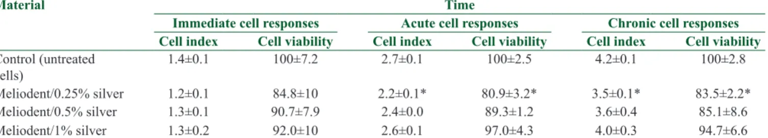
prosthesis-Table 3: Mean (±SD) cell index values and cell viability (%) of groups at immediate, acute and chronic cell response (real-time cell analysis) for 1-day eluates
Material Time
Immediate cell responses Acute cell responses Chronic cell responses
Cell index Cell viability Cell index Cell viability Cell index Cell viability
Control (untreated cells) 1.4±0.1 100±7.2 2.7±0.1 100±2.5 4.2±0.1 100±2.8 Meliodent/0.25% silver 1.2±0.1 84.8±10 2.2±0.1* 80.9±3.2* 3.5±0.1* 83.5±2.2* Meliodent/0.5% silver 1.3±0.1 90.7±7.9 2.4±0.0 89.3±1.2 3.6±0.4 85.1±8.6 Meliodent/1% silver 1.3±0.2 92.0±10 2.6±0.1 97.0±4.3 4.0±0.3 94.7±6.6
*P<0.05 versus control group at same time point. SD=Standard deviation
Table 4: Mean (±SD) of cell viability (%) of groups at acute cell response (MTT assay)
Elution period
Material Percentage of silver
0 (control) 0.25 0.5 1
Day 1 Meliodent 87.7±5.6 105.8±14.2*102.3±2.8*103.1±5.2*
Day 2 Meliodent 89.1±4.5 93.2±11.6 94.9±5.8 101.3±6.4 Day 5 Meliodent 103.8±6.6 90.7±7.2 95.0±5.2 105.0±8.0 *P<0.05 versus 0% control group at same day. SD=Standard deviation, MTT=3-[4,5-dimethylthiazol-2-yl]-2,5 diphenyl tetrazolium bromide
enhanced antifungal activity. Thus, according to the results of this study, as assessed by RTCA and MTT assays, silver microparticles have no cytotoxicity in combination with PMMA denture base material.
Acknowledgments
This study was supported by the Research Support Unit of Istanbul University (project no. 19648). The authors thank Association Prof. Ugur Aksu for performing statistical analyses of the results.
Financial support and sponsorship Nil.
Conflicts of interest
There are no conflicts of interest.
r
eferences1. Craig RG, Powers JM. Restorative Dental Materials. 11th ed. St. Louis (MO): Mosby; 2002
2. Pereira-Cenci T, Cury AA, Cenci MS, Rodrigues-Garcia RC. In
vitro Candida colonization on acrylic resins and denture liners:
Influence of surface free energy, roughness, saliva, and adhering bacteria. Int J Prosthodont 2007;20:308-10.
3. Budtz-Jörgensen E. The significance of Candida albicans in denture stomatitis. Scand J Dent Res 1974;82:151-90.
4. Figueiral MH, Azul A, Pinto E, Fonseca PA, Branco FM, Scully C. Denture-related stomatitis: Identification of aetiological and predisposing factors – A large cohort. J Oral Rehabil 2007;34:448-55.
5. Samaranayake LP, Nair RG. Oral Candidainfections – A review. Indian J Dent Res 1995;6:69-82.
6. Wright JB, Lam K, Hansen D. Burrell RE, Efficacy of topical silver against fungal burn wound pathogens. Am J Infect Control 1999;27:344-50.
7. Samuel U, Guggenbichler JP. Prevention of catheter-related infections: The potential of a new nano-silver impregnated catheter. Int J Antimicrob Agents 2004;23:Suppl 1S75-8.
8. Jain J, Arora S, Rajwade JM, Omray P, Khandelwal S, Paknikar KM. Silver nanoparticles in therapeutics: Development of an antimicrobial gel formulation for topical use. Mol Pharm 2009;6:1388-401.
9. Tolaymat TM, El Badawy AM, Genaidy A, Scheckel KG, Luxton TP, Suidan M. An evidence-based environmental perspective of manufactured silver nanoparticle in syntheses and applications: A systematic review and critical appraisal of peer-reviewed scientific papers. Sci Total Environ 2010;408:999-1006.
10. Monteiro DR, Takamiya AS, Feresin LP, Gorup LF, de Camargo ER, Delbem AC, et al. Susceptibility of Candida albicans and
Candida glabrata biofilms to silver nanoparticles in intermediate
and mature development phases. J Prosthodont Res 2015;59:42-8. 11. Li Z, Sun J, Lan J, Qi Q. Effect of a denture base acrylic resin
containing silver nanoparticles on Candida albicans adhesion and biofilm formation. Gerodontology 2014;[doi: 10.1111/ger.12142] 12. Suganya S, Ahila SC, Kumar BM, Kumar MV. Evaluation and
comparison of anti – Candida effect of heat cure poly methyl methacrylate resin enforced with silver nanoparticles and conventional heat cure resins: An in vitro study. Indian J Dent Res 2014;25:204-7.
13. Acosta-Torres LS, Mendieta I, Nuñez-Anita RE, Cajero-Juárez M, Castaño VM. Cytocompatible antifungal acrylic resin
has been suggested that toxic substances released into the medium within the 1st day are broken down over time or form complexes with other chemicals in the medium, which may alter their cytotoxic potentials.[23] Previous reports on PMMA denture base materials demonstrated a pattern of increasing cell viability with longer elution periods.[23,26-28] However, Cimpan et al.[30] found reduced cell viability in 2-day eluates compared with 1-day eluates of PMMA denture base materials. The differences in result might be due to differences in the experimental designs, such as elution conditions or cell viability assay. Baker et al.[31] investigated the levels of toxic substances in the saliva of patients with dentures and found that PMMA denture base material released toxic substances over a 1-week period after insertion. Given the pattern of elution of potential cytotoxic agents, tissue irritation that occurs soon after denture insertion may be material-specific. In order to prevent the occurrence of tissue irritation, it is recommended to be soaked in water before being delivered the PMMA denture base materials to the patient.[27] Although no groups revealed a difference in cell viability values between elution periods in this study, it may be beneficial to soak PMMA denture base material containing silver microparticles before insertion. The current study had a number of limitations. First, it analyzed the short-term effects on cytotoxicity and antifungal activity in vitro. The long-term effects of PMMA denture base material containing silver microparticles could be different, and the effects may also vary between in vitro and in vivo. Second, it is advised to evaluate the cell morphology besides viable cell number to find out more about the mechanism of cytotoxicity. However, the current study evaluated only the number of viable cells because the aim of the study was only to determine if the groups had any cytotoxicity or not. Third, the effects of microparticles on PMMA denture base material on the materials’ characterization and mechanical properties were not studied. Fourth, the color change was not measured; however, a color change that may affect clinical use was not observed during the experiments because of the small amount of silver microparticles that were added. Therefore, future studies should focus on the characterization and applications of long-term antifungal PMMA denture base material containing silver microparticles both in
vitro and in vivo.
c
onclusIonIn conclusion, PMMA denture base material containing different percentages of silver microparticles showed significant antifungal activity against C. albicans, and increasing the percentage of silver microparticles
on removing five microbial species and effects on the topographic characteristics of acrylic resin. J Prosthodont 2008;17:627-33.
25. Jorge JH, Giampaolo ET, Vergani CE, Machado AL, Pavarina AC, Carlos IZ. Effect of post-polymerization heat treatments on the cytotoxicity of two denture base acrylic resins. J Appl Oral Sci 2006;14:203-7.
26. Lefebvre CA, Knoernschild KL, Schuster GS. Cytotoxicity of eluates from light-polymerized denture base resins. J Prosthet Dent 1994;72:644-50.
27. Huang FM, Tai KW, Hu CC, Chang YC. Cytotoxic effects of denture base materials on a permanent human oral epithelial cell line and on primary human oral fibroblasts in vitro. Int J Prosthodont 2001;14:439-43.
28. Ata SO, Yavuzyilmaz H. In vitro comparison of the cytotoxicity of acetal resin, heat-polymerized resin, and auto-polymerized resin as denture base materials. J Biomed Mater Res B Appl Biomater 2009;91:905-9.
29. Xing JZ, Zhu L, Gabos S, Xie L. Microelectronic cell sensor assay for detection of cytotoxicity and prediction of acute toxicity. Toxicol In Vitro 2006;20:995-1004.
30. Cimpan MR, Cressey LI, Skaug N, Halstensen A, Lie SA, Gjertsen BT, et al. Patterns of cell death induced by eluates from denture base acrylic resins in U-937 human monoblastoid cells. Eur J Oral Sci 2000;108:59-69.
31. Baker S, Brooks SC, Walker DM, The release of residual monomeric methyl methacrylate from acrylic appliances in the human mouth: An assay for monomer in saliva. J Dent Res 1988;67:1295-9.
containing silver nanoparticles for dentures. Int J Nanomedicine 2012;7:4777-86.
14. Technical Specification ISO 7405:2008 Dentistry – Evaluation of Biocompatibility of Medical Devices Used in Dentistry. Geneva 2008. 15. Lansdown AB. Silver 2: Toxicity in mammals and how its
products aid wound repair. J Wound Care 2002;11:173-7. 16. Wei L, Tang J, Zhang Z, Chen Y, Zhou G, Xi T. Investigation
of the cytotoxicity mechanism of silver nanoparticles in vitro. Biomed Mater 2010;5:1-6.
17. AshaRani PV, Low Kah Mun G, Hande MP, Valiyaveettil S. Cytotoxicity and genotoxicity of silver nanoparticles in human cells. ACS Nano 2009;3:279-90.
18. Xiong L, Jiang X, Chen L, Tang J, Xi T. Comparison of in
vitro cytotoxicity of silver particles with different sizes. Chin J
Biomed Eng 2007;26:600-4.
19. Chen D, Xi T, Bai J. Biological effects induced by nanosilver particles: In vivo study. Biomed Mater 2007;2:S126-8.
20. Technical Specification ISO 10993-5: 2009 Biological Evaluation of Medical Devices-Part-5: Test for In Vitro Cytotoxicity. British Standard Institute 2009;
21. Kustermann S, Boess F, Buness A, Schmitz M, Watzele M, Weiser T, et al. A label-free, impedance-based real time assay to identify drug-induced toxicities and differentiate cytostatic from cytotoxic effects. Toxicol In Vitro 2013;27:1589-95.
22. Harrison A, Huggett R. Effect of the curing cycle on residual monomer levels of acrylic resin denture base polymers. J Dent 1992;20:370-4.
23. Sheridan PJ, Koka S, Ewoldsen NO, Lefebvre CA, Lavin MT. Cytotoxicity of denture base resins. Int J Prosthodont 1997;10:73-7.
24. da Silva FC, Kimpara ET, Mancini MN, Balducci I, Jorge AO, Koga-Ito CY. Effectiveness of six different disinfectants