The European Research Journal
http://www.eurj.org
Case
Report
e-ISSN: 2149-3189 DOI: 10.18621/eurj.2016.2.1.62
A viable childbirth after correction of spontaneous
uterine dehiscence
Tayfur Cift1 , Burcu Aydin2 , Pelin Ocal3 , Berk Bulut4 , Sennur Ilvan51 Department of Gynecology and Obstetrics, Bursa Yuksek Ihtisas Training and Research Hospital, Bursa, Turkey 2 Department of Gynecology and Obstetrics, Medipol University Esenler Hospital, Istanbul, Turkey
3 Department of Gynecology and Obstetrics, Istanbul University Cerrahpasa Medical School, Istanbul, Turkey 4 Department of Gynecology and Obstetrics, Okmeydani Trainig and Research Hospital, Istanbul, Turkey 5 Department of Pathology, Istanbul University Cerrahpasa Medical School, Istanbul, Turkey
ABSTRACT
We report a case of uterine dehiscence during pregnancy. Uterine dehiscence and rupture are serious complications of pregnancy. This situation takes place especially in women that prior uterine operation(s). We represent a 30-year-old woman diagnosed uterine dehiscence at 22ndgestation week. Uterine dehiscence treated
surgically and then medical treatment was given to prevent preterm labour. At 34thgestation week, the patient
was operated because of preterm labour and an alive foetus was born. The result of the pathologic examination of the placenta: Placental adhesion failure was detected. In conclusion women with prior caesarean delivery (one or more), ultrasound measurement should be recommended for measuring the lower uterine segment thickness in order to predict the possibility of uterine dehiscence and rupture.
Eur Res J 2016;2(1):62-65
Keywords: Alive foetus; dehiscence; primary repair
Introduction
Nowadays uterine complication can be seen rather frequently because of increasing rate of caesarean section (C/S) operation. Uterine dehiscence and rupture are serious complications which could be considered in a patient who had prior caesarean operation(s) during pregnancy. While uterine dehiscence is defined as a situation in which myometrial layer is separated but amniotic sac is not ruptured, and foetus does not pass into abdominal cavity. Uterine rupture is defined as a condition in which uterine cavity combines with peritoneal cavity by the opening of the whole uterine wall including
serosa and integrity of the amniotic sac fails [1]. The overall incidence of uterine rupture and dehiscence has decreased significantly with the modern practice of obstetrics, but, in contrast, increasing the risk of pathologies like implantation anomalies and scar dehiscence are related to prior caesarean section scar. The incidence of uterine rupture is 0.07% in a developed country [2].
Severe abdominal bleeding could be seen mostly in uterine rupture. Also, it could be seen uterine dehiscence [3]. Regarding foetal heart rate monitoring with cardiotocography (CTG) is not a good predictor
Address for correspondence:
Tayfur Cift, MD, Department of Gynecology and Obstetrics, Bursa Yuksek Ihtisas Training and Research Hospital, Bursa, Turkey Email: [email protected]
Received: 23.05.2015; Accepted: 27.10.2015; Published Online: 04.03.2016
Copyright © 2016 by The Association of Health Research & Strategy
of adverse outcomes, however; late and variable decelerations could result in long-term foetal bradycardia and suddenly loss of foetal heart rate. In some studies, uterine rupture cases constitute a significant portion of maternal and foetal deaths, especially in developing countries. In various studies, foetal mortality rates were reported as a range from 50% to 75 % [4].
We reported a rare case of uterine dehiscence and its management which was diagnosed and operated on 22ndgestation weeks.
Case Presentation
A 30-year-old woman who had prior caesarean section surgery was referred to a hospital with a lower side abdominal pain at 22nd gestational week.
According to the physical examination, she had nausea and vomiting which was compatible with acute abdomen. There was no vaginal bleeding. The patient was hospitalized immediately. During the clinical observation, tachycardia and hypothermia rapidly progressed. According to ultrasonographic examination, foetal heart beat existed, amniotic fluid volume was sufficient, the placenta was observed as located anterior wall of the uterus, and there was no retroplacental pathology. But there was a complex structure with the diameter of 84x68 millimetres comprising a hypo-hyper echogenic area at the right adnexial location. The patient was operated with the diagnosis of acute abdomen; firstly diagnostic laparoscopy was applied to the patient then converted to laparotomy with median incision because of large coagulum in the abdominal cavity. Approximately 5-6 centimetres dehiscence covering with coagulum was found on the front face of the uterus which matched to prior caesarean scar area. When the coagulum was cleaned, maternal side of placenta was seen easily. Defective uterine scar was primarily repaired by suturing with polyglactin. After the successful operation, intravenous tocolytic treatment (ritodrine) was applied with until 13th postoperative day then tocolytic treatment used with magnesium citrate. The patient was followed up in the hospital until fetal birth. A fetus, whose APGAR score 8 was born by C/S because of preterm labor at 34th gestation week.
Placenta, which was given purple halo seen on the isthmus of the uterus and front of the lower segment of corpus during operation. This 5-6 cm area on the
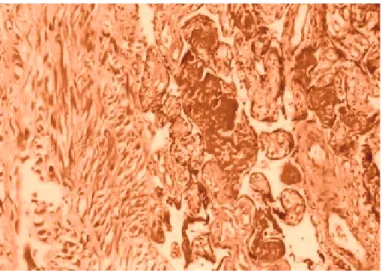
front wall of the uterus excised and primarily repaired. As a result, of pathological examination of the placenta, there was an adhesion abnormality (Figure 1). The patient was discharged from hospital without any complication.
Discussion
Uterine scar dehiscence and rupture are serious complications of pregnancy. Spontaneous uterine ruptures also observed during labor. Risk factors for uterine rupture are given in Table 1 [5]. The settlement of the placenta on the cesarean scar and the presence of placental adhesion abnormalities can be defined as the main risk factors for the formation of spontaneous rupture especially in patients with prior CS. The majority of cases of spontaneous rupture occurs in the 3rd trimester of pregnancy. Obstetrical clinicians are very sensitive to uterine rupture and dehiscence especially in pregnant women who have a history of a prior caesarean section at weeks close to last period of pregnancy. But the most important point to keep in mind is uterine rupture and dehiscence also can be seen in the early weeks of pregnancy. Cases of uterine rupture in 2ndtrimester are very rare. The main reason
is medical evacuations in the 2nd trimester due to medical reasons or fatal anomalies of the foetus. It is known that placental adhesion abnormalities such as percreta etc. constitute the majority of cases of spontaneous rupture, and mostly they are observed at 23-28 weeks of pregnancy. In the study of Chapman and colleagues in which 606 cases of medical
Eur Res J 2016;2(1):62-65 A viable childbirth & uterine dehiscence
Figure 1.Pathology of placenta. Villi adhere directly to the my-ometrium without intervening decidua (HEx200)
evacuation were examined, uterine rupture was detected between 22-24 weeks in 4 women; prior caesarean delivery was found in three of them [6]. Cases of spontaneous uterine rupture occur more frequently in adhesion abnormality such as placenta percreata. The majority of these cases occur between 23-28 weeks of gestation. Spontaneous uterine ruptures can be seen even in primigravid women beside the more common case of the previous caesarean section. There are 3 cases found in the literature [7, 8]. Radiological imaging techniques could be insufficient in diagnosing such cases so that clinical diagnosis is also crucial to determine the cases of uterine rupture and dehiscence.
In this case, during the operation, uterine dehiscence diagnosed, amniotic sac was intact, and the foetus was alive. It is critical to diagnose this situation as early as possible. Unfortunately, most of such cases are diagnosed only after the complete uterine rupture with ripped amniotic sac and foetal expulsion partially or completely placed into the abdominal cavity. Also receiving the patient to the surgery within a few hours after the onset of symptoms is a vital prognostic factor for ongoing pregnancy like in our case.
Consequently; the possibility of uterine rupture and dehiscence should be considered during each period of pregnancy. It is important especially in pregnancies with a history of prior uterine surgery, C/S, and placental adhesion abnormalities.
In women with previous caesarean delivery(ies), ultrasound measurement has been used to study the lower uterine segment thickness and predict uterine dehiscence and rupture. Gotoh et al. [9] found no difference in lower uterine segment thickness at 19 weeks between women with or without a prior caesarean delivery, but lower uterine segment
thickness was significantly lower at 27 weeks in women with a previous caesarean delivery. Another study by Rozenberg et al. [10] also concluded that the risk of rupture was related to the thickness of the lower uterine segment at 37 weeks. Using a cut-off 3.5 millimetres (mm), they found that a thin lower uterine segment had a sensitivity of 88%, specificity of 73%, positive predictive value of 11.8%, and negative predictive value of 99.3% for prediction of uterine rupture Therefore, ultrasound measurement of the lower uterine segment could be considered the standard diagnostic procedure that may assess the risk of uterine rupture and dehiscence.
On the other hand, if such cases could be diagnosed earlier and primary uterine repair could be performed on time with primary surgery can be a solution to the problem. Cases should be followed up more frequently so that it is possible to get a healthy baby as shown in our case with surgical repair.
Conclusions
Women with prior caesarean delivery (one or more), ultrasound measurement could be recommended for measuring the lower uterine segment thickness so that it may be helpful to predict the possible uterine dehiscence and rupture.
Informed Consent
Written informed consent was obtained from the patient for the publication of this case report.
Competing interests
The authors declare that they have no competing
Eur Res J 2016;2(1):62-65 Aydin et al
Table 1. The risk factors for uterine rupture
Age between 31-35 Grand multiparity Birth trauma Neglected birth Malpresentation
External or internal podalic version Breech presentation
Manual cervical dilation
Use of oxytocin or prostaglandins Placental adhesion abnormalities
Previous uterine surgery (myomectomy, cesarean section, cornual resection, hysteroscopic procedures, laparoscopic trochar injuries, abdominal penetrating injury)
interests with respect to the authorship and/or publication of this article.
References
[1] Depp R. Ceserean delivery. In: Gabbe SG, Niebyl JR,Simpson JL, eds. New York: Churchill Livingstone, 2002:580.
[2] Turner MJ. Uterine rupture. Best Pract Res Clin Obstet Gynaecol. 2002 Feb;16(1):69-79.
[3] Lichtenberg ES, Frederiksen MC. Cesarean scar dehiscence as a cause of hemorrhage after second-trimester abortion by dilation and evacuation, Contraception. 2004 Jul;70(1):61-4. [4] Williams: Obstetric hemorrage. In: Williams(ed): Obstetrics, 20.ed. Stamford.CT: Appleton&Longe, 1977:77.
[5] Dhaifalah I, Santavy J, Fingerova H. Uterine rupture during pregnancy and delivery among women attending The Al-Tthawra Hospital In Sana’a in SANA’A City Yemen Republic. Biomed Pap Med Fac Univ Palacky Olomouc Czech Repub. 2006
Nov;150(2):279-83.
[6] Chapman S, Crispens M, Owens J, Savage K. Complications of midtrimester pregnancy termination: the effect of prior ceserean delivery. Am J Obstet Gynecol. 1996 Oct;175 (4 Pt 1):889-92.
[7] Nagy PS. Placenta percreta induced uterine rupture and resulted in intraabdominal abortion. Am J Obstet Gynecol. 1989 Nov;161(5):1185-6.
[8] Imseis HM, Murtha AP, Alexander KA, Barnett BD. Spontaneous rupture of a primigravid uterus secondary to placenta percreata: A case report. J Reprod Med. 1998 Mar;43(3):233-6.
[9] Gotoh H, Masuzaki H, Yoshida A, Yoshimura S, Miyamura T, Ishimaru T. Predicting incomplete uterine rupture with vaginal sonography during the late second trimester in women with prior ceserean. Obstet Gynecol. 2000 Apr;95(4):596-600.
[10] Rozenberg P, Goffinet F, Philippe HJ, Nisand I. Ultrasonographic measurement of lower uterine segment to assess risk of defects of scarred uterus. Lancet. 1996 Feb 3;347(8997):281-4.
65