Histopathological Effects of Parylene C
(poly-chloro-p-xylylene) in the Inner Ear
Raşit Cevizci1, Mehmet Düzlü2, Pınar Göçün Uyar3, Recep Karamert2, Selin Üstün Bezgin4, Hakan Tutar2, Nebil Göksu2, Yıldırım Ahmet Bayazıt1
1Department of Otorhinolaryngology, İstanbul Medipol University School of Medicine, İstanbul, Turkey 2Department of Otorhinolaryngology, Gazi University School of Medicine, Ankara, Turkey
3Department of Pathology, Gazi University School of Medicine, Ankara, Turkey
4Department of Otorhinolaryngology, Kanuni Sultan Süleyman Training and Research Hospital, İstanbul, Turkey Original Investigation
Address for Correspondence: Raşit Cevizci E-mail: [email protected] Received Date: 11.01.2016 Accepted Date: 17.06.2016
© Copyright 2016 by Official Journal of the Turkish Society of Otorhinolaryngology and Head and Neck Surgery Available online at www.turkarchotorhinolaryngol.org DOI: 10.5152/tao.2016.1511
Abstract Objective: To assess the histopathological effects of
pa-rylene C (PC) (poly-chloro-p-xylylene) in the inner ear.
Methods: Nine adult Dunkin Hartley guinea pigs
(500–600 g) were included in the study. PC piec-es were inserted into the cochlea in the right ear of the animals (study group). The round windows were punctured in the left ears comprised the control group. After three months, the animals were sacrificed, and the dissected temporal bones were examined under a light microscope.
Results: No significant difference was revealed
be-tween the study and control groups regarding histo-pathological findings such as perineural congestion, perineural inflammation, neural fibrosis, number of ganglion cells, edema, and degeneration of ganglion cells (p>0.05).
Conclusion: PC did not cause any additional
histo-pathologic damage in the cochlea. This finding may be promising regarding the use of PC in cochlear im-plant electrodes as an alternative to silicon materials in the future.
Keywords: Cochlear implant, parylene C, silicone
Introduction
Cochlear implants have become extremely suc-cessful neural prostheses since their introduction in the 1980s. To date, thousands of people world-wide who have severe hearing loss have received cochlear implants (1). As silicon-based materials have an excellent biocompatibility, they are being used in the fabrication of a wide range of biomed-ical devices for diagnostic and therapeutic applica-tions such as in neural electrodes and implantable sensors (2). They have been used for many years in cochlear implantation. However, there are some disadvantages of silicon-based cochlear implants. These implants are relatively brittle and stiff, which may cause a break during insertion (3). In addition, failures of silicon-based cochlear implants due to inflammatory and allergic reactions have been re-ported (4-9).
Parylene technology has been used in various med-ical applications. The most common parylene ma-terials are parylene N, parylene D, and parylene C [poly(chloro-p-xylylene)] (PC). PC is hydropho-bic and is conformably deposited and chemically inert toward most common organic and biological fluids and water till up to 150°C (10). It has a high
resistance and elasticity, low dielectric constant, and the highest biocompatibility certification with United States Pharmacopeial Class VI (11, 12). In this study, we aimed to investigate the histo-pathological effects of PC in the inner ear.
Methods
Nine adult Dunkin Hartley guinea pigs (500–600 g) were included in the study. Approval for the study was obtained from the local animal ethics committee. The right and left ears of the same an-imal comprised the study and control groups, re-spectively.
Surgical procedure: General anesthesia for the
guinea pigs was achieved with 5 mL ketamine HCI (Ketalar®; Eczacıbaşı Warner Lambert, İs-tanbul, Turkey), and 4 mL xylazine (Rompun®; Bayer Vital, Leverkusen, Germany) was used for muscle paralysis. During the surgical procedure, a Carl Zeiss OPMI 9-FC® microscope (Goet-tingen, Germany) was used. Tympanic and round window membranes on the right ear (study group) were ruptured via a zero degree needle, and subsequently, 2-mm-long ribbon PC pieces
were inserted into the cochlea via the round window with a micro-ear alligator forceps. In the left ear (control group), the tympanic and round window membranes were ruptured in the same way, and no other application was performed. The ani-mals were sacrificed three months later. The temporal bones were removed to perform a histopathological examination.
Histopathological examination: Ribbon PC pieces were
re-moved from the cochlea in the study group. All temporal bones
of the guinea pigs were fixed in 10% buffered formalin for 72 hours and subsequently decalcified in formic acid for 2 weeks. Formic acid solution was replaced every other day. After de-calcification, the samples were rinsed with running tap water for 1 hour and were placed in a tissue pursuit device. Next day, 3-µ-serial sections were cut from the paraffin-embedded blocks and stained with hematoxylin–eosin H&E and trichrome dyes. The sections were examined under a light microscope (Olym-pus BX51; Tokyo, Japan). Histopathologically, five parameters were evaluated: perineural congestion and inflammation, neural fibrosis, number of ganglion cells, edema, and degeneration of ganglion cells.
Perineural congestion was scored as + (present) or − (absent). Perineural inflammation was scored from 0 to 3, with 0 indi-cating absent, 1 (mild) indiindi-cating 25% inflammation, 2 (moder-ate) indicating 26–50% inflammation, and 3 (severe) indicating >50% inflammation.
Ganglion cell count was calculated under one high magni-fication (40×) light microscopy after identifying the densest region of ganglion cells. Edema and degeneration of the gan-glion cells were scored as 0 indicating no edema or degener-ation of the cells, 1 indicating edema and degenerdegener-ation in up to 25% of the cells, 2 indicating edema and degeneration in 26–50% of the cells, and 3 indicating edema and degeneration in more than 50% of the cells.
Turk Arch Otorhinolaryngol 2016; 54: 53-7 Cevizci et al. Effects of Parylene C in the Inner Ear
54
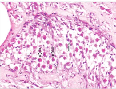
Figure 1. Perineural congestion (thin arrow) and perineural inflammation (thick arrow) shown in the nerve section (H&E, 200×)
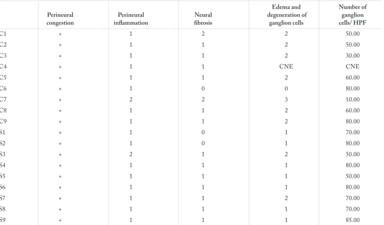
Table 1. Histopathological findings of the study group and the control group
Edema and Number of
Perineural Perineural Neural degeneration of ganglion congestion inflammation fibrosis ganglion cells cells/ HPF
C1 + 1 2 2 50.00 C2 + 1 1 2 50.00 C3 + 1 1 2 30.00 C4 + 1 1 CNE CNE C5 + 1 1 2 60.00 C6 + 1 0 0 80.00 C7 + 2 2 3 10.00 C8 + 1 1 2 60.00 C9 + 1 1 2 80.00 S1 + 1 0 1 70.00 S2 + 1 0 1 80.00 S3 + 2 1 2 50.00 S4 + 1 1 1 80.00 S5 + 1 1 1 50.00 S6 + 1 1 1 80.00 S7 + 1 1 2 70.00 S8 + 1 1 1 70.00 S9 + 1 1 1 85.00
Trichrome dye was used to evaluate neural fibrosis histochemical-ly. Sparse, thin fibers stained greenish observed in neural sections were scored as 1 (mild fibrosis), whereas denser, thicker fibers were scored as 2 (moderate fibrosis). It was scored as 0 if there was no fiber stained with trichrome dye in the nerve section.
Statistical Analysis
SPSS for Windows 22.0 (IBM Corp; Armonk, New York, USA) was used for statistical analysis. Pearson Chi-Square test was used to compare the histopathological findings. The number of ganglion cells was compared using paired sample t-test; in all tests, p<0.05 was considered to be statistically significant.
Results
The histopathological findings of both groups are shown in Table 1. Perineural congestion was observed in all animals in the study and control groups (Figure 1). There was no sig-nificant difference between the groups regarding perineural
inflammation (p=0.708) or neural fibrosis severity (p=0.526) (Figure 2). Likewise, no significant difference was revealed between the groups regarding edema and degeneration of the ganglion cells (p=0.169) (Figure 3). The mean number of gan-glion cells in the study and control groups was 69.3±13.2 and 52.5±23.7, respectively, which was not statistically significant (p=0.062).
Discussion
The causes of cochlear implant failure are extrusion and mal-positioning of the implant electrode, wound and flap prob-lems, and trauma (13-16). In addition, few authors have mentioned that allergy to silicon is one of the causes of co-chlear implant extrusion (7-9). Puri et al. (9) reported contact dermatitis developing after cochlear implantation. In a skin patch test, they diagnosed allergy to silicone LSR-30 found in the device. Shao (17) reported cochlear implant failure secondary to the restoration of silicone during device man-ufacturing.
PC has been widely used as a coating material for isolating im-plantable biomedical devices (18, 19). It has a number of fea-tures such as its high molecular weight, all-carbon structural backbone, and nonpolar entities, which prevents contaminations by most chemicals, fungi and bacteria (20). It has been reported that PC is more hemocompatible and less thrombogenic than silicon and that it has a high stability in vivo for many biological and biomedical applications (21). The biocompatibility of PC has been shown in bladder tissue (22).
The surface characteristics and cell and protein compatibility of PC is comparable with those of polystyrene, polydimeth-ylsiloxane, and glass. PC substrates preserve their hydrophilic properties over time and display a higher degree of nanoscale surface roughness (>20 nm) than other substrates (22). There-fore, PC can be a useful material for fabricating cell-based mi-crodevices (19).
Figure 2. a, b. Severity of fibrosis observed in the nerve section (a) Moderate fibrosis: connective tissue fibers widely stained green with trichrome dye (b) No fibrosis: nerve section lacking green-stained areas (trichrome, 200×)
a b
Figure 3. Mild edema and degeneration of ganglion cells. Thick arrow: Normal ganglion cells, thin arrow: degenerated ganglion cells (H&E, 400×)
The biocompatibility of polyimide or polyimide coated with amorphous aluminum oxide, amorphous carbon, parylene, poly-vinylpyrolidone (PVP), or polyethylene glycol (PEG) was eval-uated for possible use in subretinal prostheses. PEG, parylene, and PVP have been shown to produce less histologic disrup-tion than other compounds. In addidisrup-tion, there was no signif-icant difference between parylene, PEG, and the nonsurgical control group in disturbing retinal anatomy (18). In a similar study, polyimide, parylene, and silicone were evaluated as retinal prosthesis electrode array substrate materials. When compared in terms of biocompatibility, PC showed an excellent long-term performance (23).
Parylene has also been assessed as a coater for silicon in cochlear implant electrodes in a previous study. It was proposed that sili-con cochlear electrodes would be more flexible and robust after encapsulation with parylene (3).
In our study, the possible effects of PC in the inner ear were examined. No significant difference was revealed between the study and control groups regarding histopathological findings. Compared to the controls, it seems that PC did not cause additional histopathological damage to the cochlea. This find-ing is promisfind-ing for the development of a new implantable material coater to overcome silicon-based cochlear implant failures.
Conclusion
In conclusion, PC did not cause any histopathologic damage in the cochlea. This finding may be promising regarding the use of PC in cochlear implant electrodes as an alternative to silicon materials. However, further studies are needed to assess the val-ue of PC as an alternative implant coater.
Ethics Committee Approval: Ethics committee approval was received
for this study from the ethics committee of Committee for Research and Animal Ethics of Gazi University (2011).
Peer-review: Externally peer-reviewed.
Author contributions: Concept - N.G., R.C., P.U.G.; Design -
Y.A.B., R.C., M.D.; Supervision -Y.A.B., N.G.; Resource - R.C., R.K., H.T., S.U.B.; Materials - R.C., P.U.G., M.D., N.G.; Data Collection &/or Processing - N.G., R.K., R.C., P.U.G.; Analysis &/ or Interpretation - Y.A.B., N.G., R.C.; Literature Search - R.C., Y.A.B., M.D.; Writing - R.C., M.D., Y.A.B.; Critical Reviews - Y.A.B., R.C., M.D.
Conflict of Interest: No conflict of interest was declared by the authors. Financial Disclosure: This study was supported financialy by
MEDers® company.
References
1. Georgiou J, Toumazou C. A 126-mW cochlear chip for a totally implantable system. IEEE J Solid-State Circuits 2005; 40: 430-3.
[CrossRef]
2. Muthusubramaniam L, Lowe R, Fissell WH, Li L, Marchant RE, Desai TA, et al. Hemocompatibility of silicon-based substrates for biomedical implant applications. Ann Biomed Eng 2011; 39:
1296-305. [CrossRef]
3. Wang J, Gulari MN, Wise KD. A parylene-silicon cochlear elec-trode array with integrated position sensors. Conf Proc IEEE Eng
Med Biol Soc 2006; 1: 3170-3. [CrossRef]
4. Migirov L, Kronenberg J, Volkov A. Local tissue response to co-chlear implant device housings. Otol Neurotol 2011; 32: 55-7.
[CrossRef]
5. Nadol JB Jr, Eddington DK. Histologic evaluation of the tissue seal and biologic response around cochlear implant electrodes in
the human. Otol Neurotol 2004; 25: 257-62. [CrossRef]
6. Benatti A, Castiglione A, Trevisi P, Bovo R, Rosignoli M, Manara R, et al. Endocochlear inflammation in cochlear implant users: case report and literature review. Int J Pediatr Otorhinolaryngol 2013; 77: 885-93. [CrossRef]
7. Kunda LD, Stidham KR, Inserra MM, Roland PS, Franklin D, Roberson JB Jr. Silicone allergy: A new cause for cochlear implant extrusion and its management. Otol Neurotol 2006; 27: 1078-82.
[CrossRef]
8. Klykken PC, Curtis JM. Re: “Silicone allergy: a new cause for cochlear implant extrusion and its management”. Otol Neurotol 2007; 28: 1159-61. [CrossRef]
9. Puri S, Dornhoffer JL, North PE. Contact dermatitis to silicone after cochlear implantation. Laryngoscope 2005; 115: 1760-2.
[CrossRef]
10. Wahjudi PN, Oh JH, Salman SO, Seabold JA, Rodger DC, Tai YC, et al. Improvement of metal and tissue adhesion on sur-face-modified parylene C. J Biomed Mater Res A. 2009; 89: 206-14.
11. Seymour JP, Elkasabi YM, Chen HY, Lahann J, Kipke DR. The insulation performance of reactive parylene films in implantable electronic devices. Biomaterials 2009; 30: 6158-67. [CrossRef]
12. Li W, Rodger D, Weiland J, Humayun M, Tai Y. Integrated Flexi-ble Ocular Coil for Power and Data Transfer in Retinal Prostheses.
Conf Proc IEEE Eng Med Biol Soc 2005; 1: 1028-31. [CrossRef]
13. Chung D, Kim AH, Parisier S, Linstrom C, Alexiades G, Hoffman R, et al. Revision cochlear implant surgery in patients with suspect-ed soft failures. Otol Neurotol 2010; 31: 1194-8. [CrossRef]
14. Lescanne E, Al Zahrani M, Bakhos D, Robier A, Morinière S. Revision surgeries and medical interventions in young cochlear implant recipients. Int J Pediatr Otorhinolaryngol 2011; 75: 1221-4. [CrossRef]
15. Lassig AA, Zwolan TA, Telian SA. Cochlear implant failures and revision. Otol Neurotol 2005; 26: 624-34. [CrossRef]
16. Jain R, Mukherji SK. Cochlear implant failure: imaging evaluation of the electrode course. Clin Radiol 2003; 58: 288-93. [CrossRef]
17. Shao W. Cochlear implant electrode failure secondary to silicone touch-up during device manufacturing. Otol Neurotol 2013 34: e72-5. [CrossRef]
18. Montezuma SR, Loewenstein J, Scholz C, Rizzo JF 3rd. Biocom-patibility of materials implanted into the subretinal space of Yucatan pigs. Invest Ophthalmol Vis Sci 2006; 47: 3514-22. [CrossRef]
19. Chang TY, Yadav VG, De Leo S, Mohedas A, Rajalingam B, Chen CL, et al. Cell and protein compatibility of parylene-C
sur-faces. Langmuir 2007; 23: 11718-25. [CrossRef]
20. Chen PJ, Shih CY, Tai YC. Design, fabrication and characteriza-tion of monolithic embedded parylene microchannels in silicon substrate. Lab Chip 2006; 6: 803-10. [CrossRef]
Turk Arch Otorhinolaryngol 2016; 54: 53-7 Cevizci et al. Effects of Parylene C in the Inner Ear
21. Weisenberg BA, Mooradian DL. Hemocompatibility of mate-rials used in microelectromechanical systems: platelet adhesion and morphology in vitro. J Biomed Mater Res 2002; 60: 283-91.
[CrossRef]
22. Kim SJ, Lee DS, Kim IG, Sohn DW, Park JY, Choi BK, et al. Evaluation of the biocompatibility of a coating material for an
im-plantable bladder volume sensor. Kaohsiung J Med Sci 2012; 28:
123-9. [CrossRef]
23. Weiland JD, Humayun MS, Eckhardt H, Ufer S, Laude L, Basing-er B, et al. A comparison of retinal prosthesis electrode array sub-strate materials. Conf Proc IEEE Eng Med Biol Soc 2009; 2009: