CASE REPORT
Xanthelasma palpebrarum: a new side effect
of nilotinib
Irmak Sayin,
1Meltem Ayli,
2Ali Kemal O
ğuz,
1Güldane Cengiz Seval
2 1Department of InternalMedicine, Ufuk University Medical Faculty, Ankara, Turkey
2Department of Hematology,
Ufuk University Medical Faculty, Ankara, Turkey Correspondence to Dr Irmak Sayin, [email protected] Accepted 21 December 2015
To cite: Sayin I, Ayli M, Oğuz AK, et al. BMJ Case Rep Published online: [ please include Day Month Year] doi:10.1136/bcr-2015-213511
SUMMARY
Chronic myeloid leucaemia (CML) is a chronic myeloproliferative disorder characterised by a reciprocal translocation between the chromosomes 9 and 22 resulting in constitutionally active tyrosine kinase signalling. BCR-ABL tyrosine kinase inhibitors (TKIs) are highly effective molecules in the treatment of CML. Unfortunately, these novel therapeutic agents are accompanied by various side effects, and
haematological, cutaneous and metabolic abnormalities are among the most prevalent. Nilotinib, a second-generation TKI, has been shown to cause both— cutaneous lesions and lipid profile abnormalities. We present two CML cases developing xanthelasma palpebrarum while receiving nilotinib. Case 1 also acquired a lipid abnormality following the start of nilotinib therapy, while case 2 meanwhile stayed normolipidemic. In addition to a low cholesterol diet, atorvastatin was prescribed to case 1. Currently, both cases are normolipidemic and continuing their nilotinib therapy. Xanthelasma palpebrarum secondary to nilotinib therapy is new to the literature.
BACKGROUND
Chronic myeloid leucaemia (CML) is a chronic myeloproliferative disorder characterised by a recip-rocal translocation between the chromosomes 9 and 22 resulting in constitutionally active tyrosine kinase signalling. BCR-ABL tyrosine kinase inhibi-tors (TKIs) are highly effective in the treatment of CML, and imatinib mesylate was thefirst molecule in this novel family of drugs.1The appearance of intolerance and resistance to imatinib led to the for-mulation of second-generation TKIs, for example, dasatinib and nilotinib.2 While cutaneous adverse
effects are the most common non-haematological adverse reactions to TKIs, nilotinib has also been demonstrated to cause metabolic disturbances such as dyslipidaemia.3 4 We present two patients with
CML with xanthelasma palpebrarum secondary to nilotinib therapy as the unusual cause.
CASE PRESENTATION
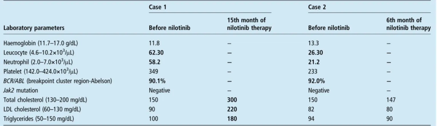
Case 1: A 40-year-old woman was referred for investigation of a significant leucocytosis finding. Results of her physical examination were unre-markable. A summary of the relevant laboratory findings are presented in table 1. The patient was diagnosed with BCR-ABL positive, chronic phase CML and started on imatinib 400 mg daily. At the end of the fourth month, as her absolute neutrophil count (ANC) was <1000/μL, the imatinib therapy was interrupted for 3 weeks, with no resolution of
her ANC, a level >1500/μL. Consequently, because of its haematological toxicity, imatinib was not rein-stated but, instead, replaced by nilotinib therapy at a dose of 400 mg twice daily. At the end of the 12th month, the patient’s BCR-ABL fusion gene levels as determined by qRT-PCR testing were negative.
Fifteen months after starting nilotinib, the patient presented with yellowish plaques around her eyelids. Physical examination revealed lesions con-sistent with xanthelasma palpebrarum (figure 1). Repeated medical history-taking documented no systemic disorders as potential causes. The patient denied smoking and consuming alcohol, and was not using any medications other than nilotinib. Her family history was also unrevealing. The patient’s repeated lipid profile is given intable 1.
Case 2: A 29-year-old woman presented for investigation of a leucocytosis finding. The results of a physical were unremarkable. Her initial labora-tory results are summarised in table 1. Subsequently, the patient was diagnosed as being BCR-ABL positive and having chronic phase CML. Imatinib therapy at a dose of 400 mg daily was started. During the fourth month of therapy, grade 3–4 haematological toxicity pertaining to cytes developed. On the occurrence of thrombo-cytopaenia with a count <50 000/μL, imatinib therapy was interrupted for a period of 2 weeks. Unfortunately, the thrombocyte count did not increase to a level >50 000μ/L and therefore imati-nib was stopped and therapy switched to nilotiimati-nib (400 mg twice daily).
During the sixth month following the start of nilotinib therapy, the patient developed yellow col-oured elevated lesions around both her eyelids. Repeated physical examination revealed findings consistent with xanthelasma palpebrarum (figure 2). Detailed medical history-taking did not document any systemic disorders, and the patient denied drinking alcohol and smoking tobacco, and was not using any medications other than nilotinib. Family history of the patient was also unremarkable. A repeated lipid profile of the patient is displayed in
table 1.
INVESTIGATIONS
While the baseline lipid profile of case 1 was normal, her lipid profile at the 15th month of nilo-tinib therapy displayed marked elevations of low-density lipoprotein (LDL-C) and total cholesterol (TC) together with triglycerides (TG). Case 2 demonstrated a normal lipid profile both initially and while she presented with lesions of
Sayin I, et al. BMJ Case Rep 2016. doi:10.1136/bcr-2015-213511 1
xanthelasma palpebrarum (at the sixth month of nilotinib therapy) (table 1). Both cases had normal fasting plasma glucose levels, normal hepatic, renal and thyroid function tests and normal pancreatic lipase/amylase levels.
DIFFERENTIAL DIAGNOSIS
The typical differential diagnosis of xanthelasma palpebrarum includes familial hyperlipidaemias (ie, types II, III and IV), dysli-pidaemias characterised by low high-density lipoprotein (HDL) cholesterol levels, uncontrolled diabetes and atypical lymphoid infiltrates. The unrevealing family histories, normal fasting plasma glucose levels and the myeloid origin of the underlying haematological neoplasm make these differential diagnoses highly unlikely.
TREATMENT
Case 1 was counselled to strictly adopt a low-cholesterol diet, perform regular aerobic exercise and start a course of 20 mg of atorvastatin daily. Nilotinib therapy (400 mg twice daily) was not interrupted. As case 2 was normolipidemic, neither a diet change nor a course of statin treatment was implemented. She also continued her nilotinib 400 mg twice daily therapy.
OUTCOME AND FOLLOW-UP
Both cases are currently normolipidemic and on nilotinib therapy. Case 1 did not experience any statin related side effects. Her creatinine kinase and alanine aminotransferase (ALT) levels were monitored and found to be within normal limits.
DISCUSSION
In recent years, several TKIs have been developed and received approval for CML treatment. The safety of these drugs is becoming an important issue. Although not life-threatening, skin problems constitute the most common untoward effects of these novel therapeutic agents.3
Skin rash associated with imatinib therapy is generally mild and is often characterised by maculopapular lesions. It has been shown that imatinib causes grade 1–2 skin rashes in 30–40% of patients, pruritus in 7.3% of patients, alopecia in 4.4% of patients, increased sweating in 3.6% of patients, stomatitis in 2.9% of patients and dry mouth in 2.2% of patients using it.5
The cutaneous side effects of dasatinib were noted from one phase I andfive phase II trials, collectively, from a total of 911 patients with CML. Dasatinib was associated with a 35% risk of cutaneous reactions. Most frequent side-effects consisted of localised/generalised erythaema, maculopapular eruptions and exfoliative rashes. Most of these were grade 1–2 lesions. Pruritus was observed in 11% of patients while mucositis and stomatitis were present in 16% of patients.6A rare side effect of
panniculitis presenting with painful subcutaneous nodules was reported in two patients with chronic phase CML receiving dasatinib treatment.7Also, very few cases of pustular rashes and acne-like eruptions associated with dasatinib therapy are described in the literature.
Likewise, patients and their clinicians frequently encounter cutaneous side-effects of nilotinib therapy. In a phase I study of nilotinib in 119 leucaemic patients, Kantarjian et al8 reported cutaneous side-effects in the form of grade 1–2 lesions. The most frequent untoward cutaneous effects were pruritus Table 1 Relevant laboratory findings of case 1 and case 2 before and after nilotinib therapy
Case 1 Case 2
Laboratory parameters Before nilotinib
15th month of
nilotinib therapy Before nilotinib
6th month of nilotinib therapy Haemoglobin (11.7–17.0 g/dL) 11.8 − 13.3 − Leucocyte (4.6–10.2×10³/μL) 62.30 − 26.30 − Neutrophil (2.0–7.0×10³/μL) 58.2 − 21.2 − Platelet (142.0–424.0×10³/μL) 349 − 233 −
BCR/ABL (breakpoint cluster region-Abelson) 90.1% − 92.0% −
Jak2 mutation Negative − Negative −
Total cholesterol (130–200 mg/dL) 150 300 150 147
LDL cholesterol (60–130 mg/dL) 90 220 82 80
Triglycerides (50–150 mg/dL) 100 180 94 90
Laboratory results falling out of the reference ranges are designated in bold LDL, low-density lipoprotein.
Figure 2 Xanthelasma palpebrarum of the superior and inferior palpebrae (case 2).
Figure 1 Xanthelasma palpebrarum of the superior and inferior palpebrae (case 1).
2 Sayin I, et al. BMJ Case Rep 2016. doi:10.1136/bcr-2015-213511
(2–15%), non-specific rashes (2–20%), dry skin (0–12%) and alopecia (0–6%), all of which appeared to be dose related. Another, this time a phase II study conducted on 280 patients, demonstrated the appearance of non-specific rashes in 28% and pruritus in 24% of the treated patients.9
Non-cutaneous adverse events resulting from nilotinib therapy included reversible constitutional symptoms, and eleva-tions in ALT, aspartate transaminase, lipase, bilirubin, fasting plasma glucose and plasma lipid concentrations.10 Hypercholesterolaemia, hyperlipidaemia and hypertriglyceridae-mia are mentioned as side effects by the manufacturer. Product instructions do suggest assessing the plasma lipid profile both prior to and during nilotinib therapy as clinically indicated. Rea et al showed a significant rise in TC within 3 months of initi-ation of nilotinib therapy in a pattern involving eleviniti-ation of both, LDL and HDL cholesterol fractions. Although the mech-anism of nilotinib-associated dyslipidaemia is not fully under-stood, it is thought to be due to increased hepatic synthesis and/ or impaired clearance from the blood.4
Xanthomas form as foam cells cluster in the connective tissues of the skin, tendons or fasciae. Foam cells are macro-phages loaded with excessive amounts of LDL particles and their oxidised forms. From a pathological viewpoint, the follow-ing factors may take part durfollow-ing the formation of xanthomas: (1) high local concentrations of lipids in the connective tissue; (2) the presence of qualitatively different lipoproteins at normal plasma lipid concentrations; (3) increased extravasation of lipids due to increased vascular permeability, increased local circula-tion or chronic inflammation; (4) lipid synthesis in situ and their deposition in histiocytes; (5) dysfunction of the reverse cholesterol transport.11 12
Xanthelasma palpebrarum is the most commonly observed form of xanthomas and most commonly identified in subjects over 50 years of age. About half of these subjects have under-lying dyslipidaemia (high LDL-C and TG, low HDL-C and apo A-1).13 The presence of xanthelasma palpebrarum should not
be underestimated in clinical practice.
With respect to cases presented, while case 1 was demonstrat-ing a markedly abnormal lipid profile, case 2 had xanthelasma palpebrarum lesions while exhibiting a normal lipid profile. Consequently, while in case 1 xanthelasma palpebrarum was associated with a nilotinib-induced dyslipidaemia, in case 2, it could be associated with a nilotinib-induced presence of qualita-tively different lipoproteins at normal plasma concentrations, or increased extravasation of lipids, or lipid synthesis in situ together with deposition in histiocytes.
Thefindings pertinent to case 2 suggest that xanthelasma pal-pebrarum may appear as a cutaneous side effect of nilotinib therapy, independent of hyperlipidaemia. To the best of our knowledge, there have been no reported cases of xanthelasma palpebrarum observed in patients treated with nilotinib. In case 2, even though the appearance of xanthelasma palpebrarum without associated hyperlipidaemia may have been an incidental co-occurrence, we strongly believe that this cutaneous untoward effect should be noted in the literature and required attention should be placed on it. However, studies including larger cohorts are surely needed to confirm xanthelasma palpebrarum as a cutaneous side effect of nilotinib.
Contributors IS contributed to the design of the work, and analysis and interpretation of the data and authored the manuscript. MA contributed to the design and interpretation, and revised the work critically for intellectual content. AKO and GCS interpreted the data and co-authored the manuscript. All the authors made substantial contributions to the conception or design of the work, or the acquisition, analysis or interpretation of data, drafting of the work or revising it critically for important intellectual content, and gavefinal approval of the version published. The authors agree to be accountable for all aspects of the work, ensuring that questions related to the accuracy or integrity of any part of the work are appropriately investigated and resolved.
Competing interests None declared. Patient consent Obtained.
Provenance and peer review Not commissioned; externally peer reviewed.
REFERENCES
1 Abernethy AP, McCrory DC. Report on the relative efficacy of oral cancer therapy for medicare beneficiaries versus currently covered therapy: part 3, Imatinib for Chronic Myeloid Leukemia (CML) [Internet]. Rockville, MD: Agency for Healthcare Research and Quality (US), 2005. AHRQ Technology Assessments.
2 Giles FJ. New directions in the treatment of imatinib failure and/or resistance.Semin Hematol2009;46:S27–33.
3 Brazzelli V, Grasso V, Borroni G. Imatinib, dasatinib and nilotinib: a review of adverse cutaneous reactions with emphasis on our clinical experience.J Eur Acad Dermatol Venereol2013;27:1471–80.
4 Rea D, Mirault T, Cluzeau T, et al. Early onset hypercholesterolemia induced by the 2nd-generation tyrosine kinase inhibitor nilotinib in patients with chronic phase-chronic myeloid leukemia.Haematologica2014;99:1197–203.
5 O’Brien SG, Guilhot F, Larson RA, et al. Imatinib compared with interferon and low-dose cytarabine for newly diagnosed chronic-phase chronic myeloid leukemia.
N Engl J Med2003;348:994–1004.
6 Shah NP, Kim DW, Kantarjian HM. Dasatinib 50mg or 70mg BID compared to 100mg or 140mg QD in patients with CML in chronic phase (CP) who are resistant or intolerant to imatinib: one-year results of CA180034. J Clin Oncol
2007;25:7004.
7 Assouline S, Laneuville P, Gambacorti-Passerini C. Panniculitis during dasatinib therapy for imatinib-resistant chronic myelogenous leukemia.N Engl J Med
2006;354:2623–4.
8 Kantarjian HM, Giles F, Wunderle L, et al. Nilotinib in imatinib-resistant CML and Philadelphia chromosome-positive ALL.N Engl J Med2006;354:2542–51.
9 Kantarjian HM, Giles F, Gattermann N, et al. Nilotinib (formerly AMN107), a highly selective BCR-ABL tyrosine kinase inhibitor, is effective in patients with Philadelphia chromosome-positive chronic myelogenous leukemia in chronic phase following imatinib resistance and intolerance.Blood2007;110:3540–6.
10 Larson R, le Coutre P, Reiffers J, et al. Comparison of nilotinib and imatinib in patients with newly diagnosed chronic myeloid leukemia in chronic phase (CML-CP): ENESTnd beyond one year. J Clin Oncol 2010;28:7s.
11 Durrington P, Sniderman A, eds. Hyperlipidaemia. Oxford, Health Press, 2000. 12 Thompson GR, ed. A handbook of hyperlipidemia. London: Current Science, 1990. 13 Kim J, Kim YJ, Lim H, et al. Bilateral circular xanthelasma palpebrarum.Arch Plast
Surg2012;39:435–7. Learning points
▸ Patients with chronic myeloid leucaemia should be screened for lipid disorders prior to and during nilotinib therapy. ▸ Patients with hyperlipidaemia secondary to nilotinib therapy
should receive dietary advice and, if needed, lipid-lowering agents should be added to the therapy.
▸ Xanthelasma palpebrarum should be considered an untoward effect of nilotinib, independent of the presence of hyperlipidaemia.
Sayin I, et al. BMJ Case Rep 2016. doi:10.1136/bcr-2015-213511 3
Copyright 2016 BMJ Publishing Group. All rights reserved. For permission to reuse any of this content visit http://group.bmj.com/group/rights-licensing/permissions.
BMJ Case Report Fellows may re-use this article for personal use and teaching without any further permission. Become a Fellow of BMJ Case Reports today and you can:
▸ Submit as many cases as you like
▸ Enjoy fast sympathetic peer review and rapid publication of accepted articles ▸ Access all the published articles
▸ Re-use any of the published material for personal use and teaching without further permission For information on Institutional Fellowships contact [email protected]
Visit casereports.bmj.com for more articles like this and to become a Fellow
4 Sayin I, et al. BMJ Case Rep 2016. doi:10.1136/bcr-2015-213511