Neuropsychological Assessment of Adult Patients
with Shunted Hydrocephalus
Emel Erdogan Bakar, M.D.,1 Bulent Bakar, M.D.2 Department of Psychology,1Ufuk University, Ankara, Turkey
Department of Neurosurgery,2Klrlkkale University Faculty of Medicine, Klrlkkale, Turkey
Objective : This study is planned to determine the neurocognitive difficulties of hydrocephalic adults.
Methods : The research group contained healthy adults (control group, n : 15), and hydrocephalic adults (n : 15). Hydrocephalic group consisted of patients with idiopathic aquaduct stenosis and post-meningitis hydrocephalus. All patients were followed with shunted hydrocephalus and not gone to shunt revision during last two years. They were chosen from either asymptomatic or had only minor symptoms without motor and sensorineural deficit. A neuropsychological test battery (Raven Standart Progressive Matrices, Bender-Gestalt Test, Cancellation Test, Clock Drawing Test, Facial Recognition Test, Line Orientation Test, Serial Digit Learning Test, Stroop Color Word Interference Test-TBAG Form, Verbal Fluency Test, Verbal Fluency Test, Visual-Aural Digit Span Test-B) was applied to all groups.
Results : Neuropsychological assessment of hydrocephalic patients demonstrated that they had poor performance on visual, semantic and working memory, visuoconstructive and frontal functions, reading, attention, motor coordination and executive function of parietal lobe which related with complex and perseverative behaviour. Eventually, these patients had significant impairment on the neurocognitive functions of their frontal, parietal and temporal lobes. On the other hand, the statistical analyses performed on demographic data showed that the aetiology of the hydrocephalus, age, sex and localization of the shunt (frontal or posterior parietal) did not affect the test results.
Conclusion : This prospective study showed that adult patients with hydrocephalus have serious neuropsychological problems which might be directly caused by the hydrocephalus; and these problems may cause serious adaptive difficulties in their social, cultural, behavioral and academic life.
KEY WORDS : Adult˙ Hydrocephalus˙ Neuropsychological assessment.
Clinical Article
INTRODUCTION
The term of hydrocephalus means that an abnormal accumulation of cerebrospinal fluid (CSF) in the cerebral ventricular system which increases the intracranial pressure
(ICP)22). Increased ICP can cause some neuropsychological
problems8). In literature, some authors suggested that
although the hydrocephalus is apparently arrested, the disorder may progress and produce neuropsychological disturbances in certain patients. Recent study results have support this thesis that these patients have persistent neuro-psychological difficulties affecting visuospatial, language
skills and motor functions; and difficulties in other non-verbal domains such as attention, memory, maths, nume-racy, and problem solving3,12,14,22). Because these problems
with memory and concentration reduce intellectual ability and decrease performance in school, these patients have serious adaptive problems in their social, cultural, beha-vioral and academic life11,12,14).
In literature, aim of most studies were constructed to des-cribe the neuropsychological difficulties of the spina bifida with hydrocephalus. Neurodevelopmental disorders affect-ing physical, cognitive, and adaptive function throughout the childhood years has been well demonstrated in those studies. However, little knowledge about the adult out-comes of the neuropsychological difficulties. Some of the cognitive disfunction in hydrocephalic young adult with spina bifida are now being identified; but their adaptive difficulties are poorly explained11,12,14,22). In these studies,
there are some questions between the aetiological factors
• Received : August 24, 2009 • Revised : January 18, 2010 • Accepted : January 31, 2010
• Address for reprints : Bulent Bakar, M.D.
Department of Neurosurgery, Klrlkkale University Faculty of Medicine, 71100-Klrlkkale, Turkey
Tel : +903182252585, Fax : +903122845870 E-mail : [email protected]
and development of the neuropsycho-logical difficulties. The purpose of this prospective study was to describe neu-ropsychological problems, and other intellectual skill difficulties of adult patients with hydrocephalus without spina bifida.
MATERIALS AND METHODS Subjects
The research group contained healthy adults (control group) and hydrocephlic adults (hydrocephalus group). Non of the patients had overt clinical signs or symptoms of in-creased intracerebral pressure.
The hydrocephalus group (n : 15) consisted of 9 male (mean age, 36.85
±
13.36 years) and 6 female (mean age, 33.88±
5.84 years) who followed with shunted hydro-cephalus and not gone to shunt revision last two years at the Hacettepe University Faculty of Medicine, Department of Neurosurgery. The hydrocephalus group subjects were chosen from adults who were either asymptomatic or had only minor symptoms (such as sporadic headache, atten-tion and memory difficulties, psychomotor slowness, etc.) without motor and sensorineural deficit. All subjects with other neurological abnormalities such as tumour, intraven-tricular haemorrhage, epilepsy or stroke were excluded. This group could be divided into two subgroups aetiologi-cally as follows : idiopathic aquaductal stenosis (n : 7), and post-meningitis hydrocephalus (n : 8). Post-meningitis hydrocephalus patients who were treated absolutely had not any infection parameter in CSF sample. Three of them had tuberculous meningitis, 2 had mumps, 3 had Haemophilus influenzae. All subjects were statistically analysed todeter-mine the effect of the aetiological factors described above on the all neuropsychological tests points used by “Indipen-dent Sample t-test”; and there was no statistically signifi-cant difference between them; and this group was therefore not divided into its subgroups.
The control group (n : 15) consisted of 9 male (mean age,
36.61
±
9.63 years) and 6 female (mean age, 35.07±
6.36years) were chosen from healthy adults. The all groups’ sex and age discrimination are shown at the Table 1.
Neuropsychological assessment
Patients underwent neuropsychological examinations which were administered in the same order by the same examiner. They were evaluated by an experienced neuro-psychologist (EEB) blinded to the groups. The examiner performed ten different neuropsychological tests battery to
all groups with its routin procedures. These tests are desc-ribed below respectively.
Raven Standart Progressive Matrices (RSPM)
It was originally developed by Raven et al.24)The test is
accepted as a culture-fair test of general intelligence (g factor) and also of visuospatial cognition (K factor)24). Raven Standart
Progressive Matrices (RSPM) performance requires working memory, category shift, mental flexibility, problem solving, abstraction and reasoning; these functions are combined under the umbrella term, “executive functions”16,19,23).
Bender-Gestalt Test (BGT)
It is first developed by Bender1). This test is used to evaluate
“visual-motor maturity”, to screen for developmental disor-ders, or to assess neurological function or brain damage. It measures perceptual motor skills, perceptual motor devel-opment, and gives an indication of neurological intactness of parietal lobes19).
Cancellation Test (CT)
It was originally developed by Weintraub and Mesulam28)
to measure a sensory component of the parietal lobes that is related to perceptual errors, a motor component that is related to visual search and scan and a motivational com-ponent that is related to expectation and affect. Cancellation Test (CT) is used in the literature as a measure of visuomotor performance and of vigilance and sustained attention15,18,19).
Clock Drawing Test (CDT)
The Clock Drawing Test (CDT) draws on several skills, including auditory comprehension, visuospatial ability, and constructional praxis which are mainly functions of the parietal lobes. Additionally, the CDT may detect deficits in executive functioning that are overlooked by other routine cognitive tests. Since executive function relates to a person’s ability to plan, initiate, sequence, monitor and stop complex behavior, it may be particularly useful when assessing pa-tients’ functional status26).
Facial Recognition Test (FRT)
This instrument consists of 13 pairs of stimulus plates, Table 1. Descriptive table of the demographic data of the all group
Group Sex N Min Max Mean SD
Hydrocephalus Famale 6 26.09 40 33.88 5.84 Male 9 22.11 60 36.85 13.36 Control Famale 6 28.08 42.1 35.07 6.36 Male 9 24.09 53.8 36.61 9.63
N : number of the patients, Min : minimum age, Max : maximum age, Mean : mean of age, SD : standart deviation
which require the patient to perform 27 visuoperceptive discriminations of unfamiliar Caucasoid faces. The task has neither memory nor speed components. It measures visuos-patial recognition function of the posterior association cortex and temporal lobe19).
Line Orientation Test (LOT)
It was originally developed by Benton et al.2)to measure
visuospatial perception and orientation. This test measures visual-perceptual/visual-spatial discrimination, and the pa-tient’s ability to analyze and synthesize visual form and spa-tial relations. It is more specific for the right hemispheric cognitive functions and sensitive especially in the injuries involving the right cerebral hemisphere and parietal lobe16,19).
Serial Digit Learning Test (SDLT)
Tasks that involve digit series are among the primary tools of neuropsychology for measuring attention, learning, and memory, since the contaminating effect of associative influ-ences is lower on digits than on words or pictures. This test has demonstrated validity and provides additional substan-tive data in the evaluation of brain-damaged patients; and performance on SDLT is dependent upon the mesial
tem-poral lobe and hippocampus16,19).
Stroop Color Word Interference Test-TBAG Form (SCWT)
It measures the ability to shift perceptual set with the changing demands, also measures to inhibit a habitual behavior pattern, and to behave in an unusual way. Defects in these abilities result in perseverations, stereotypic beha-viors, and difficulty in controlling the behaviors. These functions are mainly the functions of the frontal lobes. Higher interference score indicates poorer performance. Stroop Color Word Interference Test-TBAG Form (SCWT) also assesses the information processing rate, and parallel processing of attended and non-attended stimuli, and attention16,20).
Verbal Fluency Test (VFT) [The Initial Letter Verbal Fluency Test (FAS)]
It evaluates executive function of the frontal lobe and semantic memory store (a temporal lobe function). In a timed test (one minute), the patient generates words beginning with the letters K, A and S (“Association” score). We also provided a semantic category (animals) and scored the number of words generated (“Animals” score). Last, we combined letter fluency with category fluency in which the subject had to switch between words beginning with the letter “K” and names of animals (“Categories” score)19).
Visual-Aural Digit Span Test-B (VADS-B)
Visual-Aural Digit Span (VADS) test was originally design-ed as a diagnostic tool for assessing reading and learning disabilities which are mainly functions of the temporal lobes; and it is specifically used for measuring attention span16,17). Statistical analysis
The study results were normally distributed and the varia-tion was homogenous between groups. Therefore they were analyzed statistically using by parametric tests. Age, sex and aetiological factors were investigated using “Independent Sample t-test”; and p values less than 0.05 were considered to be significant. Effect of intelligence on test scores was in-vestigated using by the “analyses of variance test” (ANOVA); and p values less than 0.05 were considered to be significant. Neuropsychological test points of the hydrocephalus and control groups were analyzed “Multiple Analysis of Cova-riance Test” (MANCOVA); and p values less than 0.05 were considered to be significant. All data were analyzed using SPSS for Windows Release 13.0.27).
RESULTS
Demographic data was investigated using by “Indipen-dent Sample t-test”. Age, sex and aetiological factors were not statistically significant difference between hydrocephalus and control groups (p > 0.05).
In the hydrocephalus group and control group, the effect of intelligence on test scores was investigated using by ANOVA. Significant differences among the groups were found in RSPM total score (F = 16.58, df = 1, p < 0.001) and time score (F = 4.41, df = 1, p = 0.045) (Table 2, Fig. 1).
Performances of the hydrocephalus and control groups
Fig. 1. Figure shows the mean values of the raven standart progressive matrices (RSPM) total correct and RSPM duration scores. Each error bar shows the minimum and maximum of the RSPM values.
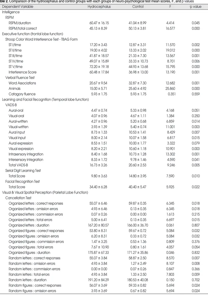
Table 2. Comparison of the hydrocephalus and control groups with each groups of neuro-psychological test mean scores, F, and p values
Dependent Variable Hydrocephalus Control F p value
Intelligence RSPM
RSPM/duration 60.47 ± 16.15 41.04 ± 8.99 4.414 0.045 RSPM/total correct 45.13 ± 8.39 50.13 ± 3.81 16.577 0.000 Executive function (frontal lobe function)
Stroop Color Word Interference Test - TBAG Form
ST I/time 17.20 ± 3.43 12.87 ± 3.31 11.570 0.002 ST II/time 19.00 ± 4.02 13.33 ± 2.02 19.012 0.000 ST III/time 41.87 ± 18.57 21.33 ± 7.30 13.567 0.001 ST IV/time 49.07 ± 15.89 33.33 ± 10.73 8.701 0.006 ST V/time 72.20 ± 19.18 44.93 ± 13.68 15.795 0.000 Interference Score 60.48 ± 17.84 36.98 ± 13.00 13.190 0.001 Verbal Fluence Test
Word Associations 20.67 ± 9.54 32.87 ± 7.30 12.682 0.001
Animals 15.00 ± 5.71 25.60 ± 4.92 25.860 0.000
Catagory fluence 5.93 ± 1.75 5.93 ± 1.75 0.351 0.559 Learning and Facial Recognition (Temporal lobe function)
VADS-B Aural-oral 4.47 ± 0.74 5.33 ± 0.98 4.168 0.051 Visual-oral 4.07 ± 0.96 4.67 ± 1.11 1.384 0.250 Aural-written 4.27 ± 0.96 5.20 ± 0.68 6.859 0.014 Visual-written 3.93 ± 1.39 5.40 ± 0.74 11.001 0.003 Aural input 8.73 ± 1.33 10.53 ± 1.41 8.429 0.007 Visual input 8.00 ± 2.14 10.07 ± 1.58 6.817 0.015 Aural expression 8.53 ± 1.51 10.00 ± 1.77 3.322 0.079 Visual expression 8.20 ± 2.21 10.60 ± 1.18 10.901 0.003 Intrasensory integration 8.40 ± 1.68 10.73 ± 1.28 13.302 0.001 Intersensory integration 8.33 ± 1.72 9.78 ± 1.46 4.590 0.041 Total VADS-B 16.73 ± 3.26 20.60 ± 2.53 9.246 0.005 Serial Digit Learning Test
Total Score 9.80 ± 3.63 14.80 ± 3.95 7.590 0.010
Facial Recognition Test
Total Score 34.40 ± 6.28 40.40 ± 5.47 5.925 0.022 Visual & Visual Spatial Perception (Parietal Lobe Function)
Cancellation Test
Organized letters : correct responses 55.07 ± 6.46 59.87 ± 0.35 6.345 0.018 Organized letters : omission errors 4.93 ± 6.46 0.13 ± 0.35 6.345 0.018 Organized letters : commission errors 0.07 ± 0.26 0.00 ± 0.00 1.613 0.215 Organized letters : total errors 5.00 ± 6.41 0.13 ± 0.35 6.697 0.015 Organized letters : duration 167.20 ± 80.57 166.00 ± 36.70 0.061 0.807 Organized figures : correct responses 53.80 ± 8.31 59.67 ± 0.72 5.084 0.032 Organized figures : omission errors 6.20 ± 8.31 0.33 ± 0.72 5.084 0.032 Organized figures : commission errors 1.47 ± 3.25 0.53 ± 1.36 0.809 0.376 Organized figures : total errors 7.67 ± 10.90 0.80 ± 1.61 4.057 0.054 Organized figures : duration 175.87 ± 67.33 171.27 ± 35.86 0.046 0.832 Random letters : correct responses 55.07 ± 3.84 58.87 ± 2.50 8.570 0.007 Random letters : omission errors 4.93 ± 3.84 1.27 ± 2.49 8.107 0.008 Random letters : commission errors 0.00 ± 0.00 0.07 ± 0.26 0.847 0.366 Random letters : total errors 4.93 ± 3.84 1.33 ± 2.50 7.803 0.009 Random letters : duration 191.20 ± 84.29 180.53 ± 40.08 0.150 0.702 Random figures : correct responses 56.07 ± 3.69 59.33 ± 0.82 5.694 0.024 Random figures : omission errors 3.93 ± 3.69 0.67 ± 0.82 5.694 0.024
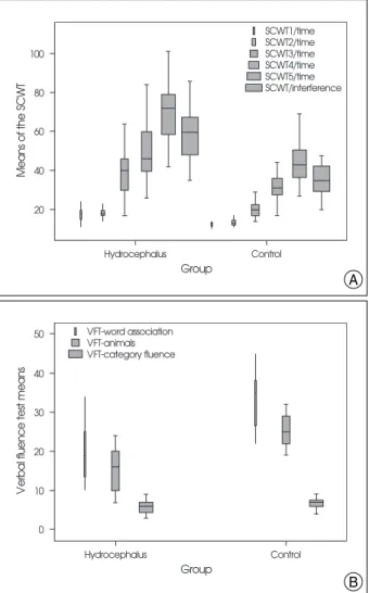
on neuropsychological tests were compared using by MAN-COVA, controlling for the effects of intelligence scores. All the time points and interference scores of the Stroop Color Word Interference Test remained significant after the IQ scores was controlled (Fig. 2A). Verbal Fluency Test Word
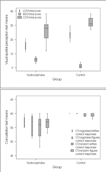
Associations (F = 12.68, df = 1, p < 0.001), Animals (F = 25.86, df = 1, p < 0.001) scores were significantly different between the hydrocephalus and control groups (Fig. 2B). There were statistically significant differences for all of the VADS-B subtest points except visual verbal and verbal expression section points. Serial Digit Learning Test (F = 7.59, df = 1, p = 0.010), Facial Recognition Test (F = 5.92, df = 1, p = 0.022) (Fig. 3), Line Orientation Test (F = 27.70, df = 1, p < 0.001), and Bender-Gestalt Test (F = 20.73, df = 1, p < 0.001) scores remained significant after the intel-ligence score was controlled (Fig. 4A). Cancellation Test organized/random letters and figures of correct responses and omission-error scores were also statistically significant (Table 2, Fig. 4B). These analytic results showed that the hydrocephalus group had poorer intelligence measured by RSPM than control group. Evenly, they had difficulties on the attention (especially sustained attention) which was measured by the CT, SCWT, SDLT, VADS-B. They also had problems on the visuospatial recognition, visual-perce-ptual/visual spatial discrimination, visuomotor
perfor-Table 2. Continued
Dependent Variable Hydrocephalus Control F p value
Random figures : commission errors 1.20 ± 3.86 0.00 ± 0.00 0.514 0.480
Random figures : total errors 5.07 ± 5.69 0.67 ± 0.82 3.998 0.056
Random figures : duration 149.20 ± 59.92 168.80 ± 38.97 1.345 0.256
Line Orientation Test
Total Score 15.00 ± 4.42 23.53 ± 3.25 27.704 0.000
Bender-Gestalt Visual Perception Test
Total Score 6.07 ± 2.96 1.47 ± 1.46 20.729 0.000
Clock Drawing Test
Total Score 26.47 ± 7.66 32.13 ± 3.78 3.503 0.072
Multiple Analysis of Covariance Test, p< 0.05. F: Measurement of distance between individual distribution. RSPM : Raven Standart Progressive Matrices, VADS-B : Visual-Aural Digit Span Test-B
Fig. 2. (A) Figure shows the mean scores of the stroop color word test (SCWT) time and SCWT interference; and (B) mean scores of the verbal fluency test for subjects. SCWT : Stroop Color Word Test, VFT : Verbal Fluency Test.
A
B
Fig. 3. Mean scores of the learning and facial recognition tests for subjects. FRT : Facial Recognition Test, SDLT : Serial Digit Learning Test, VADS-B : Visual-Aural Digit Span Test-B.
mance, reaction time and ability to analyze and synthe-size visual form and spatial relations which were measured by BGT, CT, FRT, LOT. They were unsuccessful on the executive function (i.e., ability to plan, initiate, inhibitory control, verbal fluence, perseveration and modulating im-pulsive responding) which was measured by SCWT, and VFT. They had learning and reading disabilities, and memory problems which were demonstrated by SDLT and VADS-B.
DISCUSSION
In recent literature, severeal studies have reported that raised ICP can produce mechanical stress. This stress may develope impaired cerebral metabolism by decreasing of the cerebral blood flow which may cause to reduce the concen-tration of neurotransmitters and peptide neuromodulators, and to reflect the neuronal dysfunction and neurocognitive
disruption/deficits5,6,12). Fletcher et al. (1992) showed that
the hydrocephalic children have low points at their perfor-mance tests which measure the functions of the right phere of the brain (known as the non-dominant hemis-phere). They also had low performances at their verbal tests which measure functions of the left hemisphere (known as the dominant hemisphere)9). Rourke et al.25) described the
hydrocephalus as a syndrome of non-verbal learning dis-abilities which could be described by impairments in visual and tactile perception, visual and tactile attention, visual and tactile memory, conceptualization, and problem solv-ing. They also considered that these patients has difficulties on school achivement, and has social maladaptation with maladaptive behaviour25). In our study, the statistical analyses
disclosed that all patients with shunted hydrocephalus presented poorer performance on most of the neuropsy-chological measures than the control group. Overall, neuro-psychological assessment of hydrocephalic patients demon-strated that they had poor performance on visual, semantic and working memory, visuoconstructive and frontal func-tions, reading, attention, motor coordination and executive function of parietal lobe which related with complex and perseverative behaviour. Eventually, the patients with hydrocephalus had significant impairment on the neuro-cognitive functions of their frontal, parietal and temporal lobes. On the other hand, the statistical analyses performed on demographic data showed that the aetiology of the hydrocephalus, age, sex and localization of the shunt (fron-tal or posterior parie(fron-tal) did not affect the test results, as reported in the literature12); it could therefore be said that
these difficulties may be caused by hydrocephalus itself and not the aetiological factors.
Hetherington et al.17)however, suggested that spinal
le-sion level affects adult outcome. Fletcher et al.8)also
sug-gested that regional variations in brain tissue composition in children with spina bifida-shunted hydrocephalus cor-relate with a variety of cognitive and visuomotor functions. They also observed that a higher level of spinal lesion in children with spina bifida-shunted hydrocephalus is related with more severe anomalous brain development, and this is also associated with poorer neurobehavioral outcome8). To
corroborate this thesis, they performed diffusion tensor tractography to delineate and quantify bilaterally four major association pathways (arcuate, inferior longitudinal, inferior fronto-occipital, and uncinate fasciculi) of the patient with spina bifida meningomyelocele (SBM); and they showed that these patients have abnormal develop-ment, impairment in myelination as well as abnormalities in intrinsic axonal characteristics and axonal/ extra-cellular space in their association pathways. At the end of
Fig. 4. Mean scores of the visuospatial perception tests for subjects. BGT : Bender-Gestalt Test, CDT : Clock Drawing Test, LOT : Line Orientation Test, CT : Cancellation Test.
this study they hypothesized that the differences in the diffusion metrics observed in the children with SBM are suggestive of abnormal white matter development and persistent degeneration with increased age9,10). On the other
hand, there are few data on the adult hydrocephalus with-out SBM. Donnet et al.7) concluded in their study that
most neuropsychological difficulties of adult patients with aqueduct stenosis are related with fornix damage secondary to third ventricle enlargement. These difficulties are seen especially on the immediate memory and planning and consolidation of newly learned information. They also declared that adult patients with normal pressure hydroce-phalus have prefrontal lobe disconnection and their execu-tive function is more severely impaired than patients with aqueduct stenosis7). Additionally, Mataro et al.22)
investi-gated 23 adult patients with SBM, and they found that shunt placement improves the neuropsychological difficul-ties especially verbal and visual memory and attention. But they did not compare the neuropsychological test results of these patients to the other type hydrocephalic patients (such as occurring secondary to tumour or infectious disea-ses)22). Another study demonstrated that endoscopic third
ventriculostomy substantially improves the neuropsy-chological deficit in the late onset idiopathic aqueduct stenosis. These patients achieved normal or near normal cognitive function4). However, there has been still no report
about comparison of neurocognitive function of hydroce-phalic patients whose diseases are related with different aetiologies. On the other hand, Mataró et al.22) found no
significant result of comparison of patients with active versus compensated hydrocephalus in their study, however they found association between magnitude of the ventri-cular dilatation and verbal and visual memory. They sug-gested that larger ventricles are associated with poor per-formance on verbal and visual memory, and less improve-ment after surgery in a cued recall task. However, they found after shunt surgery that the neuropyschological test results were well than preoperative results but still worse than control group22). They reviewed in 2001 that shunt
surgery could only incompletely reverse the brain damage and they said that “appropriate indications and timing for the surgery are as yet not known”21). Although our study
has not contained preoperative neuropsychological or morphological assessment of hydrocephalic patients, the results of our study compared with healthy control group results confirm the findings of Mataró et al.22). Actually, we
can speculate that the shunt surgery could not improve the neuropsychological impairment as much as control group. So, we also agree with these authors that more studies are required to resolve the pathophysiology and management
of the hydrocephalus. On the other hand Burtscher et al. (2001) showed that the deficit pattern of the patients with late onset idiopathic aqueduct stenosis (LIAS) was similar to that found in other hydrocephalic disorders, but was very dissimilar to hydrocephalic dementia known as “normal pressure hydrocephalus” in older patients; and they declared that third ventriculostomy improved the neu-ropsychological function near to normal in five of six pati-ents with LIAS4). Therefore, we think that the effectiveness
of the shunt surgery and third ventriculostomy on the neu-rocognitive function of the hydrocephalic patients should be evaluated and also compared in future studies.
There are several limitation of this study. Firstly, although this prospective study has no neuropsychological or morph-ological evaluation of hydrocephalic patients preopertively, it can demonstrate that they have serious problems on their neuropsychological performances behind neurological deficits in the postoperative period. In the light of these findings, we can hypothesize that all of these problems could produce maladaptation on the social, cultural, beha-vioral and academic areas; and this maladaptation also may produce psychiatric problems such as depressive mood12,13).
Actually, Hetherington et al.11)showed that numeracy is an
important predictor of quality of life and more relevant to personal independence such as managing money, esti-mating amounts, or shopping for oneself. They also sug-gested that in children with spina bifida and hydrocephalus reduced health related quality of life may produce psychiatric disorders11). In this way our study may open a way to
evalu-ate the psychiatric problems of the adults patients with hydrocephalus by future studies. Secondly, this study does not contain the evaluation of the memory, so memory diffi-culties of such patients should be also investigated in future studies. On the other hand, this study highlights the need to determine the effects of the developmental anomalies on the neurocognition by comparison of neuropsychological difficulties of the patients spina bifida-hydrocephalus and aqueduct stenosis-hydrocephalus. Overall, this study also highlights the need to evaluate and describe the neuroco-gnitive difficulties of the hydrocephalic adults by using more specific neuropsychological tests in future investigations.
CONCLUSION
This prospective study showed that patients with hydro-cephalus have serious neuropsychological problems which might be directly caused by the hydrocephalus; and these problems may cause serious adaptive difficulties in their social, cultural, behavioral and academic life. Although this study does not explain more details of the neurocognitive
deficits of adults with hydrocephalus; its results suggest that the hydrocephalic patients should be evaluated and detailed for their neuropsychological problems before and after shunt surgery, and they should attend rehabilitation prog-rams to improve their quality of life.
References
1. Bender L : A visual-motor Gestalt test and its clinical use. American Orthopsychiatric Association Monograph Series Number 3. New York : American Orthopsychiatric Association, 1938
2. Benton AL, Varney NR, Hamsher KS : Visuo-spatial judgment. A clinical test. Arch Neurol 35 : 364-367, 1978
3. Burmeister R, Hannay HJ, Copeland K, Fletcher JM, Boudousquie A, Dennis M : Attention problems and executive functions in child-ren with spina bifida and hydrocephalus. Child Neuropsychol 11 : 265-283, 2005
4. Burtscher J, Bartha L, Twerdy K, Eisner W, Benke T : Effect of endoscopic third ventriculostomy on neuropsychological outcome in late onset idiopathic aqueduct stenosis : a prospective study. J Neurol Neurosurg Psychiatry 74 : 222-225, 2003
5. Del Bigio MR : Hydrocephalus-induced changes in the composition of cerebrospinal fluid. Neurosurgery 25 : 416-423, 1989
6. Del Bigio MR : Neuropathological changes caused by hydrocephalus. Acta Neuropathol 85 : 573-585, 1993
7. Donnet A, Schmitt A, Dufour H, Giorgi R, Grisoli F : Differential patterns of cognitive impairment in patients with aqueductal stenosis and normal pressure hydrocephalus. Acta Neurochir (Wien) 146 : 1301-1308; discusson 1308, 2004
8. Fletcher JM, Copeland K, Frederick JA, Blaser SE, Kramer LA, Northrup H, et al. : Spinal lesion level in spina bifida : a source of neural and cognitive heterogeneity. J Neurosurg 102 : 268-279, 2005 9. Fletcher JM, McCauley SR, Brandt ME, Bohan TP, Kramer LA, Francis DJ, et al. : Regional brain tissue composition in children with hydrocephalus. Relationships with cognitive development. Arch Neurol 53 : 549-557, 1996
10. Hasan KM, Eluvathingal TJ, Kramer LA, Ewing-Cobbs L, Dennis M, Fletcher JM : White matter microstructural abnormalities in children with spina bifida myelomeningocele and hydrocephalus : a diffusion tensor tractography study of the association pathways. J Magn Reson Imaging 27 : 700-709, 2008
11. Hetherington R, Dennis M, Barnes M, Drake J, Gentili F : Functional outcome in young adults with spina bifida and hydrocephalus. Childs Nerv Syst 22 : 117-124, 2006
12. Hommet C, Billard C, Gillet P, Barthez MA, Lourmiere JM, Santini
JJ, et al. : Neuropsychologic and adaptive functioning in adolescents and young adults shunted for congenital hydrocephalus. J Child Neurol 14 : 144-150, 1999
13. Houliston MJ, Taguri AH, Dutton GN, Hajivassiliou C, Young DG : Evidence of cognitive visual problems in children with hydrocephalus : a structured clinical history-taking strategy. Dev Med Child Neurol 41 : 298-306, 1999
14. Iddon JL, Morgan DJ, Loveday C, Sahakian BJ, Pickard JD : Neuro-psychological profile of young adults with spina bifida with or without hydrocephalus. J Neurol Neurosurg Psychiatry 75 : 1112-1118, 2004 15. Karakas S : Bilnot BataryaslEl Kitabl: Nöropsikolojik Testler I˙çin Arastlrma Ve Gelistirme Çallsmalarl(2. baskl). Ankara : Eryllamaz
Ofset, 2006.
16. Karakas S, Yalln A : GISD-B Kullanlm Kllavuzu. Ankara : Eryllmaz
Ofset, 2009
17. Koppitz EM : The Visual Digit Span Test. New York : Grune and Stratton, 1977
18. Kurt M, Bekçi B, Karakas S : Hormone replacement therapy and cognitive function in postmenopausal women. Maturitas 53 : 39-48, 2006
19. Lezak MD : Neuropsychological assessment (ed 3). New York : Oxford University Press, 1995
20. MacLeod CM : Half a century of research on the Stroop effect : an integrative review. Psycholl Bull 109 : 163-203, 1991
21. Mataró M, Junqué C, Poca MA, Sahuquillo J : Neuropsychological findings in congenital and acquired childhood hydrocephalus. Neuro-psychol Rev 11 : 169-178, 2001
22. Mataró M, Poca MA, Sahuquillo J, Cuxart A, Iborra J, de la Calzada MD, et al. : Cognitive changes after cerebrospinal fluid shunting in young adults with spina bifida and assumed arrested hydrocephalus. J Neurol Neurosurg Psychiatry 68 : 615-621, 2000
23. Pennington BF, Ozonoff S : Executive functions and developmental psychopathology. J Child Psychol Psychiatry 37 : 51-87, 1996 24. Raven JC, Court JH, Raven J : Manual for Raven’s standard
progres-sive matrices. Oxford : Oxford Psychologists Press, 1992
25. Rourke BP : Nonverbal learning disabilities : The syndrome and the model. New York : Guilford Press, 1989
26. Schulman KI : Clock-drawing : is it the ideal cognitive screening test? Int J Geriatr Psychiatry 15 : 548-561, 2000
27. SPSS Inc : SPSS base 13.0 for Window’s user guide. Chicago : SPSS Inc., 2004
28. Weintraub S, Mesulam MM : Mental state assessment of young and elderly adults in behavioral neurology. In : Mesulam MM, editor. Prin-ciples of behavioral neurology. Philadelphia : F.A. Davis Company, 1985