DOI: 10.5152/EurJTher.2018.300
European Journal of Therapeutics
Diagnosis and Treatment of Bronchocentric
Granulomatosis: A Rare Case Report
Mustafa Kuzucuoğlu
1, Yeşim Alpay
2, Figen Aslan
3, Derya Aydın
41Department of Thoracic Surgery, Balıkesir University School of Medicine, Balıkesir, Turkey 2Department of Infectious Diseases, Balıkesir University School of Medicine, Balıkesir, Turkey 3Department of Pathology, Balıkesir University School of Medicine, Balıkesir, Turkey
4Clinic of Chest Diseases, Balıkesir Atatürk City Hospital, Balıkesir, Turkey
ABSTRACT
Bronchocentric granulomatosis is a necrotizing granulomatous lesion of the bronchi and bronchioles and a rare disease, which is not associated with any clinical or radiological findings. It usually affects adults and is often incidentally diagnosed on chest X-ray. The diagnosis is confirmed by histopathological evaluations of the lung biopsy specimens. The present study reports the case of a 58-year-old male patient with pulmonary nodules observed on chest X-ray performed as part of general screening. Thoracic com-puted tomography confirmed the presence of multiple nodular lesions. Both invasive and non-invasive methods failed to remove the masses, and an open lung biopsy was performed. The histopathological diagnosis reported bronchocentric granulomatosis. This case has been presented to highlight the importance of multidisciplinary management of all conditions, which helps in the accurate detection of rare diseases, such as bronchocentric granulomatosis.
Keywords: Aspergillosis, granuloma, necrotizing, resection
How to cite: Kuzucuoğlu M, Alpay Y, Aslan F, Aydın D. Diagnosis and Treatment of Bronchocentric Granulomatosis: A Rare Case Report. Eur J Ther 2019; 25(2): 150-2.
ORCID IDs of the authors: M.K. 0001-9889-0061; Y.A. 0003-2298-7531; F.A. 0002-4817-1904; D.A. 0000-0003-1534-8280.
Corresponding Author: Mustafa Kuzucuoğlu E-mail: [email protected] Received: 06.12.2017 • Accepted: 14.05.2018
Case Report
150
INTRODUCTION
Bronchocentric granulomatosis (BG), which was first described by Liebow in 1973 (1), is a destructive granulomatous lesion, which forms in response to airway damage to the bronchi and bronchioles. It is a rare condition that is not associated with any specific clinical or radiological findings, and its diagnosis can be only confirmed histopathologically. Some general clinical mani-festations include fever, cough, wheezing, and respiratory insuf-ficiency. Laboratory findings primarily include eosinophilia. In radiological imaging, non-specific signs, such as nodular lesions, consolidations, and atelectasis can be observed (2, 3).
Katzenstein et al. (4) classified BG cases into two groups: those with asthma-like symptoms and those without asthmatic symp-toms. Approximately half of the cases are associated with asth-matic findings and bronchopulmonary aspergillosis. These cases are usually observed in young men who present with clinical signs of fever, cough, and respiratory failure. Patients without asthmatic symptoms are usually older and present with non-spe-cific findings, such as fatigue (2).
Herein, we report a case of BG in a 58-year-old male patient and its treatment in the light of the previously recorded scientific literature.
CASE PRESENTATION
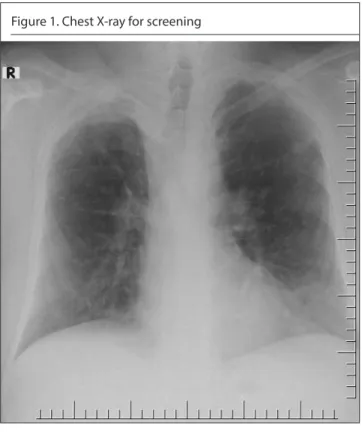
A 58-year-old male patient underwent a screening X-ray of the chest and presented with nodular lesions that were incidental-ly discovered on the radiographic images (Figure 1). He com-plained about exertional dyspnea and intermittent chest pain. His medical history revealed a 40-pack-per-year smoking his-tory without previous tuberculosis. Complete blood count and biochemical analysis results were normal. Thoracic computed tomography (CT) revealed bilateral, peripheral, mostly calcified, nodular lesions with smooth margins, the largest being 1.5 cm (Figure 2). Pulmonary function test results supported the diag-nosis of asthma. Bronchoscopy revealed no endobronchial le-sions. Acid-resistant bacilli were not detected in the bronchoal-veolar lavage fluid, and the cytology was evaluated as benign. Positron emission tomography (PET/CT), which was performed to rule out secondary malignancies, did not reveal a metastatic disease in any part of the body.
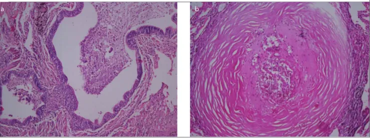
Wedge resection was performed with mini-thoracotomy for di-agnostic purposes. Pathological examination of the lung mass revealed necrotic granulomas separated by distinctive fibrous tissues. Positive-stained fungal hyphae on periodic acid-schiff (PAS) staining were observed in the field of necrosis. Mucus,
neu-Content of this journal is licensed under a Creative
trophils, eosinophils, and lymphoplasmacytic cells infiltrating the bronchiolar wall were detected in the lumen (Figure 3). The reproduction of Aspergillus niger was detected in the sputum culture. As a result of these definitive findings, the patient was diagnosed with BG.
The treatment of the disease was initiated by administering oral itraconazole with inhaled corticosteroids and bronchodilators. However, the patient was unable to tolerate oral itraconazole and was switched to intravenous (IV) amphotericin B (50 mg daily).
Af-ter 14 days of therapy, oral itraconazole was re-initiated but could not be used effectively because the patient was non-compliant with the treatment. Subsequently, IV caspofungin was initiated (50 mg daily) because the patient was clinically and radiologi-cally unresponsive and his blood urea and creatinine levels had increased during the amphotericin B treatment. Clinical recov-ery was achieved after the 14-day therapy, and the patient was discharged with the maintenance treatment of oral itraconazole. The patient did not exhibit any radiological response during the follow-up, and his clinical symptoms had markedly regressed. Written informed consent was obtained from the patient before the commencement of the procedure.
DISCUSSION
According to the BG classification system proposed by Katzen-stein et al. (4), patients with asthmatic symptoms are often ac-companied with bronchopulmonary aspergillosis; however, the underlying cause of non-asthmatic cases is often unknown. The non-asthmatic condition has been associated with chronic gran-ulomatous diseases, glomerulonephritis, influenza virus, myco-bacterial infections, and bronchogenic carcinoma. Patients with asthma-like symptoms are usually younger males aged 20-40 years, whereas those without asthmatic symptoms are relative-ly older (2, 5). In the present case, asthmatic symptoms such as cough, exertional dyspnea, and asthma were not detected, while the respiratory function test results were suggestive of asthma. Radiological evaluations reveal no specific finding of BG; howev-er, the findings are frequently observed unilaterally and located in the upper lobes. The disease usually presents with nodular lesions and may also manifest as atelectasis or pneumonic con-solidation (2, 6). Thoracic CT of idiopathic BG were reported by Umezawa et al. (7) and Li et al. (3) in a 17-year-old male and a 43-year-old female patient, respectively. In these case reports, consolidation and segmental atelectasis were detected. Howev-er, in a report published by Kılıçgün et al. (2), multiple parenchy-mal nodules were observed at the forefront in the radiographs of both a 37-and 58-year-old male patients.
Seçik et al. (8) presented a case of a 40-year-old patient, in whom radiological examination revealed a 3-cm lesion in the right lung, although there were no significant clinical symptoms of the dis-ease. The diagnosis of this patient was not confirmed by bron-choscopy or needle biopsy, instead BG was confirmed by wedge resection with thoracotomy. Because BG has no specific clinical, immunological, and radiological findings, the definite diagno-sis can be made only by performing a histopathological exam-ination of the biopsy specimens (9). Radiological imaging, bio-chemical and immunological tests, bronchoscopy, and sputum and bronchoalveolar lavage cultures have also been performed in previous studies in the literature; however, the final diagnosis was always established by histopathological examination of the open lung biopsy specimens (2, 3, 5, 7). In the previous studies, necrotizing granulomas and eosinophilia in the bronchial wall were detected histopathologically, and fungal elements were detected in some of them. In our case, we performed wedge resection with mini-thoracotomy, and the presence of necrotic
Kuzucuoğlu et al. Bronchocentric Granulomatosis Eur J Ther 2019; 25(2): 150-2
151
Figure 1. Chest X-ray for screeningFigure 2. Bilateral, multiple pulmonary nodules on thoracic computed tomography
granulomas, eosinophilia, and fungal hyphae in the lumen ob-served in the histopathological examination confirmed the diag-nosis of BG. The reproduction of A. niger was also detected in the sputum culture.
Although there is no established consensus for the treatment of BG, several studies have shown that corticosteroid therapy alone is sufficient to effectively treat this disease in non-asthmatic cases without fungal elements (3). However, antifungal therapy should be added to corticosteroids when fungal elements are detected in the histopathological evaluation or sputum culture (2, 3, 7). In the present case, we performed antifungal treatment com-bined with inhaled corticosteroid therapy because our patient was diagnosed with BG associated with aspergillosis. However, we were unable to obtain a radiological response, although the symptomatic response was achieved after treatment. There was no recurrence, progression, or additional pathology observed during follow-up.
CONCLUSION
Bronchocentric granulomatosis has no specific clinical, immuno-logical, and radiological findings, and the definite diagnosis can be made only by performing a histopathological examination. Open lung biopsy should not be avoided in this disease because achiev-ing the differential diagnosis is challengachiev-ing, and it can be difficult to distinguish BG from many diseases radiologically. It is also cru-cial to diagnose the tissue and reveal the underlying cause to tailor the treatment effectively, particularly in symptomatic patients. Informed Consent: Written informed consent was obtained from patient
who participated in this study.
Peer-review: Externally peer-reviewed.
Author Contributions: Concept - M.K., Y.A., F.A., D.A.; Design - M.K., Y.A.,
F.A., D.A.; Supervision - M.K., Y.A., F.A., D.A.; Resources - M.K., Y.A., F.A., D.A.; Materials - M.K., F.A.; Data Collection and/or Processing - M.K., Y.A. Analy-sis and/or Interpretation - M.K., Y.A., F.A., D.A.; Literature Search - M.K., Y.A., F.A.; Writing Manuscript - M.K., Y.A., F.A.; Critical Review - M.K., Y.A., F.A.
Conflict of Interest: The authors have no conflicts of interest to declare. Financial Disclosure: The authors declared that this study has received
no financial support.
REFERENCES
1. Liebow AA. The J. Burns Amberson lecture-pulmonary angiitis and granulomatosis. Am Rev Respir Dis 1973; 108: 1-18.
2. Kılıçgün A, Kurt B, Bölük M, Yılmaz F. Bronchocentric granulomatosis with multifocal lung involvement: A case report. Turk Gogus Kalp Dama 2012; 20: 649-51. [CrossRef]
3. Li H, He J, Gu Y, Zhong N. Corticosteroid monotherapy in a case of bronchocentric granulomatosis with a two-year follow-up. J Thorac Dis 2013; 5: 207-9.
4. Katzenstein AL, Liebow AA, Friedman PJ. Bronchocentric granulo-matosis, mucoid impaction and hypersensitivity reactions to fungi. Am Rev Respir Dis 1975; 111: 497-537.
5. Periwal P, Khanna A, Gothi R, Talwar D. Bronchocentric granuloma-tosis with extensive cystic lung disease in tuberculosis: An unusual presentation. Lung India 2016; 33: 320-2. [CrossRef]
6. Jeong YJ, Kim KI, Seo IJ, Lee CH, Lee KN, Kim KN, et al. Eosinophilic lung disease: a clinical, radiolagic and pathologic overview. Radio-graphics 2007; 27: 617-37. [CrossRef]
7. Umezawa H, Naito Y, Ogasawara T, Takeuchi T, Kasamatsu N, Hashi-zume I. Idiopathic bronchocentric granulomatosis in asthmatic ad-olescent. Respir Med Case Rep 2015; 16: 134-6. [CrossRef] 8. Seçik F, Dalar L, Sarıyıldız S, Özel M, Hacıibrahimoğlu G, Aydoğmuş
Ü, et al. Bir bronkosentrik granülomatozis olgusu. Turk Thorac J 2005; 6: 264-66.
9. Myers JL. Bronchocentric granulomatosis. Disease or diagnosis? Chest 1989; 96: 3-4. [CrossRef]
152
Kuzucuoğlu et al. Bronchocentric Granulomatosis Eur J Ther 2019; 25(2): 150-2
a b
Figure 3. a, b. Histopathologic examination (Hematoxylin and eosin staining, 100×). (a) Lung biopsy specimen showing a small bronchus filled with mucinous material and inflammatory cells. (b) Respiratory epithelium is replaced by granulomatous inflam-mation and luminal debris containing fungal hyphal fragments