IMMUNOREGULATORY ACTIVITIES OF NANOPARTICLE-FORMING
OLIGODEOXYNUCLEOTIDES
A THESIS SUBMITTED TO
THE DEPARTMENT OF MOLECULAR BIOLOGY AND GENETICS AND THE INSTITUTE OF ENGINEERING AND SCIENCE OF
BILKENT UNIVERSITY
IN PARTIAL FULFILLMENT OF THE REQUIREMENTS FOR THE DEGREE OF MASTER OF SCIENCE
BY
KUTAY KARATEPE AUGUST 2009
I certify that I have read this thesis and that in my opinion it is fully adequate, in scope and in quality, as a thesis for the degree of Master of Science.
Assoc. Prof. Dr. İhsan Gürsel
I certify that I have read this thesis and that in my opinion it is fully adequate, in scope and in quality, as a thesis for the degree of Master of Science.
Assist. Prof. Dr. Tamer Yağcı
I certify that I have read this thesis and that in my opinion it is fully adequate, in scope and in quality, as a thesis for the degree of Master of Science.
Assoc. Prof. Dr. Cansın Saçkesen
Approved for the Institute of Engineering and Science
Director of Institute of Engineering and Science Prof. Dr. Mehmet Baray
ABSTRACT
IMMUNOREGULATORY ACTIVITIES OF NANOPARTICLE-FORMING OLIGODEOXYNUCLEOTIDES
Kutay Karatepe
M.Sc. in Molecular Biology and Genetics Supervisor: Assoc. Prof. Dr. İhsan Gürsel
August 2009, 99 pages
Innate immune system is activated by a wide range of microbial by products leading to an immediate immune activation primarily designed to neutralize and control the invading insult. The cells of the innate immune system also instruct the development of antigen-specific adaptive immunity. While TLR9 is triggered by bacterial DNA, extended and over-exuberant immune response poses a threat since it may exacerbate cell and tissue destruction leading to organ failure. Telomeric TTAGGG conserved motifs are previously reported to antagonize TLR mediated events. The down-regulatory effect of these motifs may help to restore the desired homeostatic balance of the immune system. While CpG ODN patterned after bacterial DNA can be harnessed in different clinical settings to provide an advantage to host to resist infectious diseases, control tumor growth or alleviate allergic symptoms, the immunosuppressive telomeric motifs could be effectively applied in controlling systemic anti-inflammatory or autoimmune related disorders.
Several challenges exist in the utilization of synthetic ODNs in the clinic. The first challenge is that conventional classes of synthetic ODNs exhibit different properties. K-type ODNs are more effective in proliferation and activation of B cells and DC. D-type ODNs are in nanoparticle forms, lead to anti-viral type I IFN production and mature monocytes into DCs. Of note, the efficacy of these synthetic ODNs is reduced under physiological conditions due to premature clearance and low levels of internalization. Moreover, D-ODNs as one of the most potent IFNα inducing TLR9 ligands possess a large-scale production problem due to 3’polyG-runs, which hamper their entry into the clinic.
We have designed a novel class of ODN, designated as ODN420, devoid of polyGs that can undergo nanoparticle formation necessary for its IFNα induction. Ex vivo stimulation of mouse splenocytes and in vivo administration of ODN420 have revealed that this ODN exhibits higher immunostimulatory potential and is more stable than most commonly used ODNs due to its nanoparticle-forming ability. Another interesting finding is that ODN420 with the natural phosphodiester (PO) backbone is at least as potent as its more stable counterpart with the modified phosphorothioate backbone. Furthermore, it combines superior properties of
conventional classes of K and D-ODNs. These results have been reproduced in human peripheral blood mononuclear cells by various assays.
Next, we have analyzed whether this ODN could be utilized as a vaccine adjuvant and an anti-cancer agent with two independent experiments. Our immunization results demonstrate that ODN420 induces a higher level of Th1-mediated response than conventional ODNs and is a promising candidate as a vaccine adjuvant. This response is hampered when ODN420 is used in combination with ODN-A151. In the tumor xenograft model, ODN420 has promoted partial remission of the tumors or delayed the tumor growth. This knowledge will pave the way for more effective immunotherapeutic approaches.
Keywords: TLR, immunomodulatory DNA, nanoparticle, anti-cancer immunotherapy, vaccine
ÖZET
NANOPARÇACIK OLUŞTURAN OLİGODEOKSİNÜKLEOTİDLERİN İMMÜNMODÜLATÖR ETKİLERİ
Kutay Karatepe
Moleküler Biyoloji ve Genetik Yüksek Lisans Danışman: Doç. Dr. İhsan Gürsel
Ağustos 2009, 99 sayfa
Doğal bağışıklık sistemi öncelikle değişik mikroorganizmaların yan ürünlerini tanıyarak mikroorganizmanın yayılmasını kontrol edecek mekanizmaları harekete geçirir. Bağışıklık sisteminin bu kolundaki hücreler aynı zamanda antijene özgü kazanılmış bağışıklık sistemi hücrelerini de uyarır.
CpG-motifleri taşıyan bakteriyel DNA TLR9 almaçı tarafından tanınır. Ancak bağışıklık sisteminin uzun ve aşırı bir şekilde aktif halde olması organ hasarına kadar gidebilecek hücre ve doku yıkımına sebep olabilir. Memeli genomundaki korunmuş TTAGGG motiflerinin TLR almaçlarının sinyal yolaklarını bastırdığı daha önceden ispat edilmiştir. Bu baskılayıcı etki bağışıklık sisteminde homeostatik dengeyi tekrar eski haline getirir. Bakteriyel DNA’lardan esinlenmiş CpG-motifleri taşıyan sentetik DNA’lar bulaşıcı hastalıklar, kanser veya alerji gibi çeşitli klinik durumlarda kullanılabilirken, bağışıklık sistemini bastırıcı DNA parçacıkları antiinflamatuar etki gerektiren sistemik otoimmün hastalıklar için çözüm vaat etmektedir.
Sentetik DNA parçacıklarının (ODN) klinikte kullanılmasının önünde çeşitli engeller bulunmaktadır. Şu ana kadar kullanılmakta olan ODN’ler ait oldukları alt sınıfa göre farklı etkilere yol açarlar. K-tipi ODN’ler B hücrelerinin ve dendritik hücrelerinin çoğalması ve aktivasyonunda daha etkilidir. D-tipi ODN’ler ise nanoparçacık yapısında olup anti-viral tip I interferon üretimini ve monositlerin dendritik hücrelere farklılaşmasını tetikler. Ayrıca bu
sentetik ODN’lerin hücre içine alınmadan nükleazlar tarafından parçalanması bunların fizyolojik koşullardaki etkinliğini azatmaktadır. Son olarak, interferon-alfa üretimini tetikleyen D-tipi ODN’ler poli-G uçları içerdiğinden farkı büyüklükte parçacıklar oluşturur. Heterojen yapılar oluşturan D-tipi ODN’lerin büyük ölçekli üretimi ve klinikte kullanımı problemlidir.
Biz poli-G ucu içermeyen, interferon-alfa üretimini tetikleyebilecek homojen
nanoparçacıklar oluşturan ODN420 adını verdiğimiz yeni bir sınıf ODN geliştirdik. Fare dalak hücrelerinin ex vivo uyarımı ve bu ODN’lerin in vivo enjeksiyonu sonucunda elde ettiğimiz sonuçlar ODN420’nin daha önceden kullanılan ODN’lere göre bağışıklık sistemini daha yüksek seviyede tetiklediğini ve nanoparçacık yapısından ötürü eksonükleazlara göre daha dirençli olduğunu gösterdi. Aynı zamanda bu ODN’nin doğal fosfodiester iskelet içeren halinin daha stabil olan modifiye edilmiş fosforotioat iskeletli haliyle benzer aktivite gösterdiğini bulduk. Ayrıca, bu ODN’nin K ve D-tipi ODN’lerin üstün özelliklerini taşıdığını gösterdik. Bu sonuçlar insan kanından izole edilmiş bağışıklık sistemi hücrelerinde de tekrar gösterildi.
Sonra iki farklı hayvan deneyinde bu ODN’nin aşı adjuvanı ve anti-kanser ajanı olarak potansiyelini inceledik. Aşı deneyindeki sonuçlar ODN420’nin şu anda kullanılan ODN’lere göre daha etkin bir antijene özgü Th1 etkisi yarattığını ve bu etkinin ODN-A151 tarafından azaltıldığını bulduk. Tümör modelinde ise ODN420, tümörün büyümesini tersine çevirdi veya geciktirdi. Bu bulgular bağışıklık sisteminin manipülasyonuyla daha etkin tedavi yöntemleri bulunmasına ışık tutacaktır.
Anahtar kelimeler: TLR, immunomodülatör DNA, nanoparçacık, anti-kanser terapi, aşı
ACKNOWLEDGEMENTS
I would like to express my deepest appreciation to my advisor, Assoc. Prof. Dr. İhsan Gürsel. Not only is he an excellent supervisor and scientist but he is also a great mentor. I would like to thank him for his invaluable guidance, support, teaching and patience during my studies. He has always helped me to push my limits and he is one of the few faculty members, who work in the lab on the bench next to his students.
I would like to thank my laboratory mates Fuat, Gizem, Erdem and Tamer as wells as former lab mates Hande and Rashad for their help, precious friendship and support. I have been working in Ihsan Gursel lab for almost four years now and I can only wish I work in such exceptionally friendly academic environments, where lab feels like “home”.
Another exceptional person, who has contributed to this thesis and my personal
development as a young scientist, is Assoc. Prof. Dr. Mayda Gursel. She has never hesitated to give me a hand whenever I had a problem.
Moreover, I would like to thank to my dearest friends Emre, Elif, Nilüfer, Gurbet, Bala, Zeynep, Sumru, Ceren, Ceyhan, Ayça and Ender for their support and for always being there whenever I needed. Also, I would like to thank my roommates, Burçin and Egemen, for reminding me that I need to eat while writing this thesis. I am also thankful to Balım for her careful review of my thesis.
I would also like to thank Burcu for her help, support and patience during animal experiments and “Abdullah Abi” for solving every technical problem I have encountered.
My sincere thanks go to MBG Family for their guidance, companionship and assistance. It has been six years since I entered this department as an undergraduate student. I have learned a lot from every member of this department.
I would also like to thank to The Scientific and Technological Research Council of Turkey (TÜBİTAK) for their financial support throughout my master studies.
Without my family, none of the exceptional things in my life would have been possible. I would like to express my love and gratitude for their everlasting support in life.
TABLE OF CONTENTS SIGNATURE PAGE ... ii ABSTRACT... iii ACKNOWLEDGEMENTS... v TABLE OF CONTENTS... vi LIST OF TABLES………...viii LIST OF FIGURES………ix ABBREVIATIONS ... xi 1. INTRODUCTION ... 1 1.1. Immune System ... 1
1.2. Innate Immune System ... 3
1.2.1. Pathogen Recognition Receptors (PRR)... 6
1.2.1.1. Toll-like Receptors (TLRs)... 8 1.2.1.1.1. TLR 1, TLR2 and TLR6 ... 9 1.2.1.1.2. TLR3 ... 10 1.2.1.1.3. TLR4 ... 10 1.2.1.1.4. TLR5 ... 11 1.2.1.1.5. TLR7 and TLR8... 11 1.2.1.1.6. TLR9 ... 11 1.2.1.2. TLR Signaling Pathways ... 12 1.2.1.2.1. MyD88-Dependent Pathway... 13 1.2.1.2.2. MyD88-Independent/TRIF-Dependent Pathway... 14
1.3. A Deeper Insight into Immunostimulatory DNA Particles and TLR9 Activation 15 1.3.1. Accessory Molecules Involved in TLR9 Activation ... 16
1.3.2. Different Classes of Synthetic CpG-ODNs ... 17
1.3.2.1. A/D-type CpG ODNs... 18
1.3.2.2. K/B-type CpG ODNs... 20
1.3.2.3. C-type CpG ODNs... 21
1.3.2.4. Other Types of CpG-ODNs ... 22
1.3.3. Differential Immune Response Mediated by Particulate and Linear CpG ODN ………24
1.4. Utilization of CpG ODNs as Therapeutic Agents... 26
1.5. Synthetic DNA Containing Mammalian Telomeric Repeats with Immunosuppressive Activity as Another Class of Nanoparticle Forming ODNs ... 28
1.6. Utilization of Immunosuppressive ODNs as Therapeutic Agents... 30
2. AIM OF STUDY ... 32
3. MATERIALS & METHODS ... 34
3.1. MATERIALS... 34
3.1.1. Reagents... 34
3.1.2. Cell Culture Media, Buffers and Other Standard Solutions ... 36
3.2. METHODS ... 37
3.2.1. Cell Culture... 37
3.2.1.1. Cell Lines... 37
3.2.1.2. Single Cell Splenocyte Preparation ... 37
3.2.1.3. Peripheral Blood Mononuclear Cell (PBMC) Isolation from Whole Blood
………37
3.2.1.4. Cell Counting and Distribution... 38
3.2.1.5. Stimulation Protocols... 39
3.2.2. Fluorescence Activated Cell Sorting (FACS)... 40
3.2.2.1. Cell Surface Marker Staining... 40
3.2.2.2. Intracellular Cytokine Staining... 40
3.2.2.3. Carboxyfluorescein Diacetate Succinimidyl Ester (CFSE) Assay ... 41
3.2.3. ELISA ... 42
3.2.3.1. Cytokine ELISA... 42
3.2.3.2. IgG anti-OVA ELISA ... 43
3.2.4. Determination of Gene Expression at mRNA Level ... 43
3.2.4.1. Total RNA Isolation... 43
3.2.4.2. cDNA Synthesis... 44
3.2.4.3. PCR... 44
3.2.4.4. Agarose Gel Electrophoresis and Quantification of Band Intensities ... 48
3.2.5. In vivo Experiments ... 48
3.2.5.1. Maintenance of the Animals ... 48
3.2.5.2. Immunization Protocol with Specific ODNs and OVA... 48
3.2.5.3. Tumor Xenograft Model... 49
3.2.6. Statistical Analysis... 49
4. RESULTS ... 50
4.1. In vitro Stimulatory Potential of Dendrimeric CpG-ODNs in Mouse Splenocytes50 4.2. In vitro Stimulation Assays Using Human PBMC ... 51
4.2.1. Activation of Dendritic Cell markers and IL6 Secretion by Human PBMC as a Result of Stimulation with 1 µM NP-forming ODN Treatment... 51
4.2.2. Proliferation of Immune Cells Upon Stimulation with CpG-ODNs... 53
4.2.3. TNFα Production by pDC After Stimulation with CpG-ODNs ... 54
4.2.4. Dose-dependent Response of Human PBMC to NP-forming ODN Treatment ………56
4.2.5. Gene Expression Studies with Human PBMC After Stimulation with NP-forming ODNs at Optimal Doses... 60
4.3. In vivo Studies Utilizing Nanoparticle Forming Immunostimulatory and Immunosuppressive ODNs ... 64
4.3.1. In vivo Stimulatory Potential of Nanoparticle Forming ODNs ... 64
4.3.2. Immunization of C57BL/6 Mice with Dendrimeric ODNs and OVA in Combination With or Without Nanoparticle Forming Suppressive ODN... 68
4.3.3. Use of Nanoparticle Forming ODNs as Anti-Cancer Agents... 71
5. DISCUSSION... 75 6. FUTURE STUDIES ... 82 7. REFERENCES ... 84 8. APPENDICES ... 96 8.1. Appendix A... 96 8.2. Appendix B ... 98 vii
LIST OF TABLES
Table 1. The cardinal features of innate and adaptive immunity... 2
Table 2. Cytokines affect behavior of target cells. ... 5
Table 3. Chemokines recruit target cells to sites of infection... 5
Table 4. PRR families in mammalian cells and their associated ligands... 7
Table 5. TLR9 expression varies among mice and human... 12
Table 6. Sequences and product sizes of the primers for mouse genes. ... 45
Table 7. Sequences and product sizes of the primers for human genes... 46
Table 8. PCR reactants... 47
Table 9. PCR conditions for mouse primers... 47
Table 10. PCR conditions for human primers ... 47
Table 11. IgG anti-OVA ELISA for total IgG, IgG1, IgG2a and IgG2b subtypes in sera from primary bleeding. ... 69
Table 12. IgG anti-OVA ELISA for IgG, IgG1, IgG2a, IgG2b subtypes in sera from secondary bleeding... 70
LIST OF FIGURES
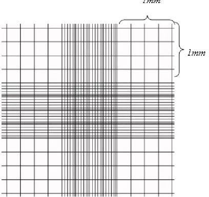
Figure 1. The TLR family members and their subcellular localizations ... 9 Figure 2. Self-DNA containing immune complexes is recognized by TLR9 and lead to the autoimmune disorder, SLE ... 13 Figure 3. G-tetrad links four D-ODN strands for higher-order structure formation... 20 Figure 4. Duplex formation through palindromic sequences by two monomeric C-ODNs . ... 22 Figure 5. Y-shaped CpG ODN... 23 Figure 6. Formation of uniform-sized nanoparticles in a novel bifunctional dendrimeric ODN, designated as ODN420... 24 Figure 7. Dichotomy of different CpG ODNs ... 26 Figure 8. Proposed mechanism of CpG ODN-mediated anti-tumor activity... 28 Figure 9. ODN420: Schematic representation of the ODN420 formation via the use of two bifunctional linkers at the 3`-end of ODN 1466 sequences... 35 Figure 10. ODN421: Schematic representation of the ODN421 formation via the use of two bifunctional linkers at the 3`-end of ODN 1471 sequences... 35 Figure 11. ODN422: Schematic representation of the ODN422 formation via the use of a bifunctional linker for G=2 and a trifunctional linker for G=3 at the 3`-ends of ODN 1466 sequences. ... 36 Figure 12. ODN423: Schematic representation of the ODN423 formation via the use of a bifunctional linker for G=2 and a trifunctional linker for G=3 at the 3` ends of ODN 1471 sequences. ... 36 Figure 13. Neubaer cell counting chamber ... 38 Figure 14. Immunization schedule for in vivo vaccination... 49 Figure 15. IL6 production by murine splenocytes in response to 0.3 µM and 1 µM
concentrations of CpG-ODN stimulation ... 51 Figure 16. IL6 secretion by human PBMC in response to 1 µM concentration of CpG-ODN stimulation... 52 Figure 17. Activation of DC markers as a result of 1 µM CpG-ODN stimulation... 53
Figure 18. Proliferating B cell percentage determined by CFSE assay and CD19 staining after stimulation with various ODNs at 1 µM concentration for 72 hours.. ... 54 Figure 19. TNFα-positive cells in BDCA2+ CD123+ pDC stimulated with 1 µM CpG-ODN in selected groups.. ... 55 Figure 20. TNFα-positive cells in BDCA2+ CD123+ pDC stimulated with 1 µM CpG-ODN in all groups... 56 Figure 21. IL6 secretion by human PBMC in response to 0.1, 0.3 and 1 µM
concentrations of CpG-ODN stimulation.. ... 57 Figure 22. IFNγ secretion by human PBMC in response to different concentrations of CpG-ODN stimulation... 57 Figure 23. Activation of DC markers upon treatment with ODN420 at 0.1, 0.3 and 1 µM concentrations.. ... 58 Figure 24. Dose-dependent activation of DC markers upon ODN treatment... 59 Figure 25. IL6 secretion by human PBMC in response to 0.1, 0.3, 1 and 3 µM
concentrations of CpG-ODN stimulation. ... 60 Figure 26. The agarose gel picture of the RT-PCR products of tnfα, ifn-related genes and endosomal tlr genes. ... 61 Figure 27. Fold induction graphs of various interferon related genes and endosomal tlr genes at mRNA level for the first sample... 64 Figure 28. The agarose gel picture of the RT-PCR products of ip10, il15, ifnγ and
endosomal tlr genes in splenocytes of mice injected with 20 µg of ODN with PS
backbone or 60 µg of ODN with PO backbone. ... 65 Figure 29. Relative band intensities of various critical genes in in vivo administration of NP-forming ODN. The expression profiles of ip10 (A), il15 (B), ifnγ (C), tlr3 (D), tlr7 (E) and tlr9 (F) in untreated and treated groups ... 67 Figure 30. The agarose gel picture of the RT-PCR products of ip10, il15, ifnγ and
endosomal tlr genes in splenocytes of mice immunized with two doses of 15 µg ODN and 7.5 µg OVA... 71 Figure 31. The effect of nanoparticle forming ODNs as anti-cancer agents. ... 73 Figure 32. Tumor xenografts in untreated and ODN420-treated mice …...………..73
ABBREVIATIONS
AFM Atomic Force Microscopy
APC Antigen presenting cell
AVA Anthrax vaccine adsorbed
bp Base pairs
BCG Bacille Calmette Guerin of Mycobacterium bovis
BCR B-cell receptor
BSA Bovine serum albumin
CD Cluster of differentiation
cDNA Complementary Deoxyribonucleic Acid
CFA Complete Freund’s adjuvant
CFSE Carboxyfluorescein Diacetate Succinimidyl Ester
CMV Cytomegalovirus
CpG Unmethylated cytosine-phosphate-guaniosine motifs
CCR Receptor specific for CC chemokine
CRP C-reactive protein
CXCL CXC-chemokine ligand
DC Dendritic cell
DMEM Dulbecco's Modified Eagle's Medium
DNA Deoxyribonucleic acid
dsRNA Double-stranded RNA
ELISA Enzyme Linked-Immunosorbent Assay
ER Endoplasmic reticulum
ERGIC Endoplasmic reticulum-Golgi intermediate
compartment
FACS Fluorescence Activated Cell Sorting
FBS Fetal Bovine Serum
HBV Hepatitis-B Virus
HIV Human Immunodeficiency Virus
HMGB High mobility group box
Ig Immunoglobulin
IκK Inhibitor kappa B kinase
IL Interleukin
iNOS Inducible nitric oxide synthase
IFN Interferon
IRAK IL-1 receptor-associated kinase
IRF3 Interferon-regulatory factor 3
LBP LPS-binding protein
LPS Lipopolysaccharide
LRR Leucine-rich repeats
LTA Lipotheicoic acid
MALP Mycoplasmal lipopeptide
MAP Mitogen-activated protein
MAPK Mitogen-activated protein kınase
MCMV Murine cytomegalovirus
MCP Monocyte chemoattractant protein
MDP Muramyl dipeptide
MHC Major histocompatibility complex
MIP Macrophage inflammatory protein
mDC Myeloid dendritic cells
MSR Macrophage scavenger receptor
MyD-88 Myeloid differentiation primary response gene 88
NF-κB Nuclear factor-kappa B
NK Natural killer
NLR Nucleotide-binding oligomerization domain like proteins
or receptors
NO Nitric oxide
NOD Nucleotide-binding oligomerization domain
ODN Oligodeoxynucleotide
OVA Ovalbumin
PAMP Pathogen associated molecular pattern
PBMC Peripheral blood mononuclear cell
PBS Phosphate buffered saline
PCR Polymerase chain reaction
pDC Plasmacytoid dendritic cells
PGN Peptidoglycan
PHA Phytohemagglutinin
pI:C Polyriboinosinic polyribocytidylic acid
PNPP Para-nitrophenyl pyro phosphate
PRR Pattern recognition receptors
PO Phosphodiester
PS Phosphorothioate
RIH RIG-like helicase
RIP Receptor-interacting protein
RNA Ribonucleic acid
RPMI Roswell Park Memorial Institute
RT Reverse transcriptase
SA-AKP Streptavidin alkaline-phosphatase
SAP Serum amyloid protein
SLE Systemic lupus erythematosus
SSCL Sterically stabilized cationic liposomes
ssRNA Single-stranded RNA
TCR T-cell receptor
TH T-helper
Th1 Cellular immunity
Th2 Humoral immunity
TIR Toll/IL-1 receptor
TIRAP Toll/IL1 receptor-associated protein
TLR Toll-like receptor
TNF Tumor necrosis factor
TRAF TNF-associated factor
TRAM TRIF-related adaptor molecules
TRIF TIR domain containing adaptor inducing IFN-β
1. INTRODUCTION
1.1. Immune SystemAll vertebrates constantly encounter potentially harmful foreign substances, known as “antigens”. In order to protect the body from foreign antigens by distinguishing them from the organism’s normal cells and tissues, the cells of the immune system are in a state of constant surveillance.
The first line of defense is formed from the physical barriers provided by our skin and mucosa. If this primary defense of line cannot prevent pathogens from entering the organism, the innate immune system is activated by the “danger signal”. According to the “danger signal” principle, cells of the innate arm of the immune system detect a specific signature molecule commonly expressed on a large verity of pathogens. These molecules, also known as Pathogen Associated Molecular Patters (PAMP) are absent on the host’s own cells and are recognized by germline encoded Pattern Recognition Receptors (PRR) of the cells of innate immunity. Upon an encounter with the “danger signal”, these cells mount a robust increase in the production of inflammatory molecules so that the invading infectious organism can be contained and eradicated. In most cases, encounters between antigens and immune cells pass by unnoticed, due to the effective protective mechanisms provided by these primary lines of defense.
In certain cases, these lines of defense are breached and the innate immune system cannot contain the infection. This results in the activation of the adaptive immune system and the cooperation between the innate immune system and adaptive immune system allows the clearance of the pathogen. This thesis focuses on manipulation of the innate immune system for therapeutic purposes and thus, the details regarding the function of the adaptive immune system are beyond the scope of this study. Therefore, briefly, adaptive immunity is mainly characterized by clonally expanded T cells and B cells. These cells make use of highly specific antigen receptors -T cell receptor (TCR) and B cell receptor (BCR), respectively- to individual antigenic peptide molecules.
Furthermore, unlike the cells of the innate immune system, they are able to retain memory for a previously encountered antigen.
Antigen presenting cells (APCs), including dendritic cells (DCs), macrophages and B cells, are found in their immature state throughout the body at the possible entry sites of pathogens such as mucosal surfaces and skin. Upon recognition of pathogens, DCs phagocytose and process the microbial antigens to their peptide content. These antigens are loaded onto major histocompatibility complex (MHC) class I and II molecules for presentation to CD8+ and CD4+ T cells, respectively. During this
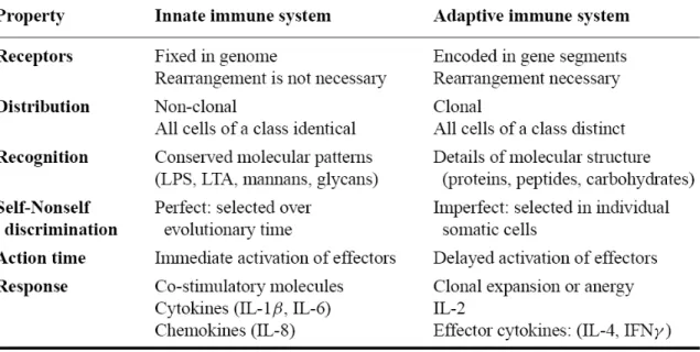
maturation process, DCs upregulate the chemokine receptor CCR7 and migrate towards the draining lymph node, the site of antigen presentation. Additionally, the expression of co-stimulatory molecules (i.e. CD80 and CD86) and cytokines required for effective activation of T cells is up-regulated and MHC molecules are translocated from the intracellular vesicles to plasma membrane. In the lymph node, upon proper antigen recognition, naïve lymphocytes become activated (Janeway, 2001; Lee and Iwasaki, 2007). When the strong and specific response generated by the adaptive immune system is dysregulated, however, it becomes responsible for allergy and autoimmunity. Adaptive immunity is also responsible for tissue rejection cases in transplantation surgeries. The major differences between innate immune system and adaptive immune system are given in Table 1.
Table 1. The cardinal features of innate and adaptive immunity. (Janeway and Medzhitov, 2002)
1.2. Innate Immune System
The innate immune system recognizes and responds to pathogens in a non-specific manner. Due to the generic mechanisms activated by innate immune system, it does not accommodate long-lasting or protective immunity to the host (Alberts, 2008). The major functions of the vertebrate innate immune system include:
• Recruitment of immune cells to sites of infection and inflammation through the production of cytokines,
• Activation of the complement cascade to identify bacteria, activate cells and to promote clearance of dead cells or antibody complexes,
• Identification and removal of foreign substances present in organs, tissues, blood and lymph by specialized white blood cells,
• Activation of the adaptive immune system through a process known as antigen presentation. (Murphy et al., 2008)
Major cell types in the innate immune system are natural killer (NK) cells, mast cells, eosinophils, basophils and phagocytes (i.e. neutrophils, monocytes/macrophages, DCs). NK cells destroy infected cells in a process known as “missing self”. As MHC I molecule, located on the surface of all hosts cells, tags the cells as “self”, the
downregulation or absence of this molecule activates NK cells for killing. Such is the case of virally infected hosts cells that down regulate cell-surface MHC I molecules (Murphy et al., 2008). NK cells also attack tumor cells by various mechanisms (Raulet and Guerra, 2009).
Mast cells release characteristic granules rich in histamine and heparin, along with various hormonal mediators and chemokines. Histamine promotes dilation of blood vessels, a characteristic sign of inflammation, and recruits other immune cells to sites of infection. Although mast cells are primarily responsible for wound healing and defense against pathogens, they are also associated with allergy and anaphylaxis. (Murphy et al., 2008)
Basophils and eosinophils are known as granulocytes along with neutrophils due to the presence of granules in their cytoplasm. Basophils release histamine and
eosinophils release a variety of toxic proteins. These products are effective in killing
pathogens. They are, however, also responsible for tissue damage during allergic reactions such as asthma (Macfarlane et al., 2000).
Phagocytes of the innate immune system include neutrophils, macrophages and DCs. Neutrophils are the most abundant type of phagocytes and comprise approximately 50-60% of total circulating leukocytes. Neutrophils are usually the first cells to arrive at the site of an infection. Their granules contain toxic substances that kill or inhibit growth of bacteria and fungi. Upon binding of bacterial molecules to receptors on neutrophils, neutrophils activate a “respiratory burst” causing the generation of reactive oxygen species such as hydrogen peroxide (H2O2). Macrophages are another type of cell that utilizes a similar “respiratory burst”. They are the most efficient phagocytes and can phagocytose a substantial number of pathogens. The precursors of these tissue-resident macrophages are blood circulating monocytes. As mentioned previously, DCs are the major cell type responsible for antigen presentation. Hematopoietic bone marrow progenitor cells transform into immature dendritic cells characterized by high endocytic activity and low T-cell activation potential. Immature DCs circulate throughout the body and upon pathogen recognition, they phagocytose pathogens, process antigenic molecules into peptides and present them to T cells, thereby activating adaptive immune system (Banchereau and Steinman, 1998). In addition to their antigen presentation function, macrophages and DCs also induce a number of cytokines and chemokines, which allow recruitment of other immune cells to sites of infection, during the onset of innate immune activation. The properties of major cytokines and chemokines are given in Table 2 and Table 3.
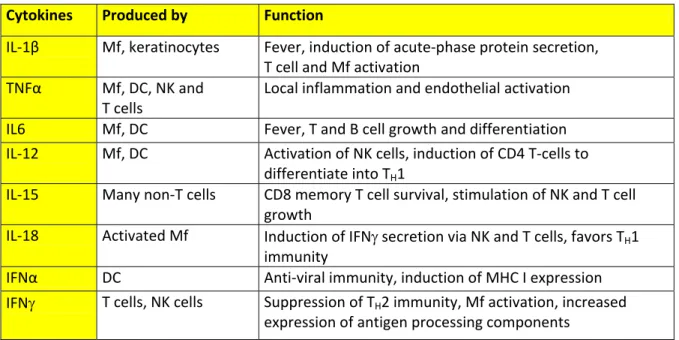
Table 2. Cytokines affect behavior of target cells. (Janeway, 2005; Murphy et al., 2008)
Mf: Macrophages, T: T cells, DC: Dendritic cells, NK: Natural Killer cells
Cytokines Produced by Function
IL‐1β Mf, keratinocytes Fever, induction of acute‐phase protein secretion, T cell and Mf activation TNFα Mf, DC, NK and T cells Local inflammation and endothelial activation IL6 Mf, DC Fever, T and B cell growth and differentiation IL‐12 Mf, DC Activation of NK cells, induction of CD4 T‐cells to differentiate into TH1
IL‐15 Many non‐T cells CD8 memory T cell survival, stimulation of NK and T cell growth
IL‐18 Activated Mf Induction of IFNγ secretion via NK and T cells, favors TH1 immunity
IFNα DC Anti‐viral immunity, induction of MHC I expression IFNγ T cells, NK cells Suppression of TH2 immunity, Mf activation, increased
expression of antigen processing components
Table 3. Chemokines recruit target cells to sites of infection. (Janeway, 2005; Murphy et al., 2008)
Chemokines Produced by Attracted cells Major effect CXCL8 (IL‐8) Monocytes, Mf, DC Neutrophils, naïve T cells Mobilization, activation and degranulation of neutrophils CCL3 (MIP‐1α) Monocytes, T, Mast cells, fibroblasts Monocyte,NK, T, basophil, DC Promotes TH1, antiviral defense, competes with HIV‐1 CCL4 (MIP‐1β) Monocytes, Mf, Neutrophils, endothelium Monocyte, NK, T, DC Competes with HIV‐1 CCL2 (MCP‐1) Monocyte, Mf, fibroblast, keratinocyte Monocyte, NK, T, basophil, DC Promotes TH2, activate Mf, histamine release from basophils CXCL10 (IP10) T, fibroblast, endothelial, monocyte, keratinocyte Resting T cells, NK, monocytes Promotes TH1, antiangiogenic, immunostimulant 5
1.2.1. Pathogen Recognition Receptors (PRR)
While T cell and B cell receptors are formed by rearrangements in gene segments and can recognize an infinite variety of antigens, the receptors of innate immune system are germline-encoded pattern recognition receptors (PRR). These receptors recognize certain signature patterns on pathogens termed as pathogen-associated molecular patterns (PAMP). These include viral nucleic acids, components of bacterial and fungal cell walls and many more. For a long time, innate immunity has been considered as a non-specific immune response characterized by engulfment and digestion of microorganisms and foreign substances by phagocytic cells (Akira et al., 2001). The discovery of PRRs, the specificity of response generated by these cells and the notion that their ligands, PAMPs, can be utilized as adjuvants has fueled interest in the previously underestimated field of innate immunity.
PRRs share common properties among themselves. First, PAMPs recognized by PRRs are essential for the survival of the microorangism and are therefore difficult for the microorganism to modify. Second, PRRs are expressed constitutively in the host and detect the pathogens regardless of their life-cycle stage. Third, PRRs are germline
encoded, nonclonal and expressed on all members of a given cell type. Furthermore, cells carrying these receptors lack immunologic memory. Finally, the basic components of innate immunity are highly conserved among species, from plants and fruit flies to mammals (Akira et al., 2006).
There are different PRR families and these are outlined in Table 4. Mannan-binding lectin (MBL), C-reactive protein (CRP), and serum amyloid protein (SAP) are secreted PRRs produced by the liver during the acute phase response at the early stages of infection. While CRP and SAP are members of the pentraxin family, MBL is a member of the collectin family. CRP and SAP bind to phosphorylcholine on bacterial surfaces and opsonize microorganisms leading to their phagocytosis. MBL binds specifically to terminal mannose residues, which are abundant on the surface of many microorganisms, it initate the lectin pathway of complement by cleaving C2 and C4 proteins.
In addition to these extracellular PRRs, many PRRs reside on the plasma
membrane. One family of PRRs, which is located on the plasma membrane, is scavenger
receptors. Macrophage scavenger receptor (MSR) has a broad specificity to a variety of ligands, such as double-stranded RNA (dsRNA), LPS, and LTA. Another scavenger receptor expressed on macrophages is MARCO, which binds to bacterial cell walls and LPS, and also mediates phagocytosis of bacterial pathogens (Janeway and Medzhitov, 2002).
NOD-like receptors (NLRs) and RIG-like helicases (RIHs) are two major families of PRRs found in the cytoplasm. NOD1 and NOD2 detect bacterial peptidoglycan, drive activation of mitogen-activated protein kinases (MAPKs) and NF-kB. The minimal structural requirement for activation of NOD2 is muramyl dipeptide, a structure present in both Gram-positive and Gram-negative bacteria. NALP proteins such as NALP1, NALP2 and NALP3 also belong to NLR family of PRRs. They have a cruicial role in activation of proinflammatory caspases through formation of a complex called the inflammasome. Retinoic-acid-inducible gene I (RIG-I) and melanoma-differentiation-associated gene 5 (MDA5) are two members of RIH family. They are activated by dsRNA during a viral infection and trigger the activation of NF-kB and IRF3/7, which cooperate in induction of antiviral type I IFNs (Meylan et al., 2006).
Table 4. PRR families in mammalian cells and their associated ligands. (Lee and Kim, 2007)
1.2.1.1. Toll-like Receptors (TLRs)
The main focus in this study is toll-like receptors (TLR), which are the most extensively studied and characterized family of PRRs. Mammalian TLRs are homologous to the Drosophila Toll receptor (Medzhitov et al., 1997). These evolutionarily conserved receptors belong to the IL-1R superfamily, characterized by an extracellular leucine-rich repeats (LRR) and an intracellular Toll/IL-1 receptor like (TIR) domain (Medzhitov, 2001). Members of TLR family differ from each other in their ligand specificities, expression patterns, and the downstream signaling pathways.
TLRs in the innate immune system serve an essential role not only in recognition of pathogen, but also in directing the course and type of innate immune response
generated following an exposure to foreign antigen (Takeda et al., 2003). TLRs have been demonstrated to have a wide array of functions including initiation of
proinflammatory responses and antiviral responses, up-regulation of costimulatory molecules on antigen presenting cells (APC), release of chemokines to induce migration of responder cells to the site of infection and cross-priming of T cells by DCs (Takeda and Akira, 2005). TLRs are responsible for the adjuvant activity that is required to initiate immune responses both in natural infection and in vaccine responses (Lien and
Golenbock, 2003). TLRs also have an essential role in shaping adaptive immune responses to pathogen. The signals for activation of adaptive immunity are mostly
provided by DCs. TLR-mediated recognition of pathogens by DCs induces the expression of costimulatory molecules such as CD80/CD86 required for the effective activation of T cells and production of inflammatory cytokines such as IL-12 (Akira et al., 2001). DC subsets can induce cellular immunity (Th1) and humoral immunity (Th2). Activation of TLR9 in DCs induces production of IL-12, thereby changing the helper T cell (Th) differentiation toward Th1 type. LPS stimulates TLR4 signaling pathway and allows DCs to support both Th1 and Th2 cell differentiation (Kaisho et al., 2002). Additionally, some pathogen-derived adjuvants such as Complete Freund’s Adjuvant (CFA), Bacille
Calmette Guerin of Mycobacterium bovis (BCG) are recognized by TLRs; TLR9 and TLR2, TLR4 respectively, which may explain the involvement of TLRs in adaptive immunity (Akira et al., 2003).
Human TLR4 is the first characterized mammalian Toll-like receptor. It is expressed in a variety of cell types, most predominantly in the cells of the immune
system, including macrophages and DCs (Medzhitov et al., 1997). TLRs can be classified according to their localization in the cells. While TLR1, 2, 4, 5, 6 and 10 are localized on the plasma membrane and recognize mainly extracellular bacterial products, TLR3, 7, 8 and 9 are located in the intracellular endosomal and/or ER compartments and recognize viral or bacterial nucleic acids (Iwasaki and Medzhitov, 2004; Latz et al., 2004).
Subcellular localizations and ligands of TLRs are given in detail in Figure 1.
Figure 1. TLR family members and their subcellular localizations (Takeda and Akira, 2005).
1.2.1.1.1. TLR 1, TLR2 and TLR6
TLR2 responds to various microbial products, including lipoproteins, Gram-positive bacterial PGN and LTA, lipoarabinomannan from mycobacteria,
glycosylphosphatidylinositol anchors from a protozoan Trypanosoma cruzi, a phenol-soluble modulin from Staphylococcus epidermis, zymosan from fungi (Takeda and Akira, 2005). The wide spectrum recognition of microbial components by TLR2 is due to its
ability to form heterodimers with other TLRs such as TLR1 and TLR6. Inflammatory response to mycoplasma-derived triacyl and diacyl lipopeptides is deficient in TLR6 and TLR1 deficient mice, respectively. This proves that TLR1 and TLR6 functionally
associate with TLR2 to discriminate between diacyl or triacyl lipopeptides. TLR2 has also shown to functionally collaborate with other distinct types of receptors such as dectin-1, a lectin family receptor for the fungal cell wall component β-glucan (Sato et al., 2000).
1.2.1.1.2. TLR3
TLR3 is located in endosomal compartments. It recognizes double-stranded RNA, which is produced by most viruses during their replication. This discovery led to the notion that TLRs may have a key role in the host defense against viruses by enhancing NF-kB and interferon (IFN)-regulatory factor 3 (IRF3) pathways (Alexopoulou et al., 2001). Upon dsRNA recognition, type I interferons (IFNα/β), which exert anti-viral and immunostimulatory activities, are induced. NK cells are the major players in anti-viral immunity and express TLR3. They are activated directly in response to synthetic dsRNA, polyriboinosinic polyribocytidylic acid (poly I:C) (Schmidt et al., 2004). Also, myeloid DCs produce IL-12 and IFN-β upon TLR3 activation (Ito et al., 2002b).
1.2.1.1.3. TLR4
As mentioned above TLR4 is the first identified mammalian Toll. This extracellular TLR is expressed in variety of cell types, most predominantly in macrophages and DCs (Medzhitov et al., 1997). The major ligand of TLR4 is
lipopolysaccharide (LPS), a major component of the outer membrane of Gram-negative bacteria (Hoshino et al., 1999). Recognition of LPS by TLR4 is complex and requires several accessory molecules. LPS is first bound to a serum protein, LPS-binding protein (LBP), which functions by transferring LPS monomers to CD14 (Wright et al., 1989). Another component of the LPS receptor complex is MD-2 (Shimazu et al., 1999). Although its precise function is not known, MD-2 is also required for LPS recognition (Schromm et al., 2001).
1.2.1.1.4. TLR5
TLR5 recognizes flagellin, the protein subunits that make up bacterial flagella. TLR5 is expressed on the basolateral side of the intestinal epithelium, where it can sense flagellin from pathogenic bacteria, such as Salmonella. Flagellin induces lung epithelial cells to induce inflammatory cytokine production (Hawn et al., 2003).
1.2.1.1.5. TLR7 and TLR8
Both TLR 7 and 8 are structurally highly conserved proteins. Although both of these TLRs are expressed in mice, mouse TLR8 appears to be nonfunctional (Akira et al., 2006). It has been revealed that murine and human TLR7, but not murine TLR8,
recognizes synthetic compounds, imidazoquinolines (R848), which are clinically used for treatment of genital warts associated with viral infection (Hemmi et al., 2002). Murine TLR7 and human TLR8 recognize guanosine or uridine-rich single-stranded RNA
(ssRNA) from viruses such as HIV, vesicular stomatitis virus and influenza virus. ssRNA is abundant in host but the endosomal localization of TLR7 and TLR8 prevent access of these receptors to self ssRNA (Lund et al., 2004).
1.2.1.1.6. TLR9
One of the most known TLRs, TLR9 is the receptor for bacterial genomic DNA, which is rich in unmethylated CpG motifs. TLR9 is primarily expressed in B cells, DCs and macrophages although its expression pattern differs between mice and humans. Human cells have a more restricted expression of TLR9 (Table 5). A single nucleotide substitution or methylation of a cytosine residue within the CpG motif completely abrogates the immunostimulatory property of bacterial DNA (Krieg et al., 1995). There are different types of synthetic CpG-DNA and their therapeutic potentials are discussed in detail in subsequent sections.
In addition to recognition of bacterial and viral CpG DNA, TLR9 is presumably involved in pathogenesis of autoimmune disorders. The immunoglobulin-G2a (IgG2a) is bound and internalized by the B cell receptor, and the chromatin, including
hypomethylated CpG motifs, is then able to engage TLR9, thereby inducing rheumatoid
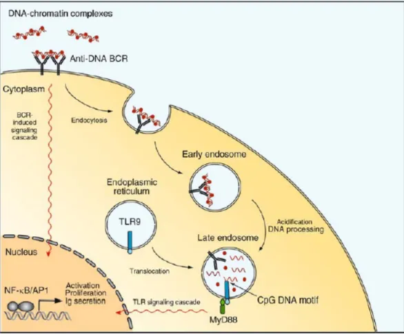
factor. Chloroquine, a chemical which blocks TLR9 dependent signaling, is widely used for treatment of autoimmune diseases such rheumatoid arthritis and systemic lupus erythematosus (SLE) (Boule et al., 2004). Proposed role of TLR9 in SLE is illustrated in Figure 2.
Table 5. TLR9 expression varies among mice and human (Krieg, 2006). B-cells pDC mDC monocyte/Mf
Human yes yes no no Mouse yes yes yes yes
pDC: plasmacytoid DC; mDC: myeloid DC; Mf: macrophage
1.2.1.2. TLR Signaling Pathways
Activation of TLRs by PAMPs leads to induction of various genes involved in host defense. These induced genes include inflammatory cytokines, chemokines, MHC molecules and co-stimulatory molecules. Mammalian TLRs also induce multiple effector molecules such as inducible nitric oxide synthase (iNOS) and antimicrobial peptides, which can directly eliminate microbial pathogens. Although both TLRs and IL-1Rs rely on TIR domains to activate NF-κB and MAP kinases and share some target genes, a growing body of evidence points to several differences in signaling pathways activated by individual TLRs. All TLR family members use a common MyD88 adaptor, except for TLR3, which recruits TRIF. TLR4 is the only family member that activates both
MYD88-dependent and TRIF-dependent signal transduction pathways (Barton and Kagan, 2009).
Figure 2. Self-DNA containing immune complexes is recognized by TLR9 and lead to the autoimmune disorder, SLE. (Rahman and Eisenberg, 2006)
.
Furthermore, activation of specific TLRs leads to slightly different patterns of gene expression profiles. While activation of TLR3 and TLR4 signaling pathways results in induction of type I IFNs (Doyle et al., 2002), TLR2- and TLR5-mediated pathways do not induce these anti-viral IFNs (Hoshino et al., 2002). TLR7, TLR8 and TLR9 signaling pathways also lead to induction of type I IFNs in a different manner (Ito et al., 2002a). 1.2.1.2.1. MyD88-Dependent Pathway
MyD88, comprised of a C-terminal TIR domain and an N-terminal death domain, associates with the TIR domain of TLRs. After stimulation, MyD88 recruits IL-1
receptor-associated kinase-4 (IRAK-4) to TLRs through the interaction of the death domains of both molecules. Following this, it facilitates IRAK-4-mediated
phosphorylation of IRAK-1. Activated IRAK-1 then associates with TRAF6, leading to the activation of two distinct signaling pathways. One pathway leads to activation of
1 transcription factors through activation of MAP kinases. Another pathway activates the TAK1/TAB complex, which enhances activity of the Inhibitor kappa B kinase (IkK) complex. Once activated, the IkK complex induces phosphorylation and subsequent degradation of IkB, which leads to nuclear translocation of transcription factor NF-kB (Klinman, 2004; Takeda and Akira, 2004). MyD88-deficient mice do not show
production of inflammatory cytokines such as TNF-α and IL-12p40 in response to any TLR ligand (Takeuchi et al., 2000). This once again proves that MyD88 is essential for inflammatory cytokine production through all TLRs. MyD88-deficient macrophages, show impaired inflammatory cytokine production in response to TLR4 and TLR2 ligands in contrast to TLR3, TLR5, TLR7 and TLR9 ligands (Yamamoto et al., 2002a).
1.2.1.2.2. MyD88-Independent/TRIF-Dependent Pathway
While TLR4 ligand-induced production of inflammatory cytokines is not observed in MyD88-knock-out macrophages, delayed NF-κB expression is observed. This shows that although TLR4 signaling relies on MyD88-dependent pathways, a MyD88-independent component exists in TLR4 signaling. TLR4-induced activation of IRF-3 leads to production of IFN-β, which in turn activates Stat1 and induces several IFN-inducible genes, like TLR3 (Alexopoulou et al., 2001; Yoneyama et al., 1998). TRIF-deficient mice exhibit impaired expression of IFN-β and IFN-inducible genes in response to TLR3 and TLR4 ligands (Yamamoto et al., 2002b). Studies with other TRIF-related adaptor molecules TRAM/TICAM-2 showed that TRAM is involved in TLR4-mediated, but not TLR3-TLR4-mediated, activation of IRF-3 and induction of β and IFN-inducible genes (Yamamoto et al., 2003). Key molecules that mediate IRF-3 activation have been revealed to be non-canonical IkKs, Tank binding kinase-1 (TBK1) and IkKi/IkKe (Fitzgerald et al., 2003). It has been recently reported that complete MyD88 and TRIF expression is required for the effective cooperation, resulting in the induction of IL-12, IL6, and IL-23 but not of TNF-α and IP-10 upon MyD88- and TRIF-dependent TLR stimulation. Downstream of MyD88, TRIF and IRF5 are identified as essential transcription factors for the synergism of IL6, IL-12, and IL-23 gene expression (Ouyang et al., 2007). TRAF6 associates with the N terminal portion of TRIF and the C-terminal portion of TRIF associates with receptor-interacting protein-1 (RIP1), thereby leading to NF-kB activation (Gohda et al., 2004; Meylan et al., 2004).
1.3. A Deeper Insight into Immunostimulatory DNA Particles and TLR9 Activation
TLR9 is the only known member of TLR family that can recognize specific DNA motifs. Five years after the identification of CpG motifs, Hemmi et. al. reported that TLR9 is responsible for the recognition of CpG DNA in mice. They showed that splenocytes, lymph node cells, dendritic cells, and macrophages from TLR9-deficient mice do not respond to CpG motif expressing oligodeoxynucleotides (CpG ODN), evident from the loss of pro-inflammatory cytokine production or cell surface maturation marker downregulation. Moreover, these mice are resistant to harmful side effects of CpG ODN (Hemmi et al., 2000). It has later been shown that human TLR9 is also
prerequisite for bacterial DNA/CpG DNA-dependent immunostimulation in both primary cells and TLR9-transfected cell lines (Bauer et al., 2001; Takeshita et al., 2001). Later findings challenged the idea that recognition of foreign DNA is restricted to CpG motifs. Non-CpG phosphodiester ODNs (PO-ODNs), which are delivered into endosomes via DOTAP complexation, and self chromatin DNA/IgG autoantibody complexes have been shown to be recognized via TLR9 (Boule et al., 2004; Yasuda et al., 2006). Recently, TLR9 has been shown to recognize 2’-deoxyribose sugar backbone (base-free) of phophodiester DNA -but not phosphorothioate (PS) modified 2’-deoxyribose, PS or PO modified 2’-ribose backbones- and activate cytokine secretion. Immunostimulatory activity and TLR9 affinity is increased further, when bases and CpG motifs are added to this backbone. PolyG addition (24 extra guanosines at 3’end) or DOTAP complexations are used here to target sugar backbone or PO-ODN into endosomes. PS-modified sugar backbones exhibit higher affinity to TLR9 and TLR7 than PO-modified counterparts both
in vitro and in vivo (Haas et al., 2008).
In unstimulated cells, TLR9 is localized to endoplasmic reticulum, which is transported to early endosomes and then lysosomes upon CpG stimulation (Latz et al., 2004). It has been suggested that the transfer from ER to endosomes is mediated by UNC93B1 protein (Kim et al., 2008). Two independent studies reveal that the ER to endosome docking is mediated by endosomal localization motifs present on TLR9 which are on opposite locations on ER and endosomes (Barton et al., 2006; Leifer et al., 2006). There exist other controversial data on TLR9 translocation from ER to endosomes. Latz
et al. reported that this translocation is mediated by non-secretory pathway (Latz et al., 2004), while Chockalingam et al. reported that secretory pathway is involved and TLR9 passes through Golgi on the way to endosomes (Chockalingam et al., 2009). In
endosomes, TLR9 exists as homodimers and the ligand unbound form represents the inactive conformation of the receptor. Upon ligand binding, seperate TIR domains gets closer and recruit MyD88 for initiation of signaling cascade (Latz et al., 2007). Fitting with this idea, it has been shown that only aggregated, multimeric forms of ODNs are stimulatory. As concentration of multimeric forms increases in ODN solution, the immunostimulatory activity is enhanced (Wu et al., 2004).
Interaction of TLR9 with bacterial/viral DNA in endosomes of wild-type cells seems to prevent recognition of self-DNA. Expression of TLR9 on the plasma membrane by generating a chimeric protein (TLR9/TLR4) makes mammalian DNA stimulatory. Viral DNA becomes inactive in this case possibly due to the coat around the DNA. Mammalian DNA with phosphodiester backbone exhibits parallelism with PO-ODNs, which become more stimulatory against surface expressed TLR9, in sensitivity to DNase (Barton et al., 2006). This finding challenges previous notion that CpG motifs are critical factors in of self/non-self discrimination. Recently, TLR9 has been shown to become activated after proteolytic cleavage of carboxy terminal fragment at endosomes via cathepsin proteases (Ewald et al., 2008; Park et al., 2008). This explains the previous observation that TLR9 signaling is blocked by inhibition of endosomal acidification (Ahmad-Nejad et al., 2002). This acidification is required for optimal activity of TLR9-cleaving proteases. These recent findings strengthen the hypothesis that self/non-self discrimination is mainly achieved by TLR9 compartmentalization as it is functional in endosomes.
1.3.1. Accessory Molecules Involved in TLR9 Activation
LL37: LL37 is an endogenous antimicrobial peptide expressed highly in psoriatic skin. During physical injury, disease condition worsens, self-DNA is released and pDC
activation occurs. LL37 forms complexes with self-DNA. These complexes are delivered to early endosomes in pDC and IFNα is induced. Otherwise, self-DNA is inactive since it cannot colocalize with TLR9 in endosomes (Lande et al., 2007).
HMBG1 and RAGE: HMBG1 (High-mobility group box 1) is another DNA-binding protein. It is involved in bending of double-helix to increase affinity of DNA to
transcription factors. HMGB1 interacts and preassociates with TLR9 in the endoplasmic reticulum-Golgi intermediate compartment (ERGIC), and hastens TLR9’s redistribution to early endosomes in response to CpG DNA (Ivanov et al., 2007). Recent work shows that HMBG1 forms complexes with A/D-type ODN (but not B/K-type ODN), thereby increasing B cell activating and IFNα-inducing potential of pDC. This stimulation also depends on the interaction of RAGE (receptor for advanced glycation end-products), a multi-ligand receptor of immunoglobulin superfamily, with HMBG1-TLR9 complex. The critical roles of HMBG1 and RAGE in IFNα induction by DNA-containing immune complexes have also been shown in lupus patients (Tian et al., 2007).
Cathepsin-K: Cathepsin-K is an osteoclast-specific protease which is involved in degradation of bone matrices. It is seen as a therapeutic target to treat diseases, such as osteoporosis and autoimmune arthritis, in which osteoclast activity abnormally increases. Recently, specific inhibition of Cathepsin-K by pharmacological inhibitor in dendritic cells, has been shown to reduce TLR9-mediated, but not TLR2 or TLR4-mediated, production of IL-12, IL-23 and upregulation of maturation markers such as CD40, CD80 and CD86 in a dose-dependent manner. Moreover, cathepsin K-/- mice are found to be not responsive to stimulation with CpG ODNs (Asagiri et al., 2008).
UNC93B1: Missense mutation in Unc93b1 gene is called 3d mutation because of the impaired signalling via TLR3, 7 and 9. Mice with 3d mutation show increased susceptibility to murine cytomegalovirus (MCMV) infection (Tabeta et al., 2006). UNC93B1 functions in delivery of TLRs from endoplasmic reticulum to endosomes. Mice with artificially expressed TLR9 on plasma membrane are not affected from 3d mutation and can respond to CpG motifs (Kim et al., 2008).
1.3.2. Different Classes of Synthetic CpG-ODNs
Oligodeoxynucleotide (ODN) sequences designed for antisense therapy have first been shown to be immunostimulatory in 1992 (Yamamoto et al., 1992). Some antisense
ODNs containing palindromic sequences have induced strong type I and/or type II IFN production and led to profound interferon mediated NK cell killing activity. Later, this activation has been attributed to the presence of unmethylated CpG motifs, which are nearly 20 times more frequently expressed in microbial DNA than mammalian DNA (Krieg et al., 1995). CpG motif expressing ODNs stimulate murine B-cell proliferation and IgM production significantly. Meanwhile, control ODNs in which cytosines are methylated or CpG is inverted to GpC does not exhibit any stimulatory effects. Minimum ODN length for stimulatory effect has been shown 8 bp. CpG ODNs stimulate BALB/c mice spleen cells to produce IL6, IL-12 and IFNγ. If a sequence contains multiple CpG motifs over a single strand, the stimulatory activity is enhanced (Klinman et al., 1996). CpG ODN also stimulates human PBMC to release TNFα, IL12, IL6 and upregulate monocyte and B-cell activation markers (Bauer et al., 1999). They induce polyclonal activation and differentiation of memory B cells into plasma cells without B-cell receptor signaling or T cell help. This makes CpG ODNs candidates as powerful adjuvant for induction of humoral immunity. However, BCR signaling is critical for activation of naive B cells (Bernasconi et al., 2002). Overall response to CpG ODNs is Th1-polarized. The immunization of mice with CpG ODN plus an antigen induces Th1 associated IFNγ, IL-12 and antigen specific IgG2a and suppresses Th2 associated IL-5 production (Chu et al., 1997).
Central hexameric sequence is Pu-Pu-C-G-Py-Py for mice and Pu-Py-C-G-Pu-Py for humans in optimal sequences. Substituting a Purine (Pu) for a Pyramidine (Py), or vice versa, significantly reduces or eliminates ODN activity (Krieg et al., 1995; Verthelyi et al., 2001). CpG ODNs designed to date can be grouped into i) A/D-type CpG, ii) B/K-type CpG, and iii) C-B/K-type CpG. Additional less well-characterized sequences, which do not fit in any of these three groups, are also covered below.
1.3.2.1. A/D-type CpG ODNs
This group of CpG ODNs is named as A-ODN by some groups and D-ODN by others. It will be referred to as D-ODN hereafter. D-ODNs contain a mixed backbone of phosphodiester and phosphorothiote linkages. Generally, a single CpG motif is present in the middle of a palindromic sequence with phosphodiester (PO) backbone. The
palindromic sequence is capped by G-runs at both ends. The linkage between the G pairs at both ends have phosphorothioate (PS) backbones, which confer increased resistance to nucleases. Minimum length of an active D-ODN is 18 bp. Representative sequences are given below: D19: 5`-GGtgcatcgatgcagGGGGG-3` D29: 5`-GGtgcacggtgcagGGGG-3` D35: 5`-GGtgcatcgatgcaggggGG-3` D no poly(G): 5`-GGtgcttcgatgcaaaaaAA-3` D3CG: 5`-GGtcgatcgatcgaggggGG-3` ODN 2216: 5’-GGgggacgatcgtcggggGG-3’
Please note that lowercase letters indicate phophodiester (PO) linkages between bases, whereas uppercase letters indicate phosphorothioate (PS) linkages between bases. Underlined bases represent unmethylated CpG dinucleotides.
Any inversion, replacement, or methylation of the CpG abolishes the stimulatory activity completely. The polyG ends and central palindromic region of the ODN also contribute to D-type activity significantly (Verthelyi et al., 2001). D-ODN has a high tendency to spontaneously assemble into higher-order structures, heterogenous particles mainly composed of globular (~50nm size) and linear structures (~100nm size), under physiological concentration. This structure formation is dependent on the presence of poly(G) motifs and on the palindromic region (Costa et al., 2004). Kerkmann suggests that monomeric D-ODN forms a duplex via Watson-Crick base pairing from its
palindromic regions. Moreover, the poly(G) ends, of which there are four, of the two duplexes are linked together via a non Watson-Crick base pairing. This is an interaction established between guanosines and is known as “Hoogstein Base Pairing”. The
interaction between the planar four Gs is called “G-tetrads” and multiple forms of these G-tetrads are named as G-quadruplexes. The formation of this structure is given in detail in Figure 3. Exposure of G-tetrads to high temperature leads back to formation of their monomeric molecules (Kerkmann et al., 2005).
Figure 3. G-tetrad links four D-ODN strands for higher-order structure formation (Kerkmann et al., 2005).
D-ODN induces high amounts of IFN-α/β directly from pDCs and IFN-γ from PBMCs. Maximum activity is reached at 3 µM. They also indirectly induce IFN-γ, IP-10 from PBMC and NK cell activation strongly. NK cell-mediated IFN-γ production by D-ODN is IFΝα/β−dependent, and IL12-independent (Krug et al., 2001; Verthelyi et al., 2001). Moderate levels of costimulatory molecules CD80, CD86 and HLA-DR on pDC and B cells are induced. This induction is lower than that of K/B type CpG ODNs (Krug et al., 2001). D-ODN stimulates monocytes to mature into CD83+/CD86+ DCs in an IFN-α-dependent manner (Gursel et al., 2002b). NK cell-mediated cytolytic activity, which is a very critical part of innate immunity against intracellular pathogens, also increases considerably (Krug et al., 2001). D-type ODN stimulates mouse splenocytes to produce moderate IL6, IL-12 and high levels of IFNγ. Interestingly, IFNγ induction by D-type ODN reaches maximal activity at 1µg/ml and this activity is reduced at higher
concentrations (Vollmer et al., 2004). The therapeutic potentials of D-type ODNs are hampered by the fact that G-rich containing sequences are problematic during synthesis. PolyG-based ODNs lack pharmaceutical attributes due to unpredictable secondary structure formation depending on the experimental conditions and nonsequence-specific protein binding (Wang et al., 2009).
1.3.2.2. K/B-type CpG ODNs
This CpG ODN class is referred to as B or K-type ODNs by different groups. It will be referred to as K-ODN hereafter. These are full phosphorothioate backbone ODNs with at least one CpG motif. This motif is highly active in humans when it is located closer to 5’ end of the sequence and at least one more base upstream of the 5`-CpG motif is required. Thymidine in the immediate 5’ position is the most favorable for humans (ODN2006). For active CpG ODNs in mice, CpG motif does not have to be at the very 5’
end (ODN1555, ODN1826). Minimum length of ODN should be 12 bases for sustained immune activation (Verthelyi et al., 2001). This type lacks poly-G tails and is believed to remain as single stranded linear sequences under physiological conditions (Costa et al., 2004). Optimal K-ODN sequences are given below:
1. ODN1826: 5`-TCCATGACGTTCCTGACGTT-3`
2. ODN1555: 5`-GCTAGACGTTAGCGT-3`
3. ODN2006: 5`-TCGTCGTTTTGTCGTTTTGTCGTT-3`
4. K23: 5`-TCGAGCGTTCTC-3`
K-ODNs trigger IL6 and IgM secretion from B-cells and induce their
proliferation. They also activate CD19+ B cells by upregulating CD69 (early activation marker) and CD25 (late activation marker) (Gursel et al., 2002a). These ODNs are superior for pDC maturation and induction of proinflammatory cytokines from pDCs than D-type ODNs (Krug et al., 2001). They can also synergize with GM-CSF and activate the maturation markers of dendritic cells; MHC II, CD40, and CD83 (Hartmann et al., 1999). Poor IFNα induction from pDCs is also characteristic of this type of ODNs, whereas it is reported that even this trace amount of IFNα is sufficient enough to induce MHC-I cross presentation in DCs (Gray et al., 2007). K-ODN induced IFNγ, IP10 secretion from PBMC is little also if compared to other ODN types. Spleen cells also secrete very high amounts of IL6 and IL-12 in response to K-type ODNs (Krug et al., 2001; Verthelyi et al., 2001). Mouse-active K-type ODN induces much higher levels of nitric oxide (NO) from mouse-macrophage cell line (RAW264.7) in comparison with D-type ODN (Utaisincharoen et al., 2002).
1.3.2.3. C-type CpG ODNs
C-type ODNs have fully phosphorothioate backbone with 5’ CpG sequences (‘TCGTCG motif’) and a 12-16 bp palindromic sequence at 3’ end for most optimal activity (Vollmer et al., 2004). TCG at 5’ end is necessary for ODN activity and IFNα-induction capacity is reduced when this motif is shifted towards 3’. Immunostimulatory activity of C-ODN depends on the ODN length, the base content and a 5’-TCG.
Minimum length requirement for optimal activity is 22 bp, where adding poly-T to the 3’ end of shorter sequences significantly increases their activity (Jurk et al., 2004).
Palindromic sequence, which is believed to allow duplex formation in an endosomal environment, is vital for the immunostimulatory activity of C-type ODN (Krieg, 2006). This structrure is illustrated in Figure 4. Immunostimulatory effect is severely reduced in similar ODNs lacking palindromic sequences, especially in the context of IFNα
induction.
Figure 4. Duplex formation through palindromic sequences by two monomeric C-ODNs ODN2395 (Krieg, 2006).
These ODNs are reported to combine the immunostimulatory activity of D-type and type ODNs. They directly activate human B cells at a level comparable with K-ODNs. Furthermore, the production of IFNα from pDC is induced at a higher level compared to K-ODNs. They also induce also higher levels of IFNγ, IP-10 from PBMC and upregulate activation markers on NK cells better than K-ODN. B-cell activation with this type of ODN is pronounced in whole human PBMC stimulation which is not the case for K type CpG ODN. IFNα production from pDC seems to have an additive effect on B-cell activation in whole PBMC stimulation. Depleting pDC from PBMC lowers B-B-cell activation by C-type while this type of reduction does not occur upon K-ODN stimulation (Hartmann et al., 2003).
To sum up, C-ODNs are somewhere between D-ODNs and K-ODNs both
structurally and immunologically. They induce lower levels of interferon than D-type and higher levels than K-type. They also directly activate B cells. Additionally, they form duplexes under physiological conditions unlike linear K-ODNs and multimeric D-ODNs.
1.3.2.4. Other Types of CpG-ODNs
Several ODNs that do not fit into the previously mentioned classes have also been designed. One of them is Y-shaped ODN, which is formed by 3 linear CpG ODNs with complementary regions. Its structure is given in Figure 5. This ODN has a phophodiester backbone and induces higher levels of IL6 and TNFα from RAW264.7 cells, a mouse
macrophage cell line, than single- and double-strands of same ODNs used at same
concentrations. It is claimed that this structure increases the efficiency of uptake into cells (Nishikawa et al., 2008).
Another work reported that 3’ extension of phophodiester linear (CpG or non-CpG ) ODNs with 24 guanine nucleotides turns these sequences into an effective IFNα inducer. Whereas shorter ODNs have reduced activity, elongation of the ODNs does not result in increased activity. Addition of a CpG motif increases the activity of this ODN. It has been shown that polyG addition confers complexation and efficient uptake into immune cells similar to the utilization of a complexation agent such as DOTAP (Haas et al., 2009).
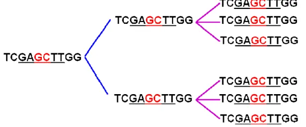
Previous research in our lab has led to the identification of a novel 70-mer dendrimeric ODN, which forms uniform-sized nanoparticles with an average size distribution of 35 ± 10 nm (Figure 6). It exhibits a higher immunostimulatory potential than its linear control. Furthermore, the substitution of CpG dinucleotide with a GpC flip completely abolishes its activity in mice (Mammadov, M.Sc. Thesis, 2009).
Figure 5. Y-shaped CpG ODN (Nishikawa et al., 2008).
Figure 6. Formation of uniform-sized nanoparticles in a novel bifunctional dendrimeric ODN, designated as ODN420. (Mammadov, M.Sc. thesis, 2009)
1.3.3. Differential Immune Response Mediated by Particulate and Linear CpG ODN
Although both D-type and K-type ODNs contain CpG motifs and initiate their effect by signaling through TLR9, the immunologic pathways induced are unique in each class. First, D-ODNs have been reported to be localized in different vesicles than those of K-ODNs. Moreover, D-ODN is taken up four-fold more efficiently than K-ODN by PBMC, where excess concentration of either ODN does not inhibit binding or uptake of the other (Gursel et al., 2002a). Collectively, these data suggest that there are different mechanisms of uptake for each of these ODNs. Poly-G tail, which confers particulate structure in ODNs, is thought to be responsible for the more efficient uptake of D-ODNs in mouse cells (Anderson et al., 2008; de Jong et al., 2007) and human pDC (Kerkmann et al., 2006). Second, interferon regulatory factor (IRF)-7 is found to be critical for IFNα induction from pDC by D-ODN, as pDCs from IRF7-/- mice are unresponsive to D-ODN. D-ODN mediated cytokine production such as IL-12 and IL6 from cDC (conventional dendritic cells) and K-ODN mediated cDC and pDC activation are not affected in IRF7-/- mice (Honda et al., 2005b). The dichotomy of D-type and K-type ODNs is illustrated in Figure 7.
Two recent works have clarified the differential activities of D- and K-type ODNs. Honda et. al. have reported that induction of different cytokines from pDCs by two types of ODNs is caused by different localization patterns of K or D-type ODNs in pDCs. D-type ODNs are retained for longer periods in the early endosomal vesicles of pDCs, whereas K-type ODNs accumulate in vesicles with lysosomal markers (Honda et al., 2005a). According to this model, D-ODN interacts with MyD88–IRF-7 complex in early endosomes, which results in IRF7-mediated IFNα production. Meanwhile, K-ODN activates MyD88-IRAK4/1-TRAF6 complex which activate NF-κB and IRF-5-mediated pro-inflammatory cytokine production (Asselin-Paturel and Trinchieri, 2005). Retention of D-ODNs in early endosomes is facilitated by their higher-order structure. Artificial targeting of K-ODNs to early endosomes can be achieved through making their complexes with DOTAP (Honda et al., 2005a). These particulate forms of K-ODNs induce IFNα levels comparable with ODN (Kerkmann et al., 2005). Recruitment of D-ODN to early endosomes and its IFNα-inducing capacity are dependent on CXCL16, a scavenger receptor only expressed on pDCs but not on B-cells (Gursel et al., 2006). Moreover, D-ODNs lacking poly-G tail cannot interact with CXCL16. Identification of CXCL16 as a specific co-receptor for D-type ODN might also explain the higher efficiency of artificially multimerized ODNs regarding uptake and immune stimulation (Haas et al., 2009; Shirota et al., 2001). To date, there is no convincing evidence proving that particulate forms of K-ODNs depend on additional recognition by other receptors. However, high immunostimulatory potential of nanoparticle-forming CpG-ODNs at low concentrations, where free CpG-ODNs are not active, strengthens this hypothesis
(Anderson et al., 2008; Gursel et al., 2001).
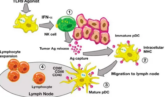
Figure 7. Dichotomy of different CpG ODN types delivered to early or late endosomes induce innate or adaptive immune responses, respectively, in pDCs. (Williams, 2006)
1.4. Utilization of CpG ODNs as Therapeutic Agents
Critical cellular target for CpG ODN therapy are pDCs. Moreover, pDCs from CpG-treated donors are able to mount protection in naive recipients. Another TLR9+ cell type is B cells, where CpG-ODNs induce production polyclonal immunoglobulins that help to eradicate pathogen. NK cells and macrophages are indirectly activated by cytokines released from pDCs (Klinman, 2004).
In an attempt to define the most optimal sequences, several thousand sequences have been synthesized and tested on PBMCs isolated from over 100 donors. It has been observed that no single ODN is optimal for all tested individuals. Two strategies are suggested to overcome this problem. The first one is to synthesize single ODNs with different CpG motifs and the second one is to use mixtures of multiple ODNs with different CpG motifs (Leifer et al., 2003). This should be taken into consideration in therapeutic approaches with CpG ODNs, which are shown to be effective against infectious diseases, cancer, allergy/asthma.