Abstract— Tm:YAP laser system at power levels up to 1.2 W at 1980 nm was established in both continuous-wave and modulated modes of operation. The fluence effect of the laser system for skin ablation was analyzed by histology analysis with Wistar rat skin tissues. Thermally altered length, thermally altered area, ablation area, and ablation depth parameters were measured on histology images of skin samples just after the laser operation and after four-day healing period. Continuous-wave mode of operation provided higher thermal effects on the skin samples. Lower fluence levels were found for efficient ablation effect.
I. INTRODUCTION
HEN laser light hits tissue, its transmission, scattering, reflection, and absorption depend primarily on the type of tissue and the wavelength of the laser light. The laser is absorbed in the target tissue by increasing its molecular vibrations and it creates a thermal effect. Infrared light is absorbed primarily by water. The laser light energy absorbed in tissue is converted into thermal energy. Different thermal effects are obtained depending on temperature range in the tissue. Some temperatures may provide tissue ablation by removing a part of biological tissue where the laser is applied.
The 1.9-µm lasers can be used in an efficient way in superficial tissue ablation with minimal coagulation depth [1]. Among the possible candidates, Thulium (Tm:YAP) is one of the most suitable candidates due to the availability of high-quality host crystals and possibility of efficient laser performance near 2 µm. In addition, Tm:YAP lasers offer easy pumping with 800-nm diode lasers, benefit from crystal durability at high powers, can be coupled to conventional
Manuscript received April 1, 2010. This work was supported by the the Türkiye Bilimsel ve Teknolojik Ara tirma Kurumu (TÜB TAK), Project No: 107E119.
T. Bilici is with the Biophotonics Laboratory, Institute of Biomedical Engineering, Boğaziçi University, Kandilli Kampüs, 34684 Çengelköy Istanbul, Turkey (phone: +90 533 434 5582; fax: +90 216 516 3479; e-mail: [email protected]).
Ö. Tabakoğlu is with the Biophotonics Laboratory, Institute of Biomedical Engineering, Boğaziçi University, Kandilli Kampüs, 34684 Çengelköy Istanbul, Turkey (e-mail: [email protected]).
H. Kalaycıoğlu is with the Institute of Material Science and Nanotechnology (UNAM), Bilkent University, Cankaya, Ankara 06800, Turkey (e-mail: [email protected]).
A. Kurt is with Teknofil Ltd. Şti., Zekeriyaköy 34450 Sarıyer Istanbul, Turkey (e-mail: [email protected]).
A. Sennaroglu is with the Laser Research Laboratory, Department of Physics, Koç University, Sarıyer, 34450, Istanbul, Turkey (e-mail: [email protected]).
M. Gulsoy is with with the Biophotonics Laboratory, Institute of Biomedical Engineering, Boğaziçi University, Kandilli Kampüs, 34684 Çengelköy Istanbul, Turkey (e-mail: [email protected]).
fibers for invasive operations, and operate in the eye-safe region [2]. In this study, a Tm:YAP laser system with power output up to 1.2 W and emission wavelength of 1980 nm was established in both continuous-wave and modulated modes of operation. The fluence effect of the laser system for skin ablation was analyzed by Wistar rat skin tissues during four-day healing period.
II. MATERIALS AND METHODS
A. Tm:YAP Laser System
The Tm:YAP laser system includes Tm:YAP laser setup, diode laser driver, water chiller for the laser setup, modulation controller unit, and software in a personal computer (PC).
Tm:YAP laser can be pumped by high power pump diode lasers that overlap with the absorption band of the crystal near 800 nm. The power performance of Tm:YAP lasers is dependent on the doping concentration of Tm:YAP crystal. Varying the active ion concentration in the crystal can change the strength of cross relaxation, reabsorption losses, and nonradiative decay rates.
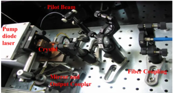
In developing the laser system, a Tm:YAP crystal (cylindrical 5 x 4 mm) with 1.5% doping was used as the laser medium. The crystal was normal cut and both faces had antireflection coatings at the lasing wavelength. The resonator consisted of a flat input mirror and a curved output coupler with a radius of 10 cm. The length of the resonator was 7.5 cm. The Tm:YAP crystal positioned near the flat mirror was wrapped in indium foil and held between copper holders which were cooled by water. The Tm:YAP resonator was end-pumped with a fiber-coupled diode array operating at a wavelength of 797 nm. An imaging telescope was used to focus the pump beam into the crystal. Output coupler of 6% transmission was used. Laser radiation was delivered by means of a 200-µm multimode optical fiber [3]. The picture of the Tm:YAP laser setup is in the Figure 1.
Figure 1: Tm:YAP laser setup and its components
Fluence of Thulium Laser System in Skin Ablation
Temel Bilici, Özgür Tabakoğlu, Hamit Kalaycıoğlu, Adnan Kurt,
Alphan Sennaroglu, and Murat Gülsoy
W
Pump diode laser Mirror and Output Coupler Pilot Beam Crystal Fiber Coupling32nd Annual International Conference of the IEEE EMBS Buenos Aires, Argentina, August 31 - September 4, 2010
The modulation microcontroller unit provides the modulation the Tm:YAP laser output. The microcontroller unit can operate the laser in pulsed mode up to frequency of 50 Hz. The user interface designed by Labview 6 software sets the duration of the emission, on cycle, off cycles, and number of cycles. In addition, the diode current and the corresponding laser output power can be set via the user interface on PC.
B. Animal Preparation
The experiments were conducted under a protocol approved by the Institutional Animal Research and Care Ethic Committee at Boğaziçi University. A total of six healthy male Wistar rats, 5–6 months old, weighing 240–250 g, were randomly selected from Psychobiology Laboratory of Bogazici University for in vivo experiments. Rats were housed in plastic cages and maintained on a 12-h-light/12-h-dark cycle in a temperature-controlled vivarium (22±2°C). Food and water were available ad libitum. Rats were anesthetized with ketamine (10% ketamidor, RichterPharma, AG, Wels, Austria) by intraperitoneal injection (1.65ml/kg). Hair at the site of application of each subject was shaved.
C. Tm:YAP Laser Application
All the laser application procedure was conducted by one operator to in vivo Wistar rat skin. Laser protective glasses were used when the laser was active. Power, duration, and mode of operation (modulated and CW) parameters were set by another operator through the user interface on the PC. Laser output power was checked with a powermeter (Newport 1918-C,Irvine, CA, USA) before each application. The laser application was sampled at a distance of 2 mm above to each spot. The laser spot diameter was measured from 2 mm as 0.6 mm by using the knife-edge technique.
D. Histology Procedure
The skin tissue samples exposed to Tm:YAP laser were dissected just after the laser operation (day0) and after four-day healing period (four-day4). The samples were then fixed in 10% formalin and processed by a tissue Processor (Leica TP1020). Dehydrated tissues were embedded (Leica EG1150H) into paraffin, and 10-micrometer-thick tissue sections were obtained via microtome (Leica RM2255). Tissue sections were obtained from removal of paraffin over tissues by inserting into 40ºC water-bath. Tissues were aligned on top of glass slides. These slides were kept in etuve overnight in order to remove remaining paraffin. The skin tissue samples were analyzed after hematoxylin&eosine histology protocol. The staining method involves application of the basic dye haematoxylin, which colors basophilic structures with blue-purple hue, and alcohol-based acidic eosin Y, which colors eosinophilic structures bright pink. Eosinophilic describes the appearance of cells and structures seen in histological sections which take up the staining dye, eosin. This is a bright pink dye that stains the cytoplasm of cells as well as extracellular proteins such as collagen. This procedure will give the idea about carbonization, infections,
and healing quality of the tissue samples under light microscopy.
The bright areas in the polarized histology pictures under light microscopy (Nikon 80i Eclipse) show the collagens whereas the holes show the fat areas in the tissues. The histology pictures were selected and sampled from tissue sections where the laser ablation was found maximum for each sample.
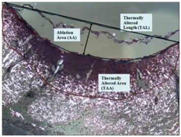
E. Ablation Analysis Parameters
Figure 2 shows the ablation analysis parameters under light microscope. In this polarized picture of the tissue sample, the bright areas include collagens of the skin. The holes in the pictures are fat areas of the skin tissue. The thermally effected area is simply distinguished by normal tissue. There is an ablation zone where the laser is applied. The area of this blue-boundary zone, ablation area (AA), can be measured by software of the microscope. Ablation depth (AD) of this zone can also be measured. In addition, thermally altered area (TAA) on the tissue is shown by red boundary. Thermally altered length (TAL) is the length on the tissue sample affected by laser application. These parameters will be used to analyze the laser-tissue interactions quantitatively under light microscopy.
Figure 2: Ablation analysis parameters III. RESULTSANDDISCUSSIONS
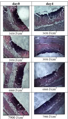
The laser parameters applied to the in vivo skin tissues were tabulated in Table 1. The Tm:YAP laser was applied in modulated mode of operation (200 ms on/off). The histology images of the skin samples at day0 and day4 were given in Figure 3.
Table 1: Modulated laser parameters applied to in vivo skin samples Laser Power (W) Average light intensity (W/cm2) Duration (s) Fluence (J/cm2) 0.99 343 10 3430 0.99 343 20 6860 1.14 395 10 3950 1.14 395 20 7900 3219
day0 day4
3430 J/cm2 3430 J/cm2
3950 J/cm2 3950 J/cm2
6860 J/cm2 6860 J/cm2
7900 J/cm2 7900 J/cm2
Figure 3: Histologic images of the tissue samples after modulated Tm:YAP laser applications in Table 1.
Figure 4: Ablation analysis parameters after the laser applications (day0)
Figure 5: Ablation analysis parameters after the laser applications (day4)
Each ablation analysis parameter was sampled by minimum eight samples for each laser parameter. Averages of each parameter were used in ablation analysis. As seen in Figure 4, TAA was increasing when duration was increased in the same power level for day0. On the other hand, AA and AD were measured smaller when the laser energy density was increasing. This shows laser parameters should be applied in less powers and energy levels for efficient ablation effect. Similar results were observed for skin samples at day4 (Figure 5). However, TAL, TAA, AA, and AD values were found higher than the values at day0. This shows that the thermal effects were spreading to the deeper layers during the healing period.
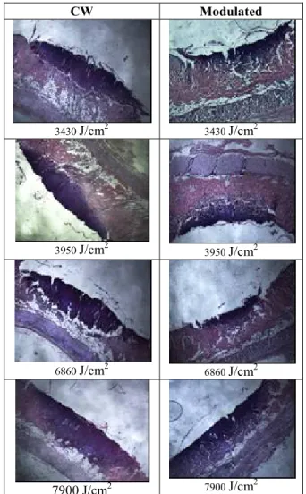
CW Modulated 3430 J/cm2 3430 J/cm2 3950 J/cm2 3950 J/cm2 6860 J/cm2 6860 J/cm2 7900 J/cm2 7900 J/cm2 Figure 6: Macro pictures of skin samples at day4.
In another group of experiment, the same method was applied to another group by using continuous-wave (CW) mode of operation of Tm:YAP laser. Then, the ablation parameters were compared among CW and modulated
modes of operations with the same laser energy levels. The macro pictures of the skin samples at day4 were given in Figure 6, whose histology images were given in Figure 7. As seen in Table 4, AA and AD were not observed after application of Tm:YAP laser in CW mode of operation at these power levels. This shows that CW mode of operation provides higher thermal effects on the skin samples. TAA and TAL values were also found higher at CW mode of operation (Figure 8). Statistical analysis of the whole study will be performed in further experiments with larger sample sizes. CW Modulated 3430 J/cm2 3430 J/cm2 3950 J/cm2 3950 J/cm2 6860 J/cm2 6860 J/cm2 7900 J/cm2 7900 J/cm2 Figure 7: Histology images of skin samples at day4.
Figure 8: Ablation analysis parameters after CW laser applications (day4)
The results of these experiments can be used in modeling of Tm:YAP laser tissue interactions at 1980 nm during 4 day healing period. The thermal effects will be compared among CW and modulated modes of operation. Due to strong water absorption, 1980-nm Tm:YAP lasers are promising efficient candidates for efficient skin tissue ablation with minimal depth compared to smaller wavelength lasers.
IV. CONCLUSION
The results of these ablation dosimetry experiments suggest that due to strong water absorption, 1980-nm Tm:YAP lasers are promising efficient candidates for efficient skin tissue ablation with minimal depth compared to smaller wavelength lasers. Whereas other types of lasers can penetrate the tissue deeply, Tm:YAP lasers interact with the tissue surface that enables to cleanly remove the unwanted part of the tissue from the healthy part or strip it layer by layer. In the future experiments, similar experiments are planned in lower power levels of Tm:YAP laser system, which will be more suitable for clinical applications. In addition, the performance of the system will be compared with the other laser systems with smaller wavelengths.
ACKNOWLEDGMENT
This work was supported by the Türkiye Bilimsel ve Teknolojik Araştirma Kurumu (TÜBITAK), Project No: 107E119.
REFERENCES
[1] A. F. El-Sherif and T. A. King, “Soft and hard tissue ablation with short-pulse high peak power and continuous thulium-silica fibre lasers”, Lasers Med. Sci., vol. 18, pp. 139-147, September 2003. [2] B. Chen, S. L. Thomsen, R. J. Thomas, J. Oliver, and A. J. Welch,
“Histological and modeling study of skin thermal injury to 2.0 µm laser irradiation”, Lasers in Surg. and Med., vol. 40, pp. 358-370, June 2008.
[3] H. Kalaycioğlu, A. Sennaroğlu, and A. Kurt, “Influence of doping concentration on the power performance of diode-pumped continuous-wave Tm3+:YAlO3 lasers”, IEEE J. of Sel.Top. in Quan. Electronics., vol. 11, pp. 667-673, 2005.