MONOCLONAL ANTIBODY PRODUCTION FOR
COILED-COIL DOMAIN CONTAINING-124 (CCDC-124) AND ITS
MOLECULAR CHARACTERIZATION
A THESIS SUBMITTED TO
THE DEPARTMENT OF MOLECULAR BIOLOGY AND GENETICS AND THE INSTITUTE OF ENGINEERING AND SCIENCE OF
BILKENT UNIVERSITY
IN PARTIAL FULFILLMENT OF THE REQUIREMENTS FOR THE DEGREE OF MASTER OF SCIENCE
BY
İREM GÜRBÜZ AUGUST 2010
ii
I certify that I have read this thesis and that in my opinion it is fully adequate, in scope and in quality, as a thesis for the degree of Master of Science.
Dr. Tamer Yagci
I certify that I have read this thesis and that in my opinion it is fully adequate, in scope and in quality, as a thesis for the degree of Master of Science.
Assist. Prof. Dr. Ayşe Elif Erson
I certify that I have read this thesis and that in my opinion it is fully adequate, in scope and in quality, as a thesis for the degree of Master of Science.
Assist. Prof. Dr. Uygar H. Tazebay
Approved for the Institute of Engineering and Science
Director of Institute of Engineering and Science Prof. Dr. Levent Onural
iii
ABSTRACT
MONOCLONAL ANTIBODY PRODUCTION FOR COILED-COIL DOMAIN CONTAINING-124 (CCDC-124) AND ITS MOLECULAR
CHARACTERIZATION
İrem Gürbüz
M.Sc in Molecular Biology and Genetics Supervisor: Assist. Prof. Uygar H. Tazebay
August 2010, 61 Pages
Coiled-coil-domain-containing-124 (CCDC-124) is a novel protein of unknown function. The Ccdc-124 gene is highly conserved among eukaryotic species and is predicted to encode a 223 amino acids long protein. Yeast-two-hybrid assays had shown that CCDC-124 interacts with RasGEF1B guanine exchange factor, which was previously identified as a specific guanine exchange factor for Rap2, a member of the Rap subfamily of Ras-like G-proteins. In order to reveal the cellular function of CCDC-124 protein, different biochemical experiments, including Western Blotting and Sub-cellular localization studies were performed. In those experiments, a polyclonal antibody against an N-terminal peptide of CCDC-124 was used. Nevertheless, due to its polyclonal nature this antibody exhibited non-specific binding. Although it was determined that the gene encodes a 33 kDa protein and that the protein has diffused cytoplasmic localization, the results were not precise. In order to detect the protein more accurately, monoclonal antibodies were generated against CCDC-124. Throughout the project, mice were injected with pure His-Tagged CCDC-124 protein. Via hybridoma technology antibodies were generated and selected for their recognition capacities. At the end, 3 positive hybridoma clones were produced: 7F7, 15C11 and 4B3. To characterize the produced monoclonal antibodies, Western Blot experiments were performed and their binding properties were compared to the polyclonal antibody. Among the three monoclones, 4B3 gave the most promising results at 33 kDa, in Western Blotting. The antibodies will be used in the determination of the protein's sub-cellular localization and in the analysis of its response to extracellular signals. These in turn will aid further analyses related to the protein's role within the cell.
iv
ÖZET
CCDC-124 PROTEİNİNE KARŞI MONO-KLONAL ANTİKOR ÜRETİMİ VE ANTİKORUN KARAKTERİZASYONU
İrem Gürbüz
Moleküler Biyoloji ve Genetik Yüksek Lisans Derecesi Tez Yöneticisi: Yrd. Doç. Dr. Uygar H. Tazebay
Ağustos 2010, 61 Sayfa
Sarılı-sarmal-bölge-içeren-124 (Coiled-coil domain containing-124; CCDC-124) henüz fonksiyonu belirlenmemiş olan yeni bir gendir. Bu gen ökaryotlar arasında korunmuştur ve genin 223 amino asitten oluşan bir protein kodladığı düşünülmektedir. Geçmişte Tazebay grup projeleri kapsamında yapılan maya ikili hibrid deneyleri, CCDC-124 proteinin RasGEF1B guanine değiştirme faktörü ile etkileşim halinde olduğunu göstermiştir. RasGEF1B’nin ise Rap2’ye özgü bir guanine değiştirme faktörü olduğu araştırmalarda gösterilmiştir. CCDC-124 proteinin hücre içindeki fonksiyonunu ortaya çıkarmak için, Western Blotlama ve hücre içindeki yer taramalarını da içeren çeşitli biyokimyasal deneyler yapılmıştır. Bu deneylerde, CCDC-124 proteininin N-uç bölgesini tanıyan, bir poliklonal antikor kullanılmıştır. Fakat, antikorun poliklonal doğası doğrultusunda, bir çok, spesifik olmayan bağlanma da görülmüştür. Yapılan deneylerde, genin 33 kDa’lık bir proteini kodladığı ve proteinin hücre içinde dağınık, sitoplazmik bir yerleşim izlediği gözlemlenmiştir. Fakat, spesifik olmayan bağlanmalar da göz önüne alındığında, bu sonuçlar kesin değildir. Proteinin daha hassas bir şekilde algılanabilmesi için, CCDC-124 proteinine karşı monoclonal antikorlar üretilmiştir. Bu proje süresince farelere His işaretli CCDC-124 proteini enjekte edilmiştir. Antikorlar hibridoma teknolojisi ile üretilmiş ve daha sonra, bağlanma kapasiteleri doğrultusunda seçilmişlerdir. Proje neticesinde 3 adet hibridoma klonu elde edilmiştir. Bunlar 7F7, 15C11 ve 4B3 klonlarıdır. Üretilen antikorları karakterize etmek amacıyla Western Blotlama deneyleri yapılmış ve antikorların bağlanma özellikleri poliklonal antikor ile karşılaştırılmıştır. 3 antikor içinde, 33 kDa’da, en net sonucu veren 4B3 klonu olmuştur. Üretilen antikorlar, ileride, proteinin hücre içindeki yerinin belirlenmesinde ve bu yerin dışarıdan gelen sinyaller doğrultusunda değişip değişmemesinin araştırılmasında kullanılacaktır. Bu tür deneyler, CCDC-124 proteininin hücre içindeki fonksiyonunun araştırılmasına ışık tutacaktır.
v
vi
ACKNOWLEDGEMENTS
First of all I would like to thank my thesis advisor Dr. Uygar Tazebay for his support throughout this study. His enthusiasm, helpfulness, and his accessibility for discussions made this project much easier for me.
And I am indebted to Dr. Tamer Yağcı for his endless guidance throughout the project. He supported me at every aspect of monoclonal antibody production and elaborated on every single detail of various experiments. I can clearly say that this project would not be possible without Dr. Yağcı.
I would also like to thank Ceyhan Ceran and Işıl Çevik for their cooperation and help during antibody generation experiments. Beside their support, they were really good friends. Likewise I would like to thank Gonca Koçancı, Burcu Cingöz İnsal and Elif Yaman for their experimental support. I am also thankful to every present and past lab member of Tazebay group for creating a cooperative and helpful atmosphere.
Last but not the least I am grateful to the members of MBG family, especially to Eylül Harputlugil, Gurbet Karahan, Nilüfer Sayar and Sinem Yılmaz Özcan for their great friendship and generous support whenever I need.
This project was supported by Bilkent University Research Funds. Additionally, throughout my M.Sc. education, I was personally supported by TÜBİTAK-BİDEB 2228 scholarship.
vii
TABLE OF CONTENTS
ABSTRACT ... III ÖZET ... IV DEDICATION PAGE ... V ACKNOWLEDGEMENTS ... VI LIST OF TABLES ... IX LIST OF FIGURES ... X 1. INTRODUCTION ... 1 1.1 General Characteristics of CCDC-124 ... 11.2 Rap G-Proteins (Rap GTPases) ... 3
1.2.1 Action mechanisms of G-proteins, Guanine Exchange Factors and GTPase activating proteins ... 4
1.2.2 Cellular functions of Rap2 G-protein ... 5
1.2.2.1 Rap2 in Wnt/ β-catenin signaling ... 5
1.2.2.2 Rap2 in Integrin-mediated cell adhesion ... 6
1.2.2.3 Rap2 in synaptic plasticity ... 7
1.3 Hybridoma Technology and Monoclonal Antibodies ... 9
2. OBJECTIVES AND RATIONALE ... 13
3. MATERIALS AND METHODS ... 14
3.1 Ingredients of Cell Culture, Work Bench, Bacterial Culture Solutions and Buffers ... 14
3.2 Production of Monoclonal Antibodies ... 16
3.2.1 Immunization of mice ... 16
3.2.2 Generation of hybridoma cells-Fusion ... 17
3.2.4 Selection and Expansion of Positive Clones ... 20
3.3 His-tagged CCDC-124 protein isolation ... 21
3.3.1 Transformation of bacteria ... 21
3.3.2 Growth of bacteria ... 21
viii
3.3.4 Lysis and Sonication ... 21
3.3.5 Ultra-Centrifugation ... 22
3.3.6 Protein Trapping ... 23
3.3.7 Washes ... 23
3.3.8 Elutions ... 23
3.3.9 Protein Isolation ... 24
3.4 Mammalian cell transfection ... 24
3.5 Protein extraction from cell lines ... 25
3.6 Cryopreservation of cell lines ... 25
3.8 Comassie Staining ... 27
3.9 Western Blotting ... 27
3. 10 4B3 monoclonal anti-Ccdc-124 Antibody purification ... 28
4. RESULTS ... 30
4.1 Production of Monoclonal Antibodies ... 30
4.2 CCDC-124 protein isolation ... 35
4.3 Use of produced monoclonal Antibodies in Western Blotting and Comparison of these Antibodies with the polyclonal anti-Ccdc-124 Antibody ... 40
4.4 4B3 monoclonal anti-Ccdc-124 Antibody purification...46
5. DISCUSSION ... 48
5.1 Production of Monoclonal Antibodies ... 48
5.2 Antigen recognition capacity of ascites fluid ... 50
5.3 CCDC-124 protein isolation ... 51
5.4 Use of produced monoclonal Antibodies in Western Blotting and Comparison of these Antibodies with the polyclonal anti-Ccdc-124 Antibody ... 52
6. FUTURE PERSPECTIVES ... 56
ix
LIST OF TABLES
Table 3.1: Resolving gel components (10%)………..26 Table 3.2: Stacking gel components (5%)………..26
x
LIST OF FIGURES
Figure 1.1: Working principle of G-proteins ... 4 Figure 1.2: Major stages of monoclonal antibody production procedure ... 10 Figure 4.1: ELISA showing antibody presence within serum isolated from three immunized mice belonging to the first group (H1, H2, H3) ... 30 Figure 4.2: ELISA showing antibody presence within serum isolated from the immunized mouse belonging to the second group (H5) ... 31 Figure 4.3: Western Blot showing antigen recognition capacity of ascites fluid (H2) as a candidate polyclonal Antibody ... 32 Figure 4.4: ELISA showing antibody presence within serum isolated from three immunized mice belonging to the third group (H6, H7, H8) ... 34 Figure 4.5: Comassie Blue staining showing the purified CCDC-124 protein (33kDa) ... 36 Figure 4.6: ELISA showing the presence of antibody of interest in H8 clones ... 37 Figure 4.7: ELISA showing the presence of antibody of interest in 4B3 and 5D3 parental clones ... 37 Figure 4.8: ELISA showing the presence of antibody of interest in 4B3 sub-clones 38 Figure 4.9: ELISA showing the presence of antibody of interest in 4B3 sub-clones and 4B3 parental clones (4B3-P) ... 38 Figure 4.10: ELISA showing the presence of antibody of interest in 4B3 sub-clones ... 39 Figure 4.11: 1st ELISA showing the presence of antibody of interest in 4B3, 7F7 and 15C11 clones ... 40 Figure 4.12: 1st Western Blot showing antigen recognition capacity of 4B3 monoclonal Antibody ... 41 Figure 4.13: 2nd Western Blot showing antigen recognition capacity of 4B3 monoclonal Antibody ... 42 Figure 4.14: 2nd ELISA showing the presence of antibody of interest in 4B3, 7F7 and 15C11 clones ... 43
xi
Figure 4.15: Western Blot showing antigen recognition capacity of 7F7 monoclonal Antibody ... 44 Figure 4.16: Western Blot showing antigen recognition capacity of 15C11 monoclonal Antibody ... 43 Figure 4.17: ELISA showing the presence of antibody of interest in 4B3 clone. ... 46 Figure 4.18: ELISA showing the antigen recognition capacity of purified 4B3 monoclonal antibody ... 46 Figure 4.19: 3rd Western Blot showing antigen recognition capacity of 4B3 monoclonal Antibody ... 47 Figure 5.1: Schematic representation of 7F7’s predicted antigen recognition property ... 54 Figure 5.2: Schematic representation of 15C11’s predicted antigen recognition property ... 55
1
1. INTRODUCTION
1.1 General Characteristics of CCDC-124
Coiled-coil domain containing protein 124, CCDC-124 (Human Genome Browser at University of California Santa Cruz; Kent. et al., 2002) was first identified in a former study, during the analysis of the sodium/ iodide symporter (NIS) gene. In this study, evolutionary conserved non-coding genomic sequences were screened with the aim of revealing cis-acting elements regulating NIS expression (Alotaibi et al., 2010). Through this former study, a conserved DNA sequence located at the 3’ position of NIS gene was recognized, which was not regulating the transcription of the analyzed gene. Further studies showed that this region encoded a conserved eukaryotic gene of unknown function (CCDC-124; Hani Alotaibi, Serap Erkek and Uygar Tazebay, personal communication).
CCDC-124 is highly conserved among eukaryotes, and it is not present in
prokaryotic species. Bioinformatics analyses show that this protein harbors 5 putative serine, 7 putative threonine and 1 putative tyrosine phosphorylation sites. Also in the Protein Knowledge Database UniProtKB (The UniProt Consortium, 2010), this protein is described as a phosphoprotein. Its conserved nature and its putative involvement in phosphorylation events point to a regulatory role of this protein within the eukaryotic cell (Serap Erkek, M.Sc. thesis, Bilkent University, 2008).
CCDC-124 gene is predicted to encode a 223 amino acid long protein in human.
Immunological analyses, performed by using a polyclonal anti-N-terminus antibody generated in rabbit, show that the protein product of this gene is 33 kDa.
2
Although several bands were detected by western blot experiments, all of which disappeared after N-terminus peptide treatment; only the band corresponding to 33 kDa was stronger in CCDC-124 transfected cells compared to non-transfected cells (Serap Erkek, M.Sc. thesis, Bilkent University, 2008).
According to the expressional analysis data, CCDC-124 gene is expressed in different breast cancer cell lines and in every human tissue analyzed, such as brain, ovary, testes and liver, and is especially showing high expression profile in the skeletal muscle (Serap Erkek, M.Sc. thesis, Bilkent University, 2008).
Using the bioinformatics tools, it was predicted that CCDC-124 localizes in the nucleus with 84% probability. However immunofluorescence analysis and immunoblotting assays both performed by using the polyclonal anti-Ccdc-124 antibody revealed that the protein of interest exhibited a diffused cytoplasmic localization (Elif Yaman and Uygar Tazebay, personal communication).
In order to detect the interacting partners of CCDC-124, yeast-two-hybrid (Y2H) assays were performed by Hybrigenics S.A., Paris, France. Y2H assay is based on detecting the interaction between two proteins, which together make up a transcription factor, positively regulating the expression of a reporter gene (Fields et
al., 1989). Hybrigenics Company detected an interaction between CCDC-124 and
Ras Guanine Exchange Factor 1B (RasGEF1B). Afterwards this interaction was validated by co-immunoprecipitation experiments (Elif Yaman and Uygar Tazebay, personal communication).
RasGEF1B is a specific guanine exchange factor for Rap2, a member of Rap family, close homolog of Ras G-proteins (Yaman et al., 2009). RasGEF1B together with RasGEF1A only acts specifically on Rap2 and does not affect the nucleotide exchange of Rap1, a G-protein which differs from Rap2 by a single amino acid in the effector domain (Bos, 1997) and is about 70% identical to Rap2. Likewise,
3
RasGEF1s have no effect on nucleotide exchange of other members of the Ras subfamily, including H-Ras, R-Ras, M-Ras, TC21, RalB and Rheb. By site-directed mutagenesis it was identified that a single residue, Phe39, in the switch I region of Rap2 is responsible for the specificity of RasGEF1s (Yaman et al. 2009).
Although CCDC-124 was found to be in interaction with RasGEF1B, in vitro studies showed that it did not affect RasGEF1B’s guanine exchange activity (Elif Yaman and Uygar Tazebay, personal communication).
1.2 Rap G-Proteins (Rap GTPases)
Ras-like GTPases superfamily has about 154 members in total, each belonging to five different families; Ras, Rab, Rho, Ran and Arf (Bos et al., 2007). G-proteins are involved in several cellular processes, such as signaling pathways regulating cell differentiation, growth and proliferation, nuclear transport events, protein synthesis, lipid vesicle formation and cytoskeletal dynamics. The Ras family is the most intensely studied one among other Ras-like GTPases superfamily members since Ras oncoproteins have a high mutation rate in many human cancers (Wennenberg et al., 2005). This family consists of three Ras proteins, five Rap proteins, two R-raslike proteins, two Ral proteins, and one Rheb protein (Bos, 1997).
The Rap subfamily of small Ras-like GTPases consists of two sub-groups, which are named Rap1 and Rap2. Rap1 is divided into two isoforms, Rap1a and Rap1b. Rap2 subgroup has three members, Rap2a, Rap2b and Rap2c. There is 95% sequence similarity between the two Rap1s and 90% similarity between the three Rap2s (Paganini et al., 2006). Rap1 and Rap2 are close relatives; they only differ from each other in few aminoacid residues located outside of the effector region. Rap1 does not transform the cells, which is a known characteristic for Ras-related proteins. In fact Rap1 is identified as a tumor suppressor working in an opposing fashion towards Ras-induced transformation (Kitayama et al. 1989). Its close relative Rap2
4
however has no transformation suppression activity, although it also does not transform cells (Bos, 1997).
1.2.1 Action mechanisms of G-proteins, Guanine Exchange Factors and GTPase activating proteins
G-proteins cycle between inactive guanosine diphosphate (GDP) bound and active guanosine triphosphate (GTP) bound states (Figure 1.1). In their active form they regulate various downstream cellular processes. Thus these proteins have important impacts on the cell fate. It is therefore reasonable to think that they are tightly regulated. Although G-proteins are very similar to each other in their effector domain, Switch I region, their regulatory proteins have very different structures and show diverse action mechanisms (Vetter et al., 2001). Guanine Exchange Factors (GEFs) and GTPase activating proteins (GAPs) play roles in the control of G-protein activity. Related to their role in activating and inactivating G-proteins, GEFs and GAPs frequently loose their function in human tumors (Bos et.al, 2007).
GTP GDP GDP GTP GTP GDP + GEFs GAPs active inactive Pi
5
G-proteins have a high, and a similar, affinity for GDP and GTP nucleotides. Hence the dissociation of the nucleotide would take a long time, which is not the case in the cellular environment. GEFs facilitate the exchange of GDP to GTP by catalyzing the dissociation of GDP. These regulatory proteins modify the nucleotide-binding domain of G-proteins and decrease their activity. As a consequence the nucleotide is released. After the nucleotide is released, the task of GEF is finished, in other words it does not favor the binding of GTP nucleotides. However since GTPs are more abundant within the cell compared to GDPs, they easily replace G-protein bound GDP nucleotides (Bos et al., 2007). Likewise a newly bound nucleotide, GTP in this case lowers the affinity of the G-protein for the GEF. So the new nucleotide becomes able to dissociate the regulatory protein and ensures its stay within the G-protein’s effector site.
G-proteins work as GTP hydrolyzing proteins, that’s why they are called GTPases. However under normal circumstances this hydrolysis reaction is very slow. At this point GAPs catalyze GTP hydrolysis and accelerate the process by several orders of magnitude (Bos et al., 2007). Their action mechanism is based on the orienteering a water molecule so that it attacks the γ-phosphate. While enabling the nucleophilic attack of the water molecule GAPs also ensure that the transition state is stabilized (Bos et al., 2007).
1.2.2 Cellular functions of Rap2 G-protein
1.2.2.1 Rap2 in Wnt/ β-catenin signaling
Wnt proteins are secreted glycoproteins and act as ligands for serpentine frizzled receptors. When bound to their receptor, Wnt proteins activate dishevelled (Dsh) protein. In its activated state Dsh inhibits β-catenin destruction complex, which is composed of Axin, APC and GSK-3-β. This phenomenon leads to the release of β-catenin and enables that it changes its sub-cellular localization by entering from
6
cytoplasm to the nucleus. In the nucleus, together with TCF/LEF transcription factors β-catenin stimulates expression of the target genes (Choi et al, 2005).
Wnt/β-catenin signaling pathway is involved in various cellular events including pattern formation in the embryo; more specifically in the dorso-ventral axis specification (Wodarz et al., 1998). Studies show that loss of β-catenin leads to problems in dorsal axis formation (Yost et al., 1998). As a regulator of β-catenin levels within the cell, Dsh plays a role in this process by moving away from the vegetal pole and accumulating in the dorsal side of the embryo (Miller et al., 1999). Following this fact, the enhanced β-catenin levels result in the establishment of the Spemann’s organizer. These results indicate that the sub-cellular localization of Dsh is important in the development.
Going one step further, Choi et al. showed that Xenopus Rap2 protein, too, is required in the dorsal axis formation during early development. It is found that Rap2 is essential for the vesicular localization of Dsh, which in turn enables its phosphorylation and activation (Choi et al., 2005). These results prove that XRap2 is involved in Wnt/β-catenin signaling pathway as a modulator of the sub-cellular localization of Dsh protein and that it is important player in the embryonic development.
1.2.2.2 Rap2 in Integrin-mediated cell adhesion
Rap1 G-proteins control cell adhesion by being involved in integrin mediated cell adhesion and cadherin mediated cell junction formation (Bos, 2005). Previous studies show that Rap1 is involved in the receptor-induced integrin activation in T cells and myeloid cells although the action mechanism is unknown (Katagiri et al. in McLeod et al., 2004). Similarly activated Rap2 can promote the activation of integrins in B cells. By increasing the expression of RapGAPII, a RapGAP which inhibits the activation of both Rap1 and Rap2, and by expressing constitutively active
7
Rap2, the scientists were able to demonstrate that integrin-dependent B cell adhesion is augmented (McLeod et al., 2004). Cell adhesion is an important process for B cell migration and B cells have to move in order to enter lymphoid organs, to come across antigens and to receive help from T cells. Therefore Rap2 G proteins as well as Rap1s play an important role in B cell fate by regulating integrin mediated cell adhesion.
1.2.2.3 Rap2 in synaptic plasticity
Recent studies point out another function for Rap proteins in neuronal cells. Several research articles confirm the important role of Rap2 protein in synaptic plasticity.
Synaptic plasticity, the ability of synapses to change their strength, is an important research area in neurosciences due to the fact of its involvement in learning and memory (Bliss et al., 1993). N-methyl-D-aspartate (NMDA) receptor opening, resulting calcium ion influx and transduction of electrical signals into the chemical ones; as well as α-amino-3-hydroxy-5-methyl-4-isoxazolepropionic acid (AMPA)-sensitive glutamate receptor trafficking (synaptic delivery and removal) at postsynaptic sites are known to affect synaptic strength (Stornetta et al., 2010), hence neurological processes such as memory formation and learning. AMPA receptors consist of two major groups, based on their subunits; subunits with long cytoplasmic termini and subunits with short cytoplasmic termini.
Long-term synaptic potentiation (LTP) is a persistent and long-lasting enhancement in neuronal transmission, and can be quickly stimulated (Brown et al., 1988). LTP is one of the mechanisms which can modify synaptic strength. LTP is induced by steering AMPA-Rs with long cytoplasmic termini into synapses. This phenomenon is enabled via the activation of MEK, ERK, PKB-Akt and PI3K pathways by certain stimuli and consequential phosphorylation of the indicated receptors (Stornetta et al., 2010).
8
Long term depression (LTD) on the other hand is a long-lasting decrease in the efficacy of neuronal transmission (Bear et al., 1996). In other words, it is the opposing process of LTP. Reduced number of postsynaptic receptors, as well as diminished presynaptic neurotransmitter release might result in LTD. Particular LTD inducing stimuli activates Rap1-p38-MAPK signaling pathway, which in turn causes phosphorylation of AMPA-Rs with short cytoplasmic termini. Then those receptors are removed from the synapses.
Another physiological mechanism underlying synaptic plasticity is depotentiation. Depotentiation is involved in the return of potentiated synapses to normal levels (Manahan-Vaughan et al., 2003). Depotentiation is a form of depression; however it only occurs at recently potentiated synapses. The small G-protein Rap2 enters the play at this point. Rap2-JNK signaling pathway receives information from activated NMDA receptors and delivers information to downstream effectors which trigger dephosphorylation of AMPA-Rs with long cytoplasmic termini. Thus Rap2 is involved in the synaptic removal of these receptors during depotentiation (Zhu et al., 2005). However the link between NMDA-Rs and Rap2-JNK is still unknown.
Furthermore Rap2 is involved in the retraction of neurites (axons and dendrites) in spiny neurons (Fu et al., 2007). It is shown that RapGAPs are able to enhance the neuronal growth by inhibiting Rap function. In addition to that it is revealed that ubiquitination of Rap2A by E3 ubiquitin ligase, Nedd4-1 inhibits Rap2A function. This incident then reduces the activity JNK and promotes dendrite growth (Kawabe
et al., 2010).
In addition to learning and memory formation, several mental disorders, including Alzheimer’s disease, Angelman Syndrome, Autism, Fragile X Syndrome, Neurofibromatosis Type 1 and Schizophrenia, are found to be linked to synaptic dysfunction (Stornetta et al., 2010). According to the above mentioned data Rap proteins play an important role in synaptic plasticity, hence aberrant Rap signaling is
9
associated with mental disorders related to cognition and adaptive behaviors. Besides, on the contrary of LTP, which is the responsible mechanism underlying memory formation, depotentiation is thought to play a role in forgetting a memory (Manahan-Vaughan et al., 2003). Additionally transgenic mice expressing constitutively active Rap2 in postnatal forebrain showed impaired loss of contextual fear (Ryu et al., 2008).
1.3 Hybridoma Technology and Monoclonal Antibodies
An immunogenic agent induces an immune response within the body. In the scope of this immune response several antibodies are generated against the same antigen. These antibodies, each arising from a different B cell clone are able to recognize one specific epitope of the same antigen. Whereas these antibodies all together are polyclonal; an antibody originating from the same B clone, thus having a unique specificity is called monoclonal antibody (Nelson et al., 2000).
Both polyclonal and monoclonal antibodies can be produced under laboratory conditions. The advantages of producing polyclonal antibodies include simplicity low cost and mass production. Additionally the fact that they recognize different epitopes of a single antigen might be useful in certain assays. However after the produced polyclonal antibody stock is finished one has to produce a new one, since there are no continuous reserves of polyclonal antibodies. As a consequence of producing a new batch, problems related to differences in reactivity and titer might arise (Nelson et al., 2000). Monoclonal antibodies on the other hand are reproducible. A continuous culture of B cell hybridomas overcomes this problem. Although it is hard and time-consuming to generate monoclonal antibodies, they are target specific and provide an everlasting supply, thus enable standard and accurate immunological analyses.
10
Monoclonal antibody production consists of four major steps (Figure 1.2):
1) Immunization 2) Fusion and Selection 3) Screening
4) Expansion
Figure 1. 2 Major stages of monoclonal antibody production procedure (Adapted from Committee on Methods of Producing Monoclonal Antibodies, 1999)
11
In order to immunize the animal, for instance Balb/c mouse, the antigen against which the antibody would be produced is used. The antigen of interest is given to mice together with an adjuvant, a substance which enhances the immune response (Nelson et al., 2000).
B cells giving rise to specific monoclonal antibodies are mortal and do not survive in cell culture for a long time. In order to surmount this problem B cells are fused with immortal cancerous cells, mostly with myeloma cells, to generate hybrid cells. Generation of B cell hybridoma cell lines secreting specific antibodies was firstly proposed by Köhler and Milstein in 1975 (Köhler et al., 1975). In this technique, spleen cells of a previously immunized animal serve as the source of antibody producing B cells and they are fused with myeloma cells of the same species (in this case mouse myeloma cells) with the help of polyethylene glycol (PEG). PEG is a chemical which causes lipid-based cell membranes to fuse (Committee on Methods of Producing Monoclonal Antibodies, 1999). SP2, NS1, NSO and X63Ag8 are myeloma cell candidates, which are all defective for an enzyme in purine metabolism, namely Hypoxanthine-guanine phosphoribosyltransferase (HGPRT) (Nelson et al., 2000). In order to synthesize nucleic acids cells depend on two pathways, de novo pathway and alternative salvage pathway. Cells lacking HGPRT enzyme could not synthesize their nucleotides via salvage pathway. As a result myeloma cells can survive as long as they have an intact de novo synthesis pathway. To stop the growth of non-fused myeloma cells hypoxanthine-aminopterin-thymidine (HAT) selection medium is used. HAT medium contains an inhibitor called aminopterin, which blocks de novo nucleic acid synthesis pathway (Committee on Methods of Producing Monoclonal Antibodies, 1999). Consequently, neither myeloma cells which lack HGPRT enzyme nor the mortal splenic cells stay alive in HAT medium. Only immortalized hybrid cells are able to reverse the expression of
12
HGPRT and to grow under these conditions (Bakay et al., 1975). After about 20 days generated hybridoma cells can be transferred to HT (hypoxanthine-thymidine) medium, since no more selection, hence aminopterin is required (Nelson et al., 2000).
Hybridoma cells secrete the antibodies they produce into the culture medium. Therefore it is possible to screen the cell supernatant for the presence of antibodies. When the cells reach approximately three quarters confluence, one can perform the screening. One of the assay systems frequently used is Enzyme-linked immunosorbent assay (ELISA). In this assay ELISA plates are coated with antigen of interest. Then the antigen is treated with primary antibodies present in the hybridoma culture supernatant, enzyme conjugated secondary antibodies and chromogenic substrates respectively. At the end positive hybridomas are detected by the presence of a colored product (Nelson et al., 2000). Especially when the antigen is in a protein mixture an alternative screening method could be Sandwich ELISA in which the ELISA plate firstly is coated with capture antibodies to increase sensitivity.
At last, positive hybridoma cells are transferred from multi-well plates to larger cell culture flasks. This expansion step is required for the well-being of the cells, additionally enables that cell number increases. The more hybridoma cells are present the more antibody containing supernatant could be isolated. It is also possible to purify the antibodies by affinity based techniques. Purified antibodies are advantageous over supernatants since they occupy less space for storage and that way it would be possible to adjust the amount of required antibody for different assays.
13
2. OBJECTIVES AND RATIONALE
Coiled-coil-domain-containing-124 (CCDC-124) is a 33 kDa long protein of unknown function. Although previous studies performed by Tazebay group revealed important data related to the functional role of the protein, in order to uncover the function of CCDC-124 within the cell more precise results obtained by using antibodies with better specificity are needed. So far, the experiments, including Western Blots and sub-cellular localization analyses were carried out by using the polyclonal Ccdc-124 antibody generated against the N-terminus of the protein. This polyclonal antibody made possible to do research at protein level. Nevertheless, the polyclonal antibody was recognizing other bands than the band belonging to CCDC-124 protein. Hence, especially in sub-cellular localization studies the precise answers regarding to the localization of CCDC-124 were still missing. With the aim of eliminating the non-specific bands seen in Western Blot studies and in order to detect the localization of the protein more accurately monoclonal antibodies were generated against CCDC-124 in this thesis. Generated monoclonal antibodies can be used in further studies to reveal the protein's role within the cell.
14
3. MATERIALS AND METHODS
3.1 Ingredients of Cell Culture, Work Bench, Bacterial Culture Solutions and Buffers
Phosphate Buffered Saline, PBS (10X, 1L) NaCl 80 g
KCl 2 g
KH2PO4 2.4 g Na2HPO4 14.4
Tris Buffered Saline, TBS (10X, 1L, pH: 8.0) 12.1 g Tris Base
87.6 g NaCl
Tris Glycine Electrophoresis Buffer (5X, 1L) 15.1 g Tris Base 94 g Glycine 50 ml 10% SDS Transfer Buffer (1L) 5.8 g Tris Base 2.9 g Glycine 0.37 g SDS 200 ml Methanol
Comassie Blue Staining Solution 0.2% Comassie Blue 7.5% Acetic Acid 50% Ethanol Destaining Solution 40% Methanol 10% Acetic Acid
15 SDS-PAGE Gel Loading Dye (5X)
250 Mm Tris-Cl (pH: 6.8) 10% SDS
0.1% Bromophenol Blue 50% Glycerol
Lysis Buffer (mammalian cells) 50 mM Tris-Cl (pH: 8.0)
250 mM NaCl 1% NP-40
1X Protease Inhibitor Coctail (Roche) DMEM Working Medium
DMEM (Dulbecco’s Modified Eagle Medium) 1X (GIBCO, Invitrogen) 4.5 g/L (high), 1g/L (low) Glucose
0.11 g/L Sodium Pyruvate L-Glutamine
Fetal Bovine Serum (GIBCO, Invitrogen) 1% penicillin-streptomycin (Biochrom AG) 1% non-essential amino acid (GIBCO, Invitrogen) LB Broth Medium (1L, pH: 7.5) 10 g Bacto-tryptone 5 g yeast extract 10 g NaCl LB Agar Medium (1L, pH: 7.5) 10 g Bacto-tryptone 5 g yeast extract 10 g NaCl 15 g agar
Lysis buffer (bacteria) 50 mM Tris-Cl (pH: 7.5) 300 mM NaCl 3 mM β-Mercapto Ethanol 0.5% NP-40 0.5% Triton X 100 Wash Buffer 1 50 mM Tris-Cl (pH: 7.5) 100 mM NaCl 3 mM β-Mercapto Ethanol
16 Wash Buffer 2 50 mM Tris-Cl (pH: 7.5) 100 mM NaCl 3 mM β-Mercapto Ethanol 20 mM Imidazol Elution Buffer 1 50 mM Tris-Cl (pH: 7.5) 100 mM NaCl 3 mM β-Mercapto Ethanol 200 mM Imidazol Elution Buffer 2 (pH : 7.5) 50 mM Tris-Cl (pH: 7.5) 100 mM NaCl 3 mM β-Mercapto Ethanol 500 mM Imidazol
Phosphate Binding Buffer (20 Mm, 500ml, pH: 7.3) 28.25 ml 0.2 M Na-Phosphate Monobasic Stock (NaH2PO4) 21.75 ml 0.2 M Na-Phosphate Dibasic Stock (Na2HPO4) 450 ml ddH2O
Citrate Buffer (pH: 3.0, 100 ml)
82 ml 0.1 M Citric Acid.anhydrous Stock (C6H807.H2O)
18 ml 0.1 M Sodium Citrate Tribasico Stock (C6H5O7Na3. 5½ H2O)
3.2 Production of Monoclonal Antibodies
3.2.1 Immunization of mice
Within the scope of this project a total number of 11 Balb/c male mice were immunized. 11 mice were grouped into three:
Group 1: 4 mice Group 2: 4 mice Group 3: 3 mice
Mice were injected with purified CCDC-124 protein. In every injection the proteins purified by Dr. Yaman were used. In the first and second group always 60 µg proteins were used per injection. For the third group however, a variable and decreasing (between 60µg and 30µg) amount of protein was used. Proteins were
17
solved in 150µl Phosphate Buffered Saline (PBS). PBS-protein solution was mixed with 150µl Freund’s adjuvant and was injected intraperitoneally. For the first injection only, complete Freund’s adjuvant (Sigma-Aldrich) was used. For every following injection incomplete Freund’s adjuvant (Sigma-Aldrich) was used. Between two regular injections there were about 20 days. Three days before the fusion one last boost injection was performed.
Prior to fusion blood was taken from mice via tail-veil sampling technique (Diehl et
al., 2001). Sera was isolated from blood samples according to the procedure stated
below and stored at -20°C for a further ELISA test, in order to detect the presence of antibodies within the blood. If the antibody titer was high spleen of the animal was removed and fusion is performed.
Serum isolation protocol: *Wait approximately 1h at RT *Centrifuge at 13000 rpm for 5 min *Prepare 10 µl aliquots of the supernatant.
*Discard the pellet and store the aliquots at -20°C
3.2.2 Generation of hybridoma cells-Fusion
For fusion mouse myeloma SP2 cells were used. SP2 cell were grown in DMEM working medium (20% FBS).
First of all an un-immunized mouse (preferentially a young female mouse) was euthanized and its spleen was taken, which serves as the source of feeder cells. After removed from the animal, the spleen was put into serum-free DMEM working medium, which was previously warmed up to 37°C. The spleen was swelled and dispersed with the help of a syringe (Cotton et al., 1980). The cell suspension was centrifuged at 1400 rpm for 3 minutes at room temperature. Afterwards the
18
supernatant was discarded and the pellet was resuspended in serum-free DMEM working medium again. Centrifugation and serum-free medium washes were repeated once again and the final pellet was resuspended in 1X HAT supplement (GIBCO, Invitrogen) containing DMEM working medium (20% FBS, high glucose). Feeder spleen cells were cultured into 10 96-well cell culture plates (Corning, 3598) and stored in 37°C, 5% CO2 cell culture incubator (Nuaire Autoflow IR direct heat CO2 incubator).
After one day, the immunized animal was euthanized and its spleen is removed. The procedure for removing and dispersing the spleen was the same as for the feeder cells. This time, after the final centrifugation of the spleen cell pellet, cells were resuspended in serum-free DMEM and counted with a hemocytometer. SP2 myeloma cells were counted, too. For fusion the ratio of myeloma cells to spleen cells should be 1:4 (This ratio could be increased in order to augment the fusion efficiency). Later the required amount of SP2 and spleen cells were mixed and centrifuged together at 1400 rpm, for 3 minutes, at room temperature. After the centrifugation, the supernatant was discarded and 1ml PEG solution (50% w/v Polyethylene glycol, Sigma-Aldrich) was added on top of the pellet very slowly and by gentle mixing. Then serum-free DMEM was added slowly and the suspension was centrifuged at 1400 rpm, for 3 minutes, at room temperature. Afterwards the supernatant was discarded and the pellet is resuspended in HAT containing DMEM working medium (20% FBS, high glucose). The resultant hybridoma cells were cultured on top of the feeder spleen cells and the plates were stored in 37°C, 5% CO2 cell culture incubator. After 10-15 days, cells were checked and grown up hybridoma cells were tested for the presence of antibodies of interest via ELISA.
19
3.2.3 Enzyme-linked immunosorbent assay (ELISA)
96 well ELISA plates (PolySorp Surface, Nunc-Immuno Plates) were coated with pure CCDC-124 protein. At the beginning proteins purified by Dr. Yaman were used. In order to test the hybridoma cells isolated from H8, CCDC-124 protein was purified again (see section 2.3). 110 ng-140 ng protein was used per well. Protein was solved in PBS and 100 µl of PBS-protein solution was added to every well. Plates were stored at 4°C overnight.
Coated plates were washed for three times with ddH2O. After the washes wells were blocked with 5% (w/v) non-fat milk powder dissolved in PBS. 350 µl of blocking solution was added per well. Plates were stored for 30-60 minutes at room temperature.
After blocking plates were washed for three times with ddH2O. 100 µl of the supernatant of grown up hybridoma cell was added in every well of the ELISA plate. 3% non-fat milk powder dissolved in PBS or DMEM working medium was used as negative control. For the positive control, serum isolated from the animal, whose spleen cells were used in fusion, was dissolved in 3% non-fat milk powder/PBS solution in 1:1000 ratio. 100 µl of positive and negative control solutions were added to the wells as duplicates. Plates were stored for 2 hours at room temperature.
After primary antibody incubation, plates were washed for six times with ddH20. Then Anti-Mouse IgG (Fc specific)-Alkaline Phosphatase secondary antibody (Sigma) dissolved in 3% non-fat milk powder/ PBS solution (1:1000) was added to wells (100 µl 2° Ab solution/ well). Plates were stored for 1hour at room temperature.
Afterwards plates were washed nine times with ddH20. Following the washes substrate was added to the wells. For the substrate incubation, firstly, SIGMA FAST
20
p-Nitrophenyl Phosphate Tablets (Sigma), 1.0 mg/ml pNPP and 0.2 M Tris Buffer, were dissolved in 20 ml ddH2O. Then, 100 µl of the substrate solution was added to every well. Plates were stored in dark at room temperature. ELISA measurements were performed in 30 minutes-3 hours with KCJunior Program (Bio-Tek Instruments) under 405 nm wavelengths. Clones which were found to be positive (more than 3X of negative control), were selected and expanded.
3.2.4 Selection and Expansion of Positive Clones
Firstly spleen of an un-immunized mouse was taken according to the protocol explained in 2.2.2. These feeder spleen cells were distributed into 96 well cell culture plates and plates were stored in 37°C, 5% CO2 cell culture incubator. The day after, the cells from the positive clone were added to HAT containing DMEM working medium (20% FBS, high glucose) and were counted with a hemocytometer. For sub-cloning 1-3 cells/ well were cultured on top of the feeder cells. The fewer cells are present in each well the more is the possibility that every well contains one single clone. Remaining cells were cultured on a different feeder spleen cell containing plate either by equal distribution or by limiting dilution method. In limiting dilution method, the number of cells decreases to the half from column 1 to column 12 and from row A to row H. Since relatively more cells are present in every well, these clones will serve as parental clones. After 10-15 days, cells were checked and grown up cells were tested for the presence of antibodies of interest via ELISA.
Parental clone cells which were identified as positives were transferred from 96-well plates to 24-well plates (Corning, 3527) and HAT containing DMEM working medium (20% FBS) was added. After the clone expands, this time, the cells were transferred from 24-well plates to 25 cm3 cell culture flasks (Corning, Canted Neck). The cells in flasks were grown in DMEM working medium (20%, without HAT) and splitted every 3-4 days. After approximately 3rd split, the cells were grown in 10% FBS containing DMEM. The same procedure was applied to the sub-clone cells.
21 3.3 His-tagged CCDC-124 protein isolation
3.3.1 Transformation of bacteria
DH5α competent bacteria (200 µl) were given to a transformation tube (Greiner bio-one PS-Tube, 14 ml) and 100 ng pQE81 Locus plasmids were added on top of bacteria. After 30 minutes incubation of bacteria on ice, heat shock was performed for 1.5 minutes in 42°C water bath. Then bacteria were put on ice again and 1 ml of LB medium was added into the transformation tube. Bacteria were incubated for 1 hour at 37°C shaker incubator. Later ~200 µl bacteria were cultured on LB agar plates containing ampicillin selective reagent and plates were stored in 37°C incubator for 16 hours.
3.3.2 Growth of bacteria
After 16 hours colonies in LB plates were picked up and given in 1 ml LB medium. 1X ampicillin was added and bacteria were incubated in 37°C shaker incubator. Approximately after 2 hours OD of bacterial solution was measured (visible light, 600 nm fixed wavelength). As a blank sample, LB medium only was used. The measurements were repeated until the OD value is 0.5.
3.3.3 Induction
Induction was performed when the OD value is 0.5. Before the induction 1ml of bacteria was isolated from culture, which will later serve as un-induced control. 100 µM IPTG was added into the bacterial culture. Both un-induced control sample and induced culture were incubated overnight at 23°C in shaker incubator.
22 3.3.4 Lysis and Sonication
After 24 hours OD values of un-induced and induced culture samples were measured with spectrophotometer (Beckman, DU 640). X µl sample is taken, where X = 500/OD value and centrifuged. The pellet was dissolved in 5% β-Mercapto Ethanol containing, 5X SDS Gel Loading dye (1/4), ddH2O (3/4) mix. These samples were loaded to SDS-PAGE Gel as un-induced and induced controls.
IPTG induced bacteria were centrifuged at 4400 rpm, for 15 minutes, at 4°C. The supernatant was discarded and the pellet was dissolved in lysis buffer. 1 mM phenylmethylsulfonyl fluoride (PMSF) protease inhibitor was added into the resuspension.
The resuspension was sonicated at 4°C for 5 minutes (Pulse: 10/10, Amplitude: 50%) with Vibra-Cell ultrasonic processor (Sonics). After the sonication 10 µl of sonicated sample was added to 20 µl, 5% β-Mercapto Ethanol containing 5X SDS Gel Loading dye and the solution was stored in order to load to SDS-PAGE Gel as crude extract.
3.3.5 Ultra-Centrifugation
Following the sonication the solution was centrifuged at 25000 rpm, for 40 minutes, at 4°C. After the ultra-centrifuge, 10 µl of supernatant sample was added to 20 µl, 5% β-Mercapto Ethanol containing 5X SDS Gel Loading dye and the solution was stored in order to load to SDS-PAGE Gel as row extract.
Meanwhile Ni-NTA Magnetic Agarose Beads (Qiagen) were centrifuged at 2000 rpm, for 4-5 minutes, at room temperature. Supernatant was discarded and beads are washed in Wash Buffer 1. Centrifuge was repeated under the same conditions. The wash procedure was repeated.
23 3.3.6 Protein Trapping
The supernatant was added on top of the beads. This mix was rotated for 1-2 hours, at 4°C, at falcon rotator. Afterwards bead-supernatant mix was centrifuged at 2000 rpm, for 3 minutes, at 4°C. 10 µl of supernatant sample was added to 20 µl, 5% β-Mercapto Ethanol containing 5X SDS Gel Loading dye and the solution was stored in order to load to SDS-PAGE Gel as flow through sample. The supernatant, too, was stored (In case of unsuccessful protein binding to beads, rotation will be repeated with the same supernatant).
3.3.7 Washes
The beads (the pellet of the previous centrifuge) were washed for 5-6 times with Wash buffer 2 and centrifuged at 2000 rpm, for 3 minutes, at 4°C. 10 µl supernatant sample each of the first and the second washes were added to 20 µl, 5% β-Mercapto Ethanol containing 5X SDS Gel Loading dye and the solutions were stored in order to load to SDS-PAGE Gel as wash 1 and wash 2 samples respectively.
3.3.8 Elutions
After the washes were complete, the supernatant was discarded. Elution Buffer 1 was added on top of the beads. This mix was rotated for 30 minutes, at 4°C, at falcon rotator. Afterwards bead-supernatant mix was centrifuged at 2000 rpm, for 3 minutes, at 4°C. 10 µl of supernatant sample was added to 20 µl, 5% β-Mercapto Ethanol containing 5X SDS Gel Loading dye and the solution was stored in order to load to SDS-PAGE Gel as elution 1 sample.
Elution Buffer 2 was added on top of the beads (the pellet of the previous centrifuge). This mix was rotated for 30 minutes, at 4°C, at falcon rotator. Afterwards bead-supernatant mix was centrifuged at 2000 rpm, for 3 minutes, at 4°C.
24
10 µl of supernatant sample was added to 20 µl, 5% β-Mercapto Ethanol containing 5X SDS Gel Loading dye and the solution was stored in order to load to SDS-PAGE Gel as elution 2 sample.
3.3.9 Protein Isolation
All samples were loaded to SDS-PAGE gel (Un-induced control, induced control, flow through, wash1 and wash 2 samples: 10 µl each; crude and row extracts: 7.5µl each; elution 1 and elution 2 samples: 20 µl each).
SDS-PAGE and Comassie Staining experiments were performed (see section 2.8). If positive results were obtained, elution 1 and elution 2 samples were centrifuged together at 4000 G, for 5-20 minutes, at 4°C. This time 10K concentrator tubes (Millipore) were used for centrifugation. After the centrifuge, the concentration of proteins was measured by NanoDrop (Thermo Scientific). The protein solution was frozen with liquid nitrogen and stored at -80°C.
3.4 Mammalian cell transfection
One vial of frozen MCF-7N cells was taken from liquid nitrogen tank and placed in 37°C water bath until about 90% of cell solution is thawed. The cell solution was washed with DMEM working medium and centrifuged at 1200 rpm, for 3 minutes, at room temperature. The supernatant was discarded and the cell pellet was resuspended in DMEM working medium (10% FBS, low glucose) (thawing procedure for hybridoma cells is the same). MCF-7N cells were cultured in one six-well cell culture plate (Greiner Bio-one, Cellstar, 657 160), so that every six-well contains about 500.000 cells. When the cells were 80-90% confluent (approximately after one day) transfection was performed. As the transfection reagent FuGENE (Roche) was used. The transfection reagent (µl) / plasmid (µg) ratio was 3:1. Firstly, required amount of FuGENE was added to pure DMEM (without serum, antibiotics,
25
aminoacids). The mixture was shook rigorously and incubated for 5 minutes at room temperature. After 5 minutes, DNA was added and the mixture is shook rigorously. This time the complete transfection mix was incubated for 30 minutes at room temperature. Following the incubation the DMEM working medium of MCF-7N cells was changed to antibiotic free DMEM (10% FBS). Then the transfection mix was added slowly on top of the cells.
3.5 Protein extraction from cell lines
Cells were washed twice with PBS. Trypsin (Trypsin-EDTA, Invitrogen) was added on top of cells. Detached cells were collected and centrifuged at 1600 rpm, for 2 minutes, at 4°C. The supernatant was discarded and lysis buffer (3X of the volume of the pellet) was added on top of the pellet. The resuspension was stored on ice by vortexing every 5-10 minutes. After the ice incubation the resuspension was centrifuged at 13000 rpm, for 30 minutes, at 4°C. The pellet was discarded and the supernatant is stored at -20°C.
3.6 Cryopreservation of cell lines
Adherent MCF-7N cells were firstly washed twice with PBS. Trypsin was added on top of cells. Detached MCF-7N cells were collected and centrifuged at 1200 rpm, for 3 minutes, at room temperature. The supernatant was discarded and the pellet was resuspended in 90% FBS, 10% DMSO (Sigma) freezing medium. 1ml of this resuspension was given into cryotubes (Corning, 430289). The tubes were stored for 30 minutes at -20°C and transferred to -80°C. After overnight storage at -80°C, the tubes were moved to liquid nitrogen tank. The resuspension hybridoma cells were neither treated with trypsin nor with PBS. The remaining cryopreservation procedure was the same for hybridoma cells.
26
3.7 Sodium Dodecyl Sulfate -Polyacrylamide Gel Electrophoresis (SDS-PAGE)
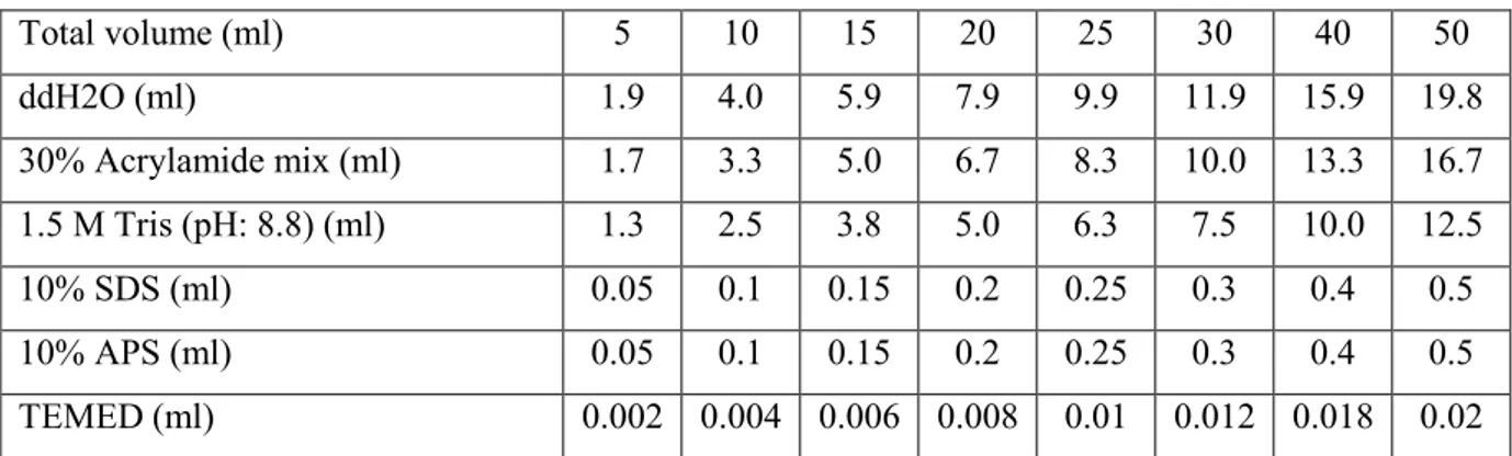
10% SDS-PAGE resolving gel (Western Blot), 12% SDS-PAGE resolving gel (Comassie Staining) and 5% stacking gel were prepared according to shown data (Table 2.1, Table 2.2).
Table 3.1 Resolving gel components (10%)
Total volume (ml) 5 10 15 20 25 30 40 50 ddH2O (ml) 1.9 4.0 5.9 7.9 9.9 11.9 15.9 19.8 30% Acrylamide mix (ml) 1.7 3.3 5.0 6.7 8.3 10.0 13.3 16.7 1.5 M Tris (pH: 8.8) (ml) 1.3 2.5 3.8 5.0 6.3 7.5 10.0 12.5 10% SDS (ml) 0.05 0.1 0.15 0.2 0.25 0.3 0.4 0.5 10% APS (ml) 0.05 0.1 0.15 0.2 0.25 0.3 0.4 0.5 TEMED (ml) 0.002 0.004 0.006 0.008 0.01 0.012 0.018 0.02
TEMED: Tetramethylethylenediamine, APS: Ammonium persulfate
Table 3.2 Stacking gel components (5%)
Total volume (ml) 1 2 3 4 5 6 8 10 ddH2O (ml) 0.68 1.4 2.1 2.7 3.4 4.1 5.5 6.8 30% Acrylamide mix (ml) 0.17 0.33 0.5 0.67 0.83 1.0 1.3 1.7 1.0 M Tris (pH: 6.8) (ml) 0.13 0.25 0.38 0.5 0.63 0.75 1.0 1.25 10% SDS (ml) 0.01 0.02 0.03 0.04 0.05 0.06 0.08 0.1 10% APS (ml) 0.01 0.02 0.03 0.04 0.05 0.06 0.08 0.1 TEMED (ml) 0.001 0.002 0.003 0.004 0.005 0.006 0.008 0.01
First of all protein amounts were quantified by Bradford Assay. In the scope of this assay a standard curve was generated by measuring the absorbance of Bovine Serum Albumin (BSA) solution (BSA/ddH2O) by spectrophotometer at 595 nm wavelength, visible light, in respect to concentration. Later absorbance of the protein samples dissolved in lysis buffer was measured and their concentration was calculated with the help of the BSA standard curve.
27
BioRad vertical gel system was set up according to the manufacturer’s instructions. Loading samples were prepared. For this, protein samples (~5 µl protein for the cell lysate samples, ~40 ng for pure CCDC-124) were mixed with 1X Loading Dye and 1X dithiothreitol (DTT) reducing agent (Fermentas). The loading samples were fulfilled to 10 µl- 15 µl with ddH2O. The samples were denatured at 100°C for 5 minutes and centrifuged at 13000 rpm for 1 minute. Later they were loaded onto gel. The electrophoresis took place at 250 V, 70 mA in 1X Tris Glycine electrophoresis buffer.
3.8 Comassie Staining
After the electrophoresis, the gel was put into commassie blue solution and incubated for 30 minutes on shaker. Later the commassie blue solution was removed and the gel was washed with either destaining solution or heated ddH2O.
3.9 Western Blotting
An alternative way for visualizing proteins is the immuno-blotting procedure. First of all proteins were transferred from gel to PVDF transfer membrane (Immobilon-P, millipore) by Trans-Blot Semi Dry transfer cell apparatus (Bio-Rad) according to the manufacturer’s instructions. For semi-dry transfer PVDF membrane has to be activated in methanol for 30 seconds. Besides Watmann papers and membrane should be wetted in transfer buffer.
After the transfer the membrane was blocked with 5% (w/v) non-fat milk powder dissolved in 0.5% Tween-20 containing TBS (TBS-T). Blocking lasts for 1 hour, at room temperature or overnight, at 4°C. Following the blocking step the membrane was incubated with primary antibodies (anti-Ccdc-124 (rabbit), anti-Flag (rabbit), monoclonal antibodies (mouse), again for 1 hour, at room temperature or overnight, at 4°C. After 3, 10 minute washes in TBS-T, the membrane was incubated with
28
Peroxidase conjugated secondary antibodies (anti-mouse or anti-rabbit, Sigma) for 1 hour. 3, 10 minute washes in TBS-T were repeated also after 2° Ab treatment. Next, the membrane was exposed to SuperSignal West Dura Extended Duration Substrate kit (Thermo Scientific) for 5 minutes. The proteins were detected on X-Ray films (18x24, blue, KODAK) by exposing the membrane to the film for 10-30 seconds in dark.
3. 10 4B3 monoclonal anti-Ccdc-124 Antibody purification
4B3 clone cell supernatants are taken and pulled down in one bottle. pH value of the supernatant solution is measured and adjusted to 7.0 with 1M Tris Base (pH: 8.0). The supernatant solution is distributed in 50 ml aliquots for a later treatment.
For Antibody purification procedure Protein A Sepharose Beads (CL-4B, GE Healthcare) are used. Lyophilized beads are treated with ddH2O. For 50 ml cell supernatant 250 µl bead medium (water treated beads) is used. The amount of required lyophilized beads is measured according to the manufacturer’s instructions. Beads are washed with ddH2O three times. Each time 50 ml water is added on top of beads and beads are centrifuged at 500 g for 15 minutes at 4°C. Following the wash step Phosphate Binding Buffer is added on top of beads (about 5X of the volume of the bead medium). Beads are washed twice with Phosphate Binding Buffer. Each time the solution is centrifuged at 500 g for 15 minutes at 4°C. (If the beads do not precipitate, the centrifuge steps might be repeated at 2000 rpm for 15 minutes at 4°C.) Finally the Phosphate Binding Buffer is removed from the beads and swollen beads are added in the supernatant solution aliquots (pH: ~7.0). This mix is rotated overnight, at 4°C, at falcon rotator.
29
Column (BIO-RAD) is washed three times with ddH2O and later twice with Phosphate Binding Buffer. Then, supernatants incubated with beads overnight are passed through the column. The flow-through is stored.
Afterwards the column is washed three times with Phosphate Binding Buffer. The flow through is stored.
Following Phosphate Binding Buffer treatment the column is washed twice with Citrate buffer. The flow through is stored in a falcon tube, in which 2 ml Tris Base was present (pH: 9.0).
Elution sample is centrifuged at 2500 G, for 15 minutes, at 4°C. This time 10K concentrator tubes (Millipore) are used for centrifugation. Then the concentrator tubes are washed twice with sterile cold PBS and centrifuged. The purified antibodies are aliquoted and stored at -20 °C.
30
4. RESULTS
4.1 Production of Monoclonal Antibodies
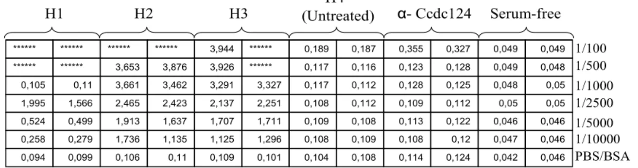
Among 11 immunized mice only 7 were used in the production of hybridoma cells. The first injection began in early January, 2009 for the first group, in February, 2009 for the second group and in late July for the third group. As the injections proceeded, one animal from the first group and three animals from the second group died due to unidentified reasons. In the last group, the amount of protein used for injections was decreased to 30 µg; nevertheless the antibody titer within the blood was high enough. The presence of antibodies in serum samples was tested with ELISA assay and it was found that all three animals in the first group were successfully immunized since the assay gave positive results for serum concentrations ranging from 1/100 to 1/10000 (Figure 4.1). Serum-free samples as well as samples with serum isolated from un-immunized animal (H4) gave negative results. Polyclonal Ccdc-124 antibody produced in rabbit (α-Ccdc-124 in Figure 4.1), gave negative results, since in this ELISA, anti-mouse secondary antibody was used. Also H5 from the second group showed high antibody titer within its blood (Figure 4.2).
0,046 0,042 0,124 0,114 0,108 0,104 0,101 0,109 0,11 0,106 0,099 0,094 0,046 0,047 0,12 0,108 0,109 0,108 1,296 1,125 1,135 1,736 0,279 0,258 0,046 0,046 0,122 0,113 0,108 0,109 1,711 1,707 1,637 1,913 0,499 0,524 0,05 0,05 0,112 0,109 0,112 0,108 2,251 2,137 2,423 2,465 1,566 1,995 0,05 0,048 0,125 0,128 0,112 0,117 3,327 3,291 3,462 3,661 0,11 0,105 0,048 0,049 0,128 0,123 0,116 0,117 ****** 3,926 3,876 3,653 ****** ****** 0,049 0,049 0,327 0,355 0,187 0,189 ****** 3,944 ****** ****** ****** ****** H1 H2 H3 H4 (Untreated) α- Ccdc124 Serum-free 1/100 1/500 1/1000 1/2500 1/5000 1/10000 PBS/BSA
Figure 4.1: ELISA showing antibody presence within serum isolated from three immunized mice belonging to the first group (H1, H2, H3). This assay was performed after the fourth injection. H4 sample, anti-Ccdc-124 antibody and serum free samples serve as negative controls. Different antibody concentrations were tested. Measurement: 30 minutes
31 0,088 0,114 0,095 0,104 0,12 0,08 2,592 0,075 0,068 0,069 0,081 0,072 0,063 1,985 0,073 0,076 0,083 0,066 0,067 1,509 0,086 0,074 0,081 0,084 0,071 1,382
H5 Serum
(-) (-) (+) (+)Figure 4.2: ELISA showing antibody presence within serum isolated from the immunized mouse belonging to the second group (H5). This assay was performed after the eighth injection. Serum sample from H1 was used as positive control. Serum was dissolved in PBS/ 5% non-fat milk powder solution in 1:1000 ratios. Measurement: 45 minutes. (-) and (+) symbolize the negative and the positive controls respectively (Section 3.2.3)
In case of high antibody titer, fusion experiment was performed. Firstly animals from the first group were sacrificed. And the fusion began with the animal giving the highest antibody titer. Since the ELISA results for H2 and H3 were similar, first fusion was performed on H2 after the fifth, boost injection. However no functional hybridomas were obtained from H2 as a result of contamination. Thus H2 hybridoma cells were discarded.
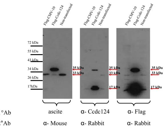
Before the spleen of H2 was isolated, it was observed that there was a fluid accumulation in the abdominal cavity of the animal. The fluid was sucked with a syringe and was stored. The presence of α-Ccdc-124 antibody in this ascites fluid was tested via ELISA assay (Figure 4.3). Since no clonal selection was performed, the ascites fluid was a candidate “poly”-clonal antibody. The ascites fluid was compared to the present polyclonal α-Ccdc-124 antibody.
32 Fla g-C cd c-124 Non -tra nsfe cted Fla g-C cd c-124 Non -tra nsfe cted Fla g-C cd c-124 Non -tra nsfe cted Fla g-C MV -10 Fla g-C MV -10 Fla g-C MV -10 72 kDa 55 kDa 43 kDa 34 kDa 26 kDa 17kDa
ascite
α- Ccdc124
α- Flag
1°Ab
2°Ab
α- Mouse α- Rabbit α- Rabbit
35 kDa 33 kDa 35 kDa 33 kDa 35 kDa 33 kDa 17 kDa 17 kDa
Figure 4.3: Western Blot showing antigen recognition capacity of ascites fluid (H2) as a candidate polyclonal antibody. The ascites fluid was tested as a candidate antibody source and its binding specificity was compared to the polyclonal α-Ccdc-124 antibody. The polyclonal Ab was dissolved in 5% non-fat milk powder/TBS-T solution in 1:500 ratio, the ascite in 1:1000 ratio and α-Flag An in 1:2000 ratio. Secondary antibodies were dissolved in 5% non-fat milk powder/TBS-T solution in 1:2000 ratios. 5 µg proteins were loaded for Flag-Ccdc-124-Transfected, CMV-Flag-CMV-10-Transfected and non-Transfected cell lysates. The α-Flag antibody was used as a control for testing the presence of transfected cell lysates. The images show 10 seconds exposure to X-ray film. CCDC-124: 33kDa
33
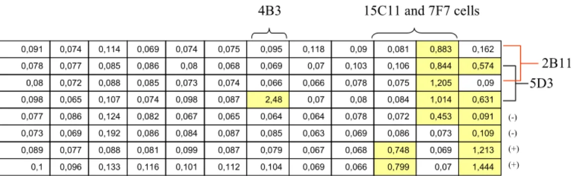
Injections continued for the remaining mice of the first and the second group. After the sixth, boost injection H3 was euthanized and its spleen was isolated. Fusion was performed as the previous one. In one ELISA, 4 clones were identified as positives. Later positive clones were tested again and one of them was found to be still positive. Sub-cloning was performed at this step. However follow-up ELISA assays showed no positive results for the clones which were previously identified as positive. Consequently H3 hybridoma cells were discarded.
After the 9th, boost injection, H1 was euthanized and its spleen was isolated for fusion. In the ELISA some positive clones were found. However there was contamination problem. Hybridoma cells were washed with newly prepared HAT medium containing additional penicillin. However contamination problem could not be overcame so that H1 hybridoma cells were discarded.
Afterwards the mouse from the other group H5 was sacrificed. One of the generated hybridoma clones was identified twice as positive in ELISA. Sub-cloning was performed. Five among H5 single clones, emerging from the former positive clone were identified as positive and no other clones founded to be positive. Thus H5 single cell plates were discarded. Five positive clones were again sub-cloned via limiting dilution method. Some of these cells gave later positive results in ELISA tests. And they were expanded in 24 well plates. However after a while positive clones were lost. As a consequence also H5 hybridoma cells were discarded.
Subsequent to the lost of previous hybridoma clones the experiments continued with the last group of mice. The presence of antibodies in serum samples was tested with ELISA assay and it was seen that all three animals were successfully immunized (Figure 4.4).
34 H8:0,961 H7:2,265 0,992 0,067 H8:0,869 H6:1,545 1,411 0,067 H7:2,141 H6:1,335
Figure 4.4: ELISA showing antibody presence within serum isolated from three immunized mice belonging to the third group (H6, H7, H8). This assay was performed after the eighth injection. Serum sample from H5 was used as positive control. Serum was dissolved in PBS/ 5% non-fat milk powder solution in 1:1000 ratios. Measurement: 30 min
Although H7 gave the highest antibody titer, spleen of H6 was isolated for the generation of hybridomas (it was a random choice). In ELISA tests one positive clone was detected (2E10). The cells of this clone were later sub-cloned and parental clones were expanded. In following assays 2 of the sub-clones were found to be positive. So these sub-clones together with parental 2E10 cells were cloned again. Although at the beginning 2E10 clone gave positive results, at the end no positive results were obtained, so H6 hybridoma cells were discarded.
Then hybridoma cells were generated by using the spleen of H7, the mouse with the highest serum antibody concentration. In the first ELISA test, 7F7 clone was identified as positive. Three other clones, 12F3, 11E8 and 15C11 were found to be positive in following ELISAs. Together with 7F7, the cells of four positive clones were sub-cloned. Parental cells were cultured on different plates via limiting dilution. In proceeding ELISA tests, 12F3 and 11E8 clone cells were shown to be negative. Therefore those plates were discarded. 7F7 cells which were again identified as positives were transferred firstly to 24 well plates, then to flasks and were expanded. Sub-clones of 7F7 and 15C11 were tested. Almost all of 7F7 sub-clones were positive, especially higher ones were expanded in 24 well plates and later stored at liquid nitrogen. However not every 15C11 sub-clone gave positive results. Only one of them was positive (15C11-3G3), which was later sub-cloned again. Positive one among 15C11-3G3 clones were expanded in 24 well plates and later in flasks. In