Protective effect of Celtis tournefortii against copper-induced toxicity in rat liver
Mehmet Ali Temiz
1, Atilla Temur
2, Yusuf Akgeyik
2, Ahmet Uyar
31Karamanoğlu Mehmetbey University, Vocational School of Technical Sciences,
Programme of Medicinal and Aromatic Plants, Karaman, Turkey
2Van Yuzuncu Yil University, Faculty of Education, Department of Mathematics and Sciences, Van, Turkey 3Hatay Mustafa Kemal University, Faculty of Veterinary Medicine, Department of Pathology, Hatay, Turkey
Received June 15, 2020 Accepted February 24, 2021
Abstract
This study aimed to investigate the antioxidant and hepatoprotective effects of Celtis tournefortii fruit extract (Ct) against copper-induced liver damage in rats. Thirty-two Wistar Albino rats were divided into four equal groups (n = 8): Control, Copper (Cu), Copper + C. tournefortii (Cu+Ct), and C. tournefortii (Ct). Superoxide dismutase (SOD), glutathione peroxidase (GPx), and catalase (CAT) activities, glutathione (GSH) concentration, malondialdehyde (MDA), total antioxidant status (TAS) and total oxidant status (TOS) were analysed in the liver tissues. Liver histopathology was also evaluated. Alanine aminotransferase and lactate dehydrogenase significantly decreased in the Cu+Ct group compared with the Cu group. Oxidative stress parameters MDA and TOS significantly increased with copper administration, whereas they decreased with C. tournefortii co-treatment compared to Cu group. GSH concentration and TAS showed significant decreases with copper administration. Celtis tournefortii co-supplementation with copper significantly enhanced antioxidant parameters such as TAS, SOD, and GPx. Celtis
tournefortii remarkably attenuated degenerative and necrotic changes caused by the oral exposure
of copper in the liver tissue of rats. Celtis tournefortii may provide amelioration of the antioxidant status and moderation of severity of liver damage against copper toxicity. The therapeutic use of
C. tournefortii may exhibit protective effects in liver injury treatment. Antioxidant, hepatoprotective, oriental hackberry, phytochemicals, TAS, TOS
Copper (Cu) exposure can occur due to pesticide residue exposure (WHO 2003) in
agriculture as well as via drinking water contaminated by environmental pollution or by
copper water pipe corrosion (Brewer 2012). Excessive copper intake may also occur
via consumption of Cu-rich foods such as liver, seafood, nuts, whole grains, and dried
fruits. The tolerable upper intake level is 10 mg/day a value which represents the limit for
protection of hepatic injury, a potentially critical side effect of excess copper ingestion (DRI
2001). Cu is a bioelement and a vital transition metal whose deficiency or excess in humans
is associated with pathological situations, especially in liver and brain tissues. However,
Cu metabolism is generally regulated by absorption and biliary excretion mechanisms.
Cu chaperones, Cu transporters, and Cu-binding proteins/enzymes maintain physiological
intracellular Cu homeostasis (Pal 2014). On the other hand, excess Cu causes production
of free radicals (e.g. reactive oxygen species [ROS]) through Haber-Weiss and Fenton
reactions, resulting in tissue damage (Valko et al. 2005). ROS-induced oxidative tissue
damage plays an important role in Cu toxicity. For example, excess Cu may lead to
peroxidative damage to membrane lipids via the reaction of lipid radicals and oxygen to
form peroxy radicals, and causes peroxidation in the membranes of hepatocyte lysosomes.
Also, Cu overload also reduces the activity of cytochrome c oxidase and impairs liver
mitochondrial respiration (Gaetke et al. 2014).
Phytochemicals are bioactive compounds in plants and they exert a protective effect
against various diseases (Zhang et al. 2015). Therefore, their nutraceutical use makes them
Address for correspondence:
Mehmet Ali Temiz
Vocational School of Technical Sciences Karamanoğlu Mehmetbey University Karaman, Turkey
Phone: +90 338 226 2000
E-mail: [email protected]; [email protected] http://actavet.vfu.cz/
important in terms of health. The antioxidant and hepatoprotective properties of plants
have underscored the importance of plants in discovery of new medications with less side
effects for use in hepatic disorder treatment. Celtis tournefortii fruit, commonly known as
the “oriental hackberry”, is palatable, nutrient-rich, and floury. Celtis tournefortii contains
flavonoid and phenolic acids such as gallic acid, vanillic acid, ellagic acid, chlorogenic
acid, ferulic acid, caffeic acid, rosmarinic acid, p-coumaric acid, myricetin, catechin, rutin,
naringin, kaempferol, resveratrol, and quercetin (Keser at al. 2017; Yıldırım et al. 2017).
Celtis tournefortii fruits are traditionally consumed for treatment of diarrhoea, dysentery,
and peptic ulcers. Some Celtis species are used in epileptic seizures, foot perspiration, and as
wound healing remedies. Celtis tournefortii seeds are used by the native population against
kidney sand, whereas its leaves are used for stomach pain, menstrual bleeding, sedative
purposes, and facilitating digestion (Keser et al. 2017). Although a few in vitro studies
(Keser et al. 2017; Yıldırım et al. 2017) have investigated the antioxidant properties of
this fruit, the protective effects against hepatotoxicity have not been investigated in vivo.
This study exhibits an authentic value due to the acquisition of primary scientific data.
Based on this assumption, we aimed to determine the antioxidant and hepatoprotective
effects of C. tournefortii fruit extract (Ct) against copper-induced toxicity in rats.
Materials and Methods
Plant material
Celtis tournefortii fruits were collected from Siirt, Turkey, in August 2016. The plant was taxonomically
identified and authenticated by Dr. Metin Armagan, Adnan Menderes University, Aydın. Fruits were dried under shade and powdered.
Celtis tournefortii fruit extraction
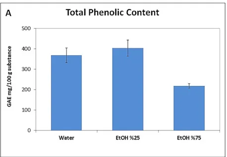
One g powdered Celtis fruit was added to a 100 ml beaker containing 50 ml solvent (water, ethanol 25%, and ethanol 75%, separately) and extracted by stirring for 3 h, at 50 °C and 750 rpm. The extract was then filtrated and centrifuged for 5 min at +4 °C, 3150 × g in a falcon tube. The obtained extract was freeze-dried for experimental study. Extraction was performed in duplicate for total phenolic and total flavonoid content analysis.
Determination of total phenolic and total flavonoid content
The total phenolic content (TPC) in the Ct extract was determined by modified Folin-Ciocalteau reagent method (Singleton and Rossi 1965) using gallic acid as a standard. The TPC was calculated as mg gallic acid equivalent for 100 g sample (mg GAE/100 g sample). The total flavonoid content (TFC) was determined by the AlCI3 method (Zhishen et al. 1999) using quercetin as a standard. The TFC was calculated as mg quercetin equivalent for 100 g sample (mg QE/100 g sample). Analyses were triplicated.
Extract prepared with ethanol 25% was used in the animal study because of the highest amounts of TPC and TFC in this extract were determined.
Acute toxicity assay (LD50)
For determining whether C. tournefortii fruit extract has acute toxicity, an LD50 study was performed on rats
for 7 days. Control and three plant extract groups (n = 6) were formed. The rats were fasted for 12 h before the plant extract was administered. The Ct was orally administered to rats with a bulbed steel needle at graded single doses (600, 1200, and 2400, mg·kg-1body weight [b.w.]), so a total amount of 24 animals were used. Rats were
anaesthetized and sacrificed at the end of the toxicity assay. Then their bloods were collected for liver function test.
Animals and experimental protocol
Experiments on the effects of the Ct were performed on 32 Wistar Albino male rats (150-250 g and 2 months old) obtained from Experimental Application and Research Center, Yuzuncu Yil University (Turkey). Rats were kept at 22°C, 12:12 h light/dark cycle in separate stainless cages. They were fed ad libitum standard chow and tap water for 28 days. The experimental groups (n = 8) were randomly formed as follows:
Control group received 1 ml saline via gavage daily for 28 days.
Group 2 of rats (CS group) were administered 10 mg/kg b.w. copper sulphate via intragastric tube twice a week and received 1 ml saline via gavage on other days for 28 days based on the modified method by Temiz et al. (2018).
Group 3 of rats (CS + Ct group) were administered 10 mg/kg b.w. copper sulphate via intragastric tube twice a week and received 10 mg/kg b.w. Ct via gavage daily for 28 days.
Group 4 of rats (Ct group) received 10 mg/kg bw Ct via gavage daily for 28 days.
The present study was approved by Yuzuncu Yil University Animal Researches Local Ethics Committee (no. 27552122-604.01.02-E.7837) and procedures complied with the Guidelines for the Care and Use of Laboratory Animals.
Sample collection
Rats were anaesthetized with ketamine + xylazine at the completion of the experiment. Blood was taken from the heart of each rat by an injector and transferred to serum-separating tubes (SSTs). The tubes were centrifuged at 850 × g for 10 min for serum enzymes including aspartate aminotransferase (AST), alanine aminotransferase (ALT), alkaline phosphatase (ALP), and lactate dehydrogenase (LDH) which were analysed in an auto-analyser (Roche Modular PP, Roche Diagnostics, Mannheim, Germany). Serum copper levels were analysed with atomic absorption spectrometer (iCE 3000 Series, Thermo Fisher Scientific, MA, USA) (y = 0.0857x + 0.0082, R2 = 0.9997). Liver tissues were dissected and rinsed by saline. Tissue samples for biochemical evaluation were
stored at −80 °C until analysis. Biochemical analyses
Liver tissues were homogenized in ice-cold phosphate buffered saline (PBS) (pH 7.4) using titanium probe homogenizer (Sonopuls HD 2200, Bandelin, Berlin, Germany) for 3 min and centrifuged at 8,570 × g for 30 min at +4 °C. The obtained supernatants of liver tissues of rats were used to analyse glutathione concentration (GSH) (Rizzi et al. 1988) and lipid peroxidation (malondialdehyde, MDA) by measuring thiobarbituric acid reactivity (Slater 1984), glutathione peroxidase (GPx) (Paglia and Valentine 1967), superoxide dismutase (SOD) (McCord and Fridovich 1969), and catalase (CAT) activity (Aebi 1984). Protein quantification was performed by modifying the method by Lowry. The total antioxidant status (TAS) and the total oxidant status (TOS) were performed using commercially available kit (Rel Assay Diagnostic, Turkey) as described by Erel (2004, 2005). The TAS method is based on antioxidants in the sample converting the ABTS+ radical
(2,2′-Azino-bis[3-ethylbenzothiazoline-6-sulfonic acid]) into the ABTS form (Erel 2004). The TOS method is based on conversion of ferrous (Fe+2) ion complexes of the oxidants present in the sample to the ferric (Fe+3) form by
oxidation (Erel 2005). Oxidative stress index (OSI) is the ratio of TAS and TOS parameters used to express the status of oxidative stress in tissues. OSI was calculated as per the following formula:
OSI (arbitrary unit) = (TOS/TAS) × 100 Histopathology
Liver tissues were taken from the rats and fixed in ice-cold, freshly prepared 10% formaldehyde for 72 h. Routine paraffin embedding and staining with haematoxylin-eosin were then performed on the liver tissue. The stained sections were evaluated by imaging with a light microscope (80i-DS-Ri2, Nikon, Tokyo, Japan). Histopathological results were evaluated semi-quantitatively according to the degree of the lesion as (-): none; (+): mild; (++): moderate; (+++): severe. Z ratio test was performed for the importance of the difference between the groups (n = 8) (Minitab 14 statistical software package).
Statistical analysis
Data were expressed as mean ± standard deviation. Significant differences between groups were assessed using one-way analysis of variance followed by Tukey’s test and Tamhane’s T2 (SPSS 18 statistical software package).
P values ≤ 0.05 were accepted as significant.
Results
The TPC and TFC results of C. tournefortii fruit extracts are presented in Fig. 1A-B
(Plate IX). The highest amounts of TPC and TFC in the extract were determined in extract
prepared with ethanol 25% (25% ethanol/75% water) compared with pure water and
ethanol 75% (ethanol 75%/25% water). This extract with ethanol 25% was then chosen for
the treatment of experimental animals.
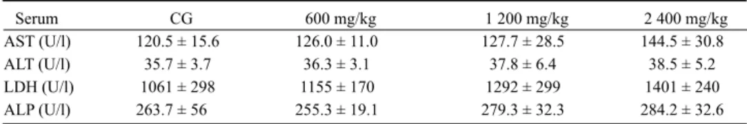
In the lethal dose (LD
50) study, no Ct group exhibited mortality. There was no reduction
in the physical activity of the rats, no change in their colour of the fur, and no molting. No
unusual changes observed in eye coloration, urine colour (bloody urine, dark brown colour
etc.), and stool of all treated rats. In the LD
50toxicity study, dose-dependent increases
in AST, ALT, LDH, and ALP levels were not significantly seen (Table 1).
Liver serum enzyme levels were used as biochemical markers for early acute hepatic
damage (Table 2). Oral exposure of rats to copper significantly increased liver serum
enzyme levels compared with control (P < 0.05). However, significantly lower values
of ALT and LDH were noted in the Ct co-treatment with copper when compared to the
Cu group. The amount of serum copper decreased in the Cu+Ct group compared to the
Cu group (P < 0.05).
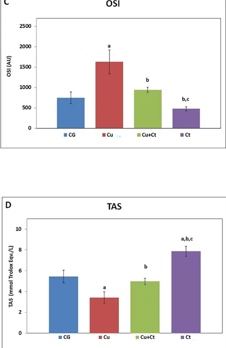
The liver MDA content, TOS, and OSI as oxidative stress parameters were significantly
increased after Cu administration to rats regarding the control group, indicating the liver
ROS generations and oxidative stress induction. Conversely, Ct co-supplementation with
copper significantly restored these indices (Plate X-XI, Fig. 2A-C). However, TOS increased
in the Cu+Ct group in comparison with control, despite being significantly lower than in
the Cu group (P < 0.05). While liver TAS decreased in the Cu group, it increased with
Ct treatment (Plate XI, Fig. 2D). The TAS was significantly higher in the Ct group compared
to all groups.
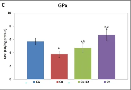
Subacute copper exposure to rats showed severe inhibitory response on the liver’s
antioxidant status (Plate XII-XIII, Fig. 3). Liver GSH content and SOD, GPx, and CAT
activities were significantly decreased in Cu-exposed rats than in control group, indicating
suppressed liver antioxidant defence against ROS. Ct co-treatment with copper significantly
recuperated liver SOD and GPx activities compared to the Cu group. Treatment with only
C. tournefortii showed significant increases of antioxidant enzyme activities and GSH than
Cu+Ct group (Plate XII-XIII, Fig. 3A-D).
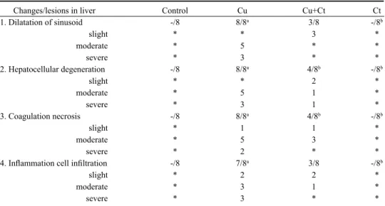
Results of histopathological changes and lesions for both control and treated rats are
shown in Table 3. No histopathological findings were observed in microscopic examination
of the liver tissue of the rats in the control group or with Ct alone. Hepatocytes and portal
areas had normal appearance, and remark cord around vena centralis was found regularly.
The sinuses between the remark cords were normal (Plate XIV, Fig. 4A,D). Conversely,
disseminated centrilobular hepatocellular degeneration and vacuolar degeneration were
observed in the liver of Cu-treated rats. Mononuclear cell infiltration was detected in portal
areas where lymphocytes with inflammatory cells were predominant. Because of dilatation
Table 2. Serum indicators of rats.
Serum CG Cu Cu+Ct Ct AST (U/l) 119.0 ± 3.9 153.4 ± 18.0a 132.5 ± 11.5a 112.9 ± 13.2b ALT (U/l) 34.6 ± 2.4 41.0 ± 1.5a 35.9 ± 4.5b 34.5 ± 2.6b LDH (U/l) 1445 ± 318 1783.5 ± 268a 1589 ± 145b 1227 ± 129b ALP (U/l) 272.5 ± 26 295.0 ± 69.4a 273.4 ± 56.3 219.3 ± 27.1b Cu (mg/l) 1.048 ± 0.076 1.491 ± 0.278a 0.965 ± 0.188b 0.475 ± 0.097a,b
CG - control group; Cu - copper group; Cu+Ct – copper + Celtis group; Ct - Celtis group; AST - aspartate aminotransferase; ALT - alanine aminotransferase; LDH - lactate dehydrogenase; ALP - alkaline phosphatase; Cu - copper.
a - significantly different from the control group (P < 0.05); b - different from the Cu group (P < 0.05).
Table 1. Serum indicators of LD50 toxicity of rats.
Serum CG 600 mg/kg 1 200 mg/kg 2 400 mg/kg
AST (U/l) 120.5 ± 15.6 126.0 ± 11.0 127.7 ± 28.5 144.5 ± 30.8
ALT (U/l) 35.7 ± 3.7 36.3 ± 3.1 37.8 ± 6.4 38.5 ± 5.2
LDH (U/l) 1061 ± 298 1155 ± 170 1292 ± 299 1401 ± 240
ALP (U/l) 263.7 ± 56 255.3 ± 19.1 279.3 ± 32.3 284.2 ± 32.6
CG - control group; AST - aspartate aminotransferase; ALT - alanine aminotransferase; LDH - lactate dehydrogenase; ALP - alkaline phosphatase.
in the sinusoid, dissociation was remarkable in the remark cord structure. The cytoplasm
of some hepatocytes had eosinophilic, pyknotic, and karyolytic nuclei. Focal coagulation
necrosis was detected, especially in periacinar regions, where partial proliferation in
Kupffer cells was observed (Plate XIV, Fig. 4B). Mild degenerative changes were observed
in the livers of rats treated with Ct and copper. However, lymphocytic mononuclear cell
infiltrations were rarely seen (Plate XIV, Fig. 4C).
Discussion
Presently, the use of phytochemicals is preferred to synthetic supplements (vitamins,
minerals, fibre etc.) due to their beneficial pharmacological effects, as they are considered
safer and more reliable (Kioukia-Fougia et al. 2016; Scott et al. 2020). These effects
could be attributed to the presence of valuable constituents (Zhang et al. 2015; Keser
et al. 2017; Yıldırım et al. 2017). In this study, antioxidant and hepatoprotective effects
of C. tournefortii fruit were investigated against hepatic damage caused by Cu-toxicity in
rats. This study exhibits an authentic value due to the acquisition of primary scientific data.
As regards to the TPC and TFC results, the 25% hydroalcoholic solvent revealed
considerable amount of polyphenolic compounds in the extract compared to pure water
and 75% ethanol. Yıldırım et al. (2017) found similar results for TPC in the methanolic
extract. However, TFC was higher in the present study than the results of Yıldırım et al.
(2017). The time, temperature and solvent preference in the extraction process may have
affected the amount of compounds in the TFC (Temiz and Temur 2017). In addition, the
sampling site, the quality of soil, the season etc. may have probably had an effect.
High-dose Ct extract did not cause death in the LD
50study, nor did it alter any physical
condition and activity on the rats. However, a non-significant dose-dependent increase in
serum parameters was detected (Table 1). It has been estimated that oral LD
50value was
> 2400 mg/kg b.w. Ntchapda et al. (2008) stated that the LD
50dose of Celtis durandii
Table 3. Histopathological changes in the control, Cu, Cu+Ct, and Ct groups of rats.
Changes/lesions in liver Control Cu Cu+Ct Ct
1. Dilatation of sinusoid -/8 8/8a 3/8 -/8b slight * * 3 * moderate * 5 * * severe * 3 * * 2. Hepatocellular degeneration -/8 8/8a 4/8b -/8b slight * * 2 * moderate * 5 1 * severe * 3 1 * 3. Coagulation necrosis -/8 8/8a 4/8b -/8b slight * 1 1 * moderate * 5 3 * severe * 2 * *
4. Inflammation cell infiltration -/8 7/8a 3/8 -/8b
slight * 2 2 * moderate * 3 1 *
severe * 3 * *
Cu - copper group; Cu+Ct – copper + Celtis group; Ct - Celtis group.
* - none; a - significantly different from control (P < 0.05); b - significantly different from the Cu group (P < 0.05); c - significantly different from the Cu+Ct group (P < 0.05).
leaf extract were 14.10 g/kg with a mortality rate of 42%. In accordance with the current
study, the Celtis durandii leaf extract caused an increase in both ALT and AST serum levels
(Ntchapda et al. 2008). Another study conducted with a Celtis iguanaea (Jacq.) Sarg.
leaf extract considered LD
50to be higher than 2,000 mg/kg and lower than 5,000 mg/kg
(Gonçalvez et al. 2015). Therefore, high doses of C. tournefortii fruit extract may have
low toxicity.
Studies have indicated that various forms of copper such as ion, compound, micro and
nanoparticle lead to liver damage and cause increased liver serum enzymes including AST,
ALT, and ALP (Lee et al. 2016; Arafa et al. 2017; Khalid et al. 2018; Temiz et al. 2018).
Transaminase enzyme levels rise in serum as biomarkers of hepatotoxicity when the liver
cell integrity is disrupted and parenchymal cells degenerated. However, Celtis treatment of
Cu-induced liver-damaged rats contributed to recovery. There is limited data on Celtis spp.
use for liver injury. Reportedly, administration of 100 mg/kg aqueous ethanolic leaf extract
of Celtis integrifolia exhibited significant reduction in serum biochemical indicators such
ALT and ALP (Geidam and Adole 2014). Polyphenols may not only act as antioxidants
terminating free radical chain reactions but also as effective chelators of redox-active
metals (Jomova and Valko 2011). Studies on flavonoids such as quercetin, catechin,
and rutin have been conducted for discrimination between its antioxidant versus metal ion
chelating properties in the red blood cell haemolysate system in vitro. The results showed
that flavonoids maintained their efficiency to chelate redox-active metals (Cherrak
et al. 2016). Serum copper levels dramatically reduced with C. tournefortii treatment
concomitantly with Cu. Celtis contains flavonoids which, like a chelator, may have
assisted the reduction of copper concentration in the serum by increasing copper excretion.
Flavonoids are capable to form stable metal complexes through their multiple OH groups
and the carbonyl moiety. For instance, quercetin which is characterized by three potential
bidentate binding sites (α-hydroxy-carbonyl, β-hydroxy-carbonyl or catechol), can lead to
stable metallic complexes. Previous investigation demonstrated that flavonoids are able to
complex Cu
2+. The Cu-quercetin complexation was suggested to occur via the 4-keto group
of the C-ring with additional involvement of the 3OH or 5OH group (Cherrak et al. 2016).
In the current study, MDA as well as TOS and OSI, which are an indicator of oxidant/
antioxidant imbalance, exhibited a significant increase in Cu-administered rats (Fig. 2).
Besides, TAS is formed by internal and external antioxidant molecules of the cell which
act synergistically. Therefore, measurement of the total antioxidant capacity gives more
valuable information than separate measurements. Findings of this study exhibited that Ct
co-treatment with Cu reduced oxidation and increased the antioxidant status compared to the
Cu group. TAS was significantly higher in the Ct group in comparison with all groups (Fig.
2D). This result showed better the antioxidant effect of Ct administered alone. In previous
studies, the ethanolic extract of C. australis and C. occidentalis leaves (El-Alfy et al.
2011) and the hydromethanolic extract of C. australis leaves (Filali-Ansari et al. 2015)
significantly reduced in vitro MDA formation. The present findings were consistent with
Zanchet et al. (2018) who reported that a C. iguanaea supplement significantly decreased
lipid peroxidation in cholesterol-fed rats by increasing SOD activity. Moreover, Arafa
et al. (2017) reported that in Cu-induced liver, the elevated hepatic ROS and suppressed
total antioxidant capacity improved with quercetin treatment. Celtis tornefortii also contains
quercetin and other flavonoids as well as other aforementioned phenolic antioxidants
(Keser et al. 2017).The decrease in lipid peroxidation and TOS in the treatment with Ct
may be due to the presence of scavenger compounds. Furthermore, animals co-treated
with Ct showed a significant improvement in the GSH content, activities of SOD and GPx
compared with Cu-intoxicated rats (Fig. 3). These improvements in antioxidant enzymes
conform to previous studies and can be attributed to antioxidant properties of the Celtis
species (Dasari et al. 2013; Fall et al. 2017).
The present and previous studies have demonstrated that Cu induces severe histological
changes and hepatic damage (Li et al. 2008; Ibrahim et al. 2015). Li et al. (2008)
stated that Cu can induce liver damage through an up-regulation of apoptosis regulator
B-cell lymphoma-2 associated protein X (Bax) expression. Conversely, co-treatment
with Ct ameliorated liver injury, showing less degeneration and necrotic changes. Celtis
co-treatment with copper may prevent liver injury or lead to the recuperation of damaged
liver parenchyma. Phenolic and flavonoid compounds in herbs may have beneficial
hepatoprotective effects. Studies indicated that many phytochemicals such as quercetin
against Pb (Liu et al. 2013), gallic acid against NaF (Nabavi et al. 2013), and rutin against
Cd (Mirani et al. 2012) effectively prevented hepatic damage against metal intoxication.
Mechanisms underlying the effects of these compounds may be attributed to inhibiting
the Fenton-like reaction, inhibiting the formation of highly reactive hydroxyl radicals
by acting as a radical scavenger donating hydrogen (Ibrahim et al. 2015). Moreover,
phytochemicals can induce the expression of endogenous antioxidants, and prevent
ROS-mediated oxidative stress on DNA by exerting their antioxidant and anti-apoptotic
effects (Ibrahim et al. 2015). Therefore, these phytochemicals may be responsible for the
hepatoprotective effects of the Celtis extract. Polyphenolic compounds may synergistically
modulate fibrosis and necrosis in the liver. Herbal medicines are mixtures of various
phytochemicals that exert synergistically their full beneficial effect in total extracts (Zhang
et al. 2019).
In conclusion, the hepatotoxicity of Cu was corroborated by the data obtained with
increasing oxidative stress markers and hepatic degeneration and necrotic changes. However,
it was revealed that C. tournefortii may have hepatoprotective effects against Cu-induced
liver damage due to mitigated oxidative stress indicators and hepatocellular degeneration as
well as on an enhanced antioxidant status. The use of C. tournefortii as nutraceutical for the
maintenance of oxidant/antioxidant balance for liver damage may be efficacious.
Acknowledgement
This research was supported by the Van Yuzuncu Yil University Scientific Research Projects Foundation (grant number FYL-2016-5479).
References
Aebi H 1984: Catalase in vitro. Methods Enzymol 105: 121-126
Arafa AF, Ghanem HZ, Soliman MS, El-Meligy E 2017: Modulation effects of quercetin against copper oxide nanoparticles-induced liver toxicity in rats. Egypt Pharma J 16:78-86
Brewer GJ 2012: Metals in the causation and treatment of Wilson’s disease and Alzheimer’s disease, and copper lowering therapy in medicine. Inorganica Chim Acta 393: 135-141
Cherrak SA, Mokhtari-Soulimane N, Berroukeche F, Bensenane B, Cherbonnel A, Merzouk H, Elhabiri M 2016: In vitro antioxidant versus metal ion chelating properties of flavonoids: A structure-activity investigation. PLOS ONE 11:1-21
Dasari R, Sathyavati D, Belide SK, Reddy PJ, Abbulu K 2013: Evaluation of antioxidant activity of two important memory enhancing medicinal plants Celtis timorensis and Vanda spathulata. Asian J Pharm Clin Res
6: 153-155
DRI (Dietary Reference Intakes) 2001: Dietary reference intakes for vitamin A, vitamin K, arsenic, boron, chromium, copper, iodine, iron, manganese, molybdenum, nickel, silicon, vanadium, and zinc. Institute of Medicine (US) Panel on Micronutrients. Washington (DC), National Academies Press (US)
Erel Ö 2004: A novel automated direct measurement method for total antioxidant capacity using a new generation, more stable ABTS radical cation. Clin Biochem 37: 277-285
Erel Ö 2005: A new automated colorimetric method for measuring total oxidant status. Clin Biochem 38:
1103-1111
El-Alfy TS, El-Gohary HMA, Sokkar NM, Hosny M, Al-Mahdy DA 2011: A new flavonoid c-glycoside from Celtis australis L. and Celtis occidentalis L. leaves and potential antioxidant and cytotoxic activities. Sci Pharm 79: 963-975
Fall AD, Dieng SIM, Diatta-Badji K, Diatta W, Bassene E 2017: Phytochemical screening, phenol content and antioxidant studies of ethanol leaf extract of Celtis toka (Forssk.) Hepper& J.R.I. Wood. J Pharmacogn Phytochem 6: 488-492
Filali-Ansari N, El Abbouyi A, El Khyari S 2015: Antioxidant properties of leaves and seeds hydromethanolic extracts from Celtis australis. J Chem Biol Phys Sci B 5: 2834-2843
Gaetke LM, Chow-Johnson HS, Chow CK 2014: Copper: Toxicological relevance and mechanisms. Arch Toxicol
88: 1929-1938
Geidam MA, Adole OS 2014: Effects of the aqueous ethanolic leaves extract of Celtis integrifolia on liver function of wister strain albino rats. Int J Sci Res Manag 2: 713-718
Gonçalves NZ, Lino Júnior RS, Rodrigues CR, Rodrigues AR, Cunha LC 2015: Acute oral toxicity of Celtis
iguanaea (Jacq.) Sargent leaf extract (Ulmaceae) in rats and mice. Rev Bras Plantas Med 17: 1118-1124
Ibrahim MA, Khalaf AA, Galal MK, Ogaly HA, Hassa AHM 2015: Ameliorative influence of green tea extract on copper nanoparticle-induced hepatotoxicity in rats. Nanoscale Res Lett 10: 2-9
Jomova K, Valko M 2011: Advances in metal-induced oxidative stress and human disease. Toxicology 283: 65-87
Keser S, Keser F, Kaygili O, Tekin S, Turkoglu I, Demir E, Turkoglu S, Karatepe M, Sandal S, Kirbag S 2017: Phytochemical compounds and biological activities of Celtis tournefortii fruits. Anal Chem Let 7: 344-355
Khalid S, Afzal N, Khan JA, Hussain Z, Qureshi AS, Hafeez A, Jamil Y 2018: Antioxidant resveratrol protects against copper oxide nanoparticle toxicity in vivo. Naunyn-Schmiedeberg’s Arch Pharmacol 391: 1053-1062
Kioukia-Fougia N, Georgiadis N, Tsarouhas K, Vasilaki F, Fragkiadaki P, Meimeti E, Tsitsimpikou C 2016: Synthetic and natural nutritional supplements: Health “allies” or risks to public health? Recent Pat Inflamm Allergy Drug Discov 10: 72-85
Lee IC, Ko JW, Park SH, Lim JO, Shin IS, Moon C, Kim S, Heo J, Kim J 2016: Comparative toxicity and biodistribution of copper nanoparticles and cupric ions in rats. Int J Nanomedicine 11: 2883-2900
Li YW, Wang XH, Nin Q, Luo XP 2008: Excessive copper induces hepatocyte apoptosis and affects Bax and Bcl-2 expression in rat liver. Chin J Contemp Pediatr 10: 42-46
Liu CM, Zheng GH, Ming QL, Sun JM, Cheng C 2013: Protective effect of quercetin on lead-induced oxidative stress and endoplasmic reticulum stress in rat liver via the IRE1/JNK and PI3K/Akt pathway. Free Radic Res
47: 192-201
McCord JM, Fridovich I 1969: Superoxide dismutase. An enzymic function for erythrocuprein (hemocuprein). J Biol Chem 244: 6049-6055
Mirani N, Ashraf JA, Siddique J, Rub A 2012: Protective effect of rutin against cadmium induced hepatotoxicity in Swiss albino mice. J Pharmacol Toxicol 7: 150-157
Nabavi SF, Nabavi SM, Habtemariam S, Moghaddam AH, Sureda A, Jafari M, Latifi AM 2013: Hepatoprotective effect of gallic acid isolated from Peltiphyllum peltatum against sodium fluoride-induced oxidative stress. Ind Crops Prod 44: 50-55
Ntchapda F, Dimo T, Mbongué G, Atchade AT, Kamtchouing P, Enow G 2008: Acute Toxic Effects of the Aqueous Leaf Extract of Celtis durandii Engler (Ulmaceae) on Mice. West Afr J Pharmacol Drug Res 24: 1-7
Paglia DE, Valentine WN 1967: Studies on the quantitative and qualitative characterization of erythrocyte glutathione peroxidase. J Lab Clin Med 70: 158-169
Pal A 2014: Copper toxicity induced hepatocerebral and neurodegenerative diseases: An urgent need for prognostic biomarkers. NeuroToxicology 40: 97-101
Rizzi R, Caroli A, Bolla P, Acciaioli A, Pagnacco G 1988: Variability of reduced glutathione levels in massese ewes and its effect on daily milk production. J Dairy Res 55: 345-353
Scott SE, Rozin P, Small DA 2020: Consumers prefer “natural” more for preventatives than for curatives. J Consum Res 47: 454-471
Singleton VL, Rossi JA 1965: Colorimetry of total phenolics with phosphomolybdic-phosphotungustic acid reagents. Am J Enol Viticult 16: 144-158
Slater TF 1984: Overview of methods used for detecting lipid peroxidation. Methods Enzymol 105: 283-305
Temiz MA, Temur A 2017: Effect of solvent variation on polyphenolic profile and total phenolic content of olive leaf extract. YYU J Agr Sci 27: 43-50
Temiz MA, Temur A, Kaval Oguz E 2018: Antioxidant and hepatoprotective effects of vitamin E and melatonin against copper-induced toxicity in rats. Trop J Pharm Res 17: 1025-1031
Valko M, Morris H, Cronin MT 2005: Metals, toxicity and oxidative stress. Curr Med Chem 12: 1161-1208
WHO 2003: Copper in drinking-water. Background document for preparation of WHO guidelines for drinking-water quality. World Health Organization, Geneva
Yıldırım I, Uğur Y, Kutlu T 2017: Investigation of antioxidant activity and phytochemical compositions of Celtis
tournefortii. Free Rad Antiox 7: 160-165
Zanchet B, Gomes DB, Corralo VS et al. 2018: Effects of hydroalcoholic extract of Celtis iguanaea on markers of cardiovascular diseases and glucose metabolism in cholesterol-fed rats. Rev Bras Farmacogn 28: 80-91
Zhang YJ, Gan RY, Li S, Zhou Y, Li AN, Xu DP, Li HB 2015: Antioxidant phytochemicals for the prevention and treatment of chronic diseases. Molecules 20: 21138-21156
Zhang L, Virgous C, Si H 2019: Synergistic anti-inflammatory effects and mechanisms of combined phytochemicals. J Nutr Biochem 69: 19-30
Zhishen J, Mengcheng T, Jianming W 1999: The determination of flavonoid contents in mulberry and their scavenging effects on superoxide radicals. Food Chem 64: 555-559
Temiz M. A.. et al.: Protective ... pp. 91-98
Fig. 2. The MDA content (A), TOS (B), OSI (C) and TAS (D) levels of groups.
MDA: malondialdehyde, TOS: total oxidant status, TAS: total antioxidant status, CG: control group, Cu: copper group, Cu+Ct: copper + Celtis group, Ct: Celtis group.
a - significantly different from the control group (P < 0 .05); b - significantly different from Cu (P < 0.05); c - significantly different from the Cu+Ct group (P < 0.05).
Fig. 3. The GSH content (A) and SOD (B), GPx (C), CAT (D) activities of groups.
GSH: glutathione, SOD: superoxide dismutase, GPx: glutathione peroxidase, CAT: catalase, CG: control group, Cu: copper group, Cu+Ct: copper + Celtis group, Ct: Celtis group.
a - significantly different from the control group (P < 0.05); b - significantly different from the Cu group
Fig. 4. A: control group, normal histological appearance of the liver; B: Cu group, disseminated centrilobular hepatocellular degeneration and coagulation necrosis (arrows), inflammatory cell infiltration in the portal area (asterisk), dilatation in sinusoid (arrow-heads); C: Cu+Ct group, centrilobular hepatocellular degeneration (asterisk); D: Ct group, normal histological appearance of the liver. H+E × 10. Cu: copper group, Cu+Ct: copper + Celtis group, Ct: Celtis group.