296 Turkish J Thorac Cardiovasc Surg 2009;17(4):296-298 Türk Göğüs Kalp Damar Cerrahisi Dergisi
Turkish Journal of Thoracic and Cardiovascular Surgery
Chylopericardium following double valve replacement
İkili kapak replasmanını izleyen şiloperikardiyum
Tekin Yıldırım,1 Özer Selimoğlu,1 Cihan Çevik,5 İrade Öztürk,3 Fatma Mine Öz,4 Nuri Kurtoğlu,2 Noyan Temuçin Oğuş1
Department of 1Cardiovascular Surgery, 2Cardiology, 3Radiology, 4Anaesthesiology and Reanimation, Avrupa Şafak Hospital, İstanbul; 5Department of Cardiology, Medicine Faculty of Dumlupınar University, Kütahya
Şiloperikardiyum açık kalp cerrahisi sonrasında nadir görülen bir komplikasyondur. Şiloperikardiyum ameli-yat sırasında duktus torasikusa drene olan küçük peri-kardiyal kanalların ve/veya timus bezi içi kanalların istenmeyen biçimde hasarı sonucunda oluşmaktadır. Şiloperikardiyumun ilk tedavisinde diyet düzenlenmesi vardır. Bu yazıda çift kapak replasmanı yaptığımız 24 yaşındaki kadın hastada gelişen şiloperikardiyum olgu-su olgu-sunuldu. Hastamızda ilk iki hafta orta zincirli trig-liserid ve proteinden zengin diyet uygulandı ve üçüncü hafta oral alım durduruldu, total paranteral beslenme başlandı. Hastada üçüncü haftanın sonunda drenajda azalma olmayınca, ikinci eksploratif girişim uygulandı. Sternotomi sonrası timus etrafında 3x4 cm şiloz birikim tespit edildi. Timus dokusu ve perikard kenarları 4/0 prolenle devamlı tarzda dikildi. Revizyon ameliya-tından bir hafta sonra ekokardiyografik ve radyolojik inceleme sonucu perikardiyal birikim olmayan hasta taburcu edildi, üçüncü ay kontrolünde hasta tamamen normaldi. Açık kalp cerrahisini izleyen üçüncü haftanın sonunda devam eden şilöz drenajda eksploratif cerrahi tavsiye edilebilir.
Anah tar söz cük ler: Şilöz drenaj; kalp kapak cerrahisi; cerrahi
girişim.
Chylopericardium is a rare complication following open heart surgery. Chylopericardium occurs because of the unin-tentional damage of the small pericardial lymphatic ducts, draining to thoracic duct, and/or thymic intraglandular ducts during the operation. Initial treatment of chylopericardium is dietary modification. In this article we present a 24-year-old female with chylopericardium who underwent aortic and mitral valve replacement. In our patient diet was arranged as high-protein with medium chained triglycerides in her first two weeks and in the third week oral intake was ceased and total parenteral nutrition was initiated. Since there was a non-decreasing continuous leakage after three weeks of operation, we performed an exploratory second surgical intervention. After sternotomy, 3x4 cm of chylous collection was detected around thymus. Thymus tissue and pericardial edges were sutured continuously with 4/0 polypropylene. Pericardial collection was not detected after echocardio-graphic and radioechocardio-graphic evaluation and the patient was dis-charged one week after surgical revision. In the postoperative third month, she was completely normal. In the cases of con-tinuous chylous drainage following open heart surgery after three weeks, exploratory surgery can be recommended.
Key words: Chylous drainage; heart valve surgery; surgical
intervention.
Received: November 7, 2005 Accepted: December 5 2005
Correspondence: Tekin Yıldırım, M.D. Göztepe Şafak Hastanesi, Kalp ve Damar Cerrahisi Kliniği, 34732 Göztepe, İstanbul, Turkey. Tel: +90 216 - 399 97 50 e-mail: [email protected]
Chylopericardium is a very rare complication of heart surgery observed following a median sternotomy (dur-ing intrapericardial procedures). It can be detected after repairs of congenital heart diseases, valve surgeries or even isolated coronary artery bypass operations.[1-3] Although rare, it may increase hospital costs and the duration of hospital stay seriously by causing malnutri-tion and necessitating further intervenmalnutri-tion or extended parenteral nutrition.
CASE REPORT
A twenty four-year-old female patient was admitted to our cardiology clinic with symptoms of dyspnea, pal-pitation and fatigue for four years. After her physical examination and echocardiographic evaluation, a rheu-matic heart valve disease was detected. The transthoracic echocardiography revealed a serious mitral stenosis (mitral valve area: 1.2 cm2) with grade 4 regurgitation; serious aortic regurgitation with stenosis (mean aortic
Yıldırım ve ark. İkili kapak replasmanını izleyen kiloperikardiyum
Türk Göğüs Kalp Damar Cer Derg 2009;17(4):296-298 297
transvalvular gradient: 38 mmHg) and serious tricuspid regurgitation. The patient was prepared for open heart surgery. A central venous catheter through the right internal jugular vein was placed and the patient was monitored from the right radial artery after the appli-cation of the standard open heart surgical anesthesia. A median sternotomy was performed and the pericar-dium was dissected vertically. The two pleural spaces remained intact. The thymus was divided medially using electrocauterization and without extracting the gland. The cardiopulmonary bypass was initiated following aortic-arterial and bicaval venous cannulation.
The superior and inferior vena cava were snared with tapes. The pulmonary artery was separated from the ascending aorta with a sharp dissection. Following the cross-clamping of aorta, an aortotomy was performed and diastolic arrest was achieved by isothermic blood cardioplegia from the coronary ostia. Isothermic blood cardioplegia was used for myocardial protection and it was delivered continuously through retrograde route. The mitral valve was replaced with a 31 mm Omnicarbon bileaflet mechanical prosthesis and the posterior leaflet was preserved. The aortic annulus was narrow and it was enlarged with a glutaraldehyde-treated pericardial patch at the level of the non-coronary cusp (without dissecting the mitral annulus, the modified Nick’s pro-cedure) and a 21 mm Omnicarbon bileaflet mechanical prosthesis was implanted in tilting position. The aortic cross-clamp was removed and a tricuspid valve Kay annuloplasty was performed on the beating heart. The patient was extubated four hours postoperatively. In her first day, 950 ml of hemorrhagic drainage was obtained from the mediastinal drain. In her second day, 650 ml of sero-hemorrhagic fluid was obtained. Later, the property of the drainage fluid was completely chylous and the patient was followed with a milky-white chylous drainage for 21 days, producing 330-750 ml of drainage daily (mean: 610 ml). The biochemical evaluation of the
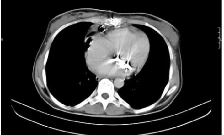
drainage fluid revealed triglyceride 789 mg/dl, protein 4.3 g/dl. In the microscopic evaluation, the leukocytes were detected with a domination of lymphocytes. The transthoracic echocardiographic examination revealed a mild pericardial effusion. The computerized tomo-graphical (CT) analysis showed a hypodense collection around the thymus area (Fig. 1). Her diet was arranged as a high-protein one containing medium chained trig-lycerides in her first two weeks and the oral food intake was ceased and total parenteral nutrition was initiated in the third week. During this time, the total blood protein level had decreased from 6.8 g/dl to 5.5 g/dl, the lym-phocyte count from 34% to 18% and the body weight had fallen from 53 kg to 48.5 kg. Since the patient did not respond to conservative measures, a revision opera-tion was performed on the 21st day.
After the sternotomy, an elliptoid, gelatinous, 3x4 cm chylous collection was detected around the thymus. The probable drainage place was suspected to be the thymus tissue, which was sutured in a horse-shoe shape continuously with 4/0 polypropylene suture. Lymphatic drainage was excluded following the control of the aor-tic annulus, the inferior and superior vena cava areas. The pericardial edges were sutured continuously with 4/0 polypropylene and controlled for any leakages. The sternum was closed again with a drain left in the mediastinum. No pericardial collection was detected after the echocardiographic and radiographic evalua-tions and the patient was discharged one week after the revision operation. At the postoperative third month, she was completely normal in her physical and echocardio-graphic examination.
DISCUSSION
Chylopericardium and chylothorax can be detected with an incidence of 0.89% and 0.12% after pediatric cardiovascular heart surgery.[4,5] Unlike adults; the thy-mic area is rich with lymphoid tissue in children, mak-ing this potentially lethal complication much less com-mon in adults. An injury to the thoracic duct directly within the pericardium is impossible. In a study of 35 human cadavers, the parietal pericardial lymphatics were demonstrated either directly or indirectly using Indian ink.[6] The lymphatic drainage from the parietal pericardium and surrounding tissues come together on the lateral and posterior face with the lymphatics of the mediastinal pleura. This anatomy sheds light on the mechanism of the normal pericardial fluid found in the pericardial sac and the chylopericardium after the pericardial incision.
Chylopericardium occurs because of the inevitable damage to the small pericardial lymphatic ducts that drain into the thoracic duct later and/or the thymic
Fig. 1. Thoracal computed tomography imaging showing the hy-podense collection 20x30x80 mm in size in thymic localization in the anterior mediastinum.
Yıldırım et al. Chylopericardium following double valve replacement
Turkish J Thorac Cardiovasc Surg 2009;17(4):296-298 298
intraglandular ducts during the operation. Leakage from the thymic tissue area occurs more commonly into the anterior mediastinum. Our patient had a major chylous collection around the thymic area that made it the possi-ble mechanism of chylopericardium. We had also used a pericardial patch for the enlargement of the aortic root.
Less commonly, the chylopericardium develops from the posterior mediastinal tissues whilst passing the tape around the ascending aorta and the inferior and superior vena cava. We had controlled these areas and found no collection pointing to a source of lymphoid leakage. Another very rare cause of chylopericardium is an injury of the thoracic duct at its junction with the innominate vein during the central venous access to the left internal jugular or the left subclavian veins or a pos-sible obstructing thrombus in these central veins.
Diagnosis and treatment approaches to the patients who develop a chylopericardium varies. The chylous pericardial fluid is a sterile, milky, odorless fluid con-taining microscopical fat droplets is alkaline with a density of 1010-1020 mg/dl.[7,8]
The CT assessment of the mediastinum and the detection of the collection with a fat density, the lymphangiographical localization of the thoracic duct and its relation with the pericardial lymphatics are the other important diagnostic methods for chylopericardium.[9,10]
The initial treatment of chylopericardium is a dietary modification (triglyceride diet with medium chained fatty acids) or total parenteral nutrition.[11] Also, soma-tostatin infusion has been shown to decrease the drain-age.[12] Another management strategy of non-decreasing continuous leakage after three weeks is a pleuroperito-neal shunt operation.[13] For a few patients in whom the surgery is ineffective, a direct clipping of the thoracic duct either through a thoracotomy or a video-assisted thoracoscopic surgery may be performed.[14] Since there was a non-decreasing continuous leakage after three weeks of operation in our patient, we chose to perform an exploratory second surgical intervention.
In conclusion, chylopericardial effusions are poten-tially lethal postoperative complications. The ideal strategy for management is controlling the chylous effusion during the first operation. Electrocauterization or coagulation of the thin-walled lymphatic vessels may not be safe enough. This problem can be solved by a surgical ligation of the thymic vascular tissues. Chylopericardium is less common in heart surgery with sternotomy without entering the thoracal spaces.
The management strategy for non-responding patients is a dietary modification, and later a total parenteral nutrition regimen within the first three weeks. An exploratory surgery is recommended in the cases with continued drainage after three weeks. Ligation of the thoracic duct is the final choice of treatment for non-responding patients to the other strategies.
REFERENCES
1. Papaioannou Y, Vomvoyannis A, Andritsakis G. Combined chylopericardium and chylothorax after total correction of Fallot’s tetralogy. Thorac Cardiovasc Surg 1984;32:115-6. 2. Bar-El Y, Smolinsky A, Yellin A. Chylopericardium as a
com-plication of mitral valve replacement. Thorax 1989;44:74-5. 3. Sharpe DA, Pullen MD, McGoldrick JP. A minimally
inva-sive approach to chylopericardium after coronary artery surgery. Ann Thorac Surg 1999;68:1062-3.
4. Büttiker V, Fanconi S, Burger R. Chylothorax in chil-dren: guidelines for diagnosis and management. Chest 1999;116:682-7.
5. Densupsoontorn NS, Jirapinyo P, Wongarn R, Thamonsiri N, Nana A, Laohaprasitiporn D, et al. Management of chylotho-rax and chylopericardium in pediatric patients: experiences at Siriraj Hospital, Bangkok. Asia Pac J Clin Nutr. 2005; 14:182-7.
6. Eliskova M, Eliska O, Miller AJ. The lymphatic drainage of the parietal pericardium in man. Lymphology 1995;28:208-17. 7. Akamatsu H, Amano J, Sakamoto T, Suzuki A. Primary
chylopericardium. Ann Thorac Surg 1994;58:262-6. 8. Bendayan P, Glock Y, Galinier M, Lesbordes JL, Fauvel JM,
Haguenauer G, et al. Idiopathic chylopericardium. Apropos of a new case. Review of the literature. Arch Mal Coeur Vaiss. 1991 Jan;84(1):127-30. [Abstract]
9. Svedjeholm R, Jansson K, Olin C. Primary idiopathic chylo-pericardium-a case report and review of the literature. Eur J Cardiothorac Surg 1997;11:387-90.
10. Kannagi T, Osakada G, Wakabayashi A, Kawai C, Matsuda M, Miki S. Primary chylopericardium. Chest 1982;81:105-8. 11. Nguyen DM, Shum-Tim D, Dobell AR, Tchervenkov CI.
The management of chylothorax/chylopericardium following pediatric cardiac surgery: a 10-year experience. J Card Surg 1995;10:302-8.
12. Rimensberger PC, Müller-Schenker B, Kalangos A, Beghetti M. Treatment of a persistent postoperative chylothorax with somatostatin. Ann Thorac Surg 1998;66:253-4.
13. Scholten C, Staudacher M, Girsch W, Wolf G, Grimm M, Binder T, et al. A novel therapeutic strategy for the man-agement of idiopathic chylopericardium and chylothorax. Surgery 1998;123:369-70.
14. Wurnig PN, Hollaus PH, Ohtsuka T, Flege JB, Wolf RK. Thoracoscopic direct clipping of the thoracic duct for chy-lopericardium and chylothorax. Ann Thorac Surg 2000; 70:1662-5.