INTRODUCTION
Lipoma of the alimentary tract is uncommon, with an overall incidence of 4.1%, but that of the esop-hagus is extremely rare with an incidence of only 0.4% (1). We report herein a patient with dyspha-gia and odynophadyspha-gia due to compression by an esophageal lipoma. We present a rare cause for esophageal resection in patients suffering from esophageal lipoma and suggest a treatment moda-lity for inadvertent opening of the lumen of the esophagus.
CASE REPORT
A 55-year-old man was seen three months before in the Gastroenterology Department in another hospital, with long-duration dysphagia and ody-nophagia (2 years). A large intraluminal mass in
the esophagus was determined in the upper gast-rointestinal endoscopy.
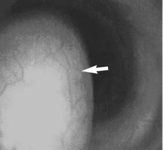
He was admitted to our clinic. His clinical findings included weight loss, mild fever and mild leukocy-tosis. The general physical examination was nor-mal. The erythrocyte sedimentation rate was 89 mm and tumor markers were in normal range. En-doscopy was reperformed and showed a large int-raluminal mass, occupying the proximal half of the esophageal lumen, covered by normal mucosa and arising on the posterior wall (Figure 1). Computed tomography and abdominal ultrasonog-raphy were performed because of endoscopic sus-picion of submucosal tumor. The mass was confir-med to be a lipoma in the wall of the esophagus. Turk J Gastroenterol 2006; 17 (2): 110-112
Manuscript received: 31.05.2005 Accepted: 13.10.2005 Address for correspondence: Cem ALGIN
Department of General Surgery, Dumlup›nar University Hospital, 43270, Kütahya, Turkey
Phone: +90 274 265 22 30 • Fax: +90 274 265 22 77 E-mail: [email protected]
Esophagectomy in esophageal lipoma: Report of a case
Özofagus lipomunda özofajektomi: Vaka takdimi
Cem ALGIN1, Alper HACIO⁄LU1, Tayfun AYDIN2, Enver ‹HT‹YAR3
Departments of 1General Surgery and 2Anesthesiology, Dumlup›nar University Hospital, Kütahya
Department of 3General Surgery, Osmangazi University, School of Medicine, Eskiflehir
Lipom; insidans› %4.1 olan sindirim sisteminin nadir görülen benign bir tümörüdür, fakat özofagusta %0.4 gibi son derece düflük oranda oldu¤u saptanm›flt›r. Bu makalede bir özofagus lipomu vakas› takdim edilmektedir. 55 yafl›nda bir erkek has-tada 2 y›ld›r disfaji ve odinofaji mevcuttu. Üst gastoinestinal system endoskopisinde özofagus duvar›nda, lümeni daraltan kitle saptand›. Endoskopik olarak submukozal bir tumor flüp-hesi oldu¤u için hastaya bilgisayarl› tomografi ve abdominal ultasonografi yap›ld› ve kitlenin özofagus lipomu oldu¤u do¤-ruland›. Bu kitle torasik yaklafl›mla cerrahi olarak eksize edil-di. Hastan›n flikayetleri ameliyattan sonra tamamen gerileedil-di. Özofagus lipomunun tercih edilen tedavi flekli cerrahi olarak tümörün enükleasyonla eksizyonudur, fakat ifllem s›ras›nda özofagus mukozas›n›n aç›lmas› bu hastal›kta özofagus rezeksi-yonu gerektirebilen nadir bir nedendir.
Anahtar kelimeler: Özofagus, lipom, özofajektomi Lipoma is an uncommon benign tumor of the alimentary tract
and its overall incidence is 4.1%, but that of the esophagus is extremely rare, with an incidence of only 0.4%. We present a ca-se of esophageal lipoma. A 55-year-old man had a two-year his-tory of dysphagia and odynophagia. Upper gastrointestinal sys-tem endoscopy showed a mass in the wall of the esophagus, oc-cupying the lumen, and causing obstruction. Computed tomog-raphy and abdominal ultrasonogtomog-raphy were performed because of endoscopic suspicion of submucosal tumor, and the mass was confirmed to be a lipoma in the wall of the esophagus. It was re-moved surgically by a thoracic approach. His symptoms resol-ved after the operation. Surgical excision by enucleation of the tumor is the preferred treatment of esophageal lipoma, but ope-ning of the esophageal mucosa during this procedure is a rare cause for esophageal resection.
Gross examination of the specimen showed a well-circumscribed encapsulated mass, which measu-red 4.5×3×2 cm. The cut surface was soft and bright yellow, resembling lipoma. Histological examination revealed large mature lipocytes. The-re was neither incThe-reased mitotic activity nor lipob-lasts (Figure 2).
The post-operative course of the patient was uneventful. His symptoms resolved after the operation.
DISCUSSION
Lipomas of the esophagus commonly present with dysphagia (2). There may be symptoms of odynop-hagia, recurrent melena (3) and mechanical comp-ression of the upper respiratory tract (4). In adults, most esophageal lipomas are pedunculated and located in the cervical esophagus (5), which commonly assume giant proportions occupying a significant length of the esophagus (6) and may be regurgitated and cause death by suffocation (7, 8). Esophageal lipomas may have malignant differen-tiation because of the vascularity of large benign polyps (9), and ulceration may complicate the problem (6). The diagnosis depends on endoscopic examination and computed tomography, to evalu-ate the origin, extent, surface and consistency of the mass (10).
Surgical excision by enucleation is the preferred treatment of esophageal lipoma. Oral route (7, 8), cervical esophagotomy (5), and endoscopic remo-val (11) are available options. If the tumor is in the thoracic esophagus, thoracotomy is advocated as an invasive, but safe technique (5). In our case, the tumor was in the thoracic esophagus and therefo-re was therefo-removed by right-side thoracotomy. Opera-tive treatment was planned by enucleation of the mass, but despite careful dissection, the esophage-al lumen was opened. Since primary repair of the esophageal wall carries high incidence of obstruc-tion and leakage and subsequent mediastinitis, segmental esophagectomy and esophagogastros-tomy were performed despite the invasiveness and morbidity. Symptoms of the patient resolved after operation. Surgical excision by enucleation of the tumor is the preferred treatment of esophageal li-poma, but opening of the esophageal mucosa du-ring this procedure is a cause for esophageal resection.
Lipoma of the esophagus 111
Computed tomography demonstrated an intralu-minal pedunculated mass, with a consistency sug-gesting lipoma; abdominal ultrasonography sho-wed no remarkable lesion in the abdomen. The tumor was removed by right-side thoraco-tomy. Enucleation of the mass was the planned operative strategy. During dissection of the mass, the lumen of the esophagus was opened. Therefo-re, segmental esophagectomy and esophagogast-rostomy were performed.
Figure 1. Endoscopy of lipoma of the esophagus. Upper
gastro-intestinal endoscopy shows a large, lobulated intraluminal mass within proximal half of the esophageal lumen, covered by nor-mal mucosa, which arose on the posterior wall (arrow = lipoma of the esophagus)
Figure 2. Light microscopy of lipoma of the esophagus.
Histo-pathological examination of the specimen shows lobules of mature adipose tissue (hematoxylin and eosin, ×100; arrow = mature adipose tissue)
ALGIN et al. 112
In conclusion, we suggest esophagectomy in ca-ses with inadvertent opening of the esophageal lumen during enucleation of the lipoma, due to safety of the procedure and improvement in the patient’s symptoms.
ACKNOWLEDGEMENTS
Written consent was obtained from the patient for the publication of his details. We thank him for providing written permission to publish this study.
REFERENCES
1. Mayo CW, Pagtalunan RJG, Brown DJ. Lipoma of alimen-tary tract. Surgery 1963; 53: 598-603.
2. Sossai P, De Bernardin M, Bissoli E, et al. Lipomas of the esophagus: a new case. Digestion 1996; 57: 210-2. 3. Zschiedrich M, Neuhaus P. Pedunculated giant lipoma of
the esophagus. Am J Gastroenterol 1990; 85: 1614-6. 4. Hasan N, Mandhan P. Respiratory obstruction caused by
lipoma of the esophagus. J Pediatr Surg 1994; 29: 1565-6. 5. Akiyama S, Kataoka M, Horisawa M, et al. Lipoma of the
esophagus-report of a case and review of the literature. Jpn J Surg 1990; 20: 458-62.
6. Patel J, Kieffer RW, Martin M, et al. Giant fibrovascular polyp of the esophagus. Gastroenterology 1984; 87: 953-6.
7. Taff ML, Schwartz IS, Boglioli LR. Sudden asphyxial death due to prolapsed esophageal fibrolipoma. Am J Forensic Med Pathol 1991; 12: 85-8.
8. Allen MS Jr, Talbot WH. Sudden death due to regurgitati-on of a pedunculated esophageal lipoma. J Thorac Cardiovasc Surg 1967; 54: 756-8.
9. Bak YT, Kim HJ, Kim JG, et al. Liposarcoma arising in a giant lipomatous polyp of the esophagus. Korean J Intern Med 1989; 4: 86-9.
10. Kang JY, Chan-Wilde C, Wee A, et al. Role of computed to-mography and endoscopy in the management of alimentary tract lipomas. Gut 1990; 31: 550-3.
11. Yu JP, Luo HS, Wang XZ. Endoscopic treatment of submu-cosal lesions of the gastrointestinal tract. Endoscopy 1992; 24: 190-3.