Morphometric investigations on the vertebral column of the rock partridges (Alectoris
graeca) and pheasants (Phasianus colchicus)
Mehmet Fatih Yüksel, Sadettin Tıpırdamaz* Özet
Yüksel MF, Tıpırdamaz S. Kaya kekliği (Alectoris graeca)
ve sülünlerin (Phasianus colchicus) columna vertebralisinin morfometrik incelenmesi. Eurasian J Vet Sci, 2012, 28, 1,
05-09
Amaç: Bu çalışmanın amacı kaya kekliği (Alectoris graeca)
ve sülünlerin (Phasianus colchicus) columna vertebralisini oluşturan cervical, thoracal, lumbal ve sacral omurların sa-yılarını ve makroanatomisini belirlemektir.
Gereç ve Yöntem: 10 adet Kaya kekliği ve 10 adet sülün
materyal olarak kullanıldı. Columna vertebralis üzerindeki deri ve kaslar diseke edilerek ayrıldı. Açığa çıkarılan kemik-ler %8’lik amonyak çözeltisinde 1.5 saat süre ile kaynatıldı. Daha sonra kemik üzerindeki kas, tendo ve benzeri gibi tüm oluşumlar temizlendi. Omurlarla ilgili ölçümler digital kum-pas ile yapıldı.
Bulgular: Kaya kekliğinin vertebra cervicalis’ini oluşturan
kemiklerin morfometrik ölçüleri değerlendirildiğinde; C7 ve C8’in en uzun, C13’ün en yüksek, C2 ve C3’ün en geniş, C8, C9, C10, C11, C12’nin kanal yüksekliği en fazla, C13’ün kanal genişliği en fazla olan omurlar olduğu, proc. articu-laris cranialis ve proc. articuarticu-laris caudalis genişliği en fazla olan omurların ise sırasıyla C13 ve C3, C7 olduğu gözlendi. Sülünde vertebra cervicalis’i oluşturan kemiklerinin morfo-metrik ölçüleri değerlendirildiğinde; C8’in en uzun, C13’ün en yüksek, C3’ün en geniş, C8, C10 ve C12’nin kanal yük-sekliği en fazla, C13’ün kanal genişliği en fazla olan omurlar olduğu, proc. articularis cranialis ve proc. articularis cauda-lis genişliği en fazla olan omurların ise sırasıyla C13 ve C8 olduğu gözlendi.
Öneri: Kaya kekliği ve sülüne ait vertebra’ların
morfomet-rik değerlendirilmesi sonucunda diğer kanatlılara benzedi-ği ifade edilebilir.
Abstract
Yuksel MF, Tipirdamaz S. Morphometric investigations on
the vertebral column of the rock partridges (Alectoris
grae-ca) and pheasants (Phasianus colchicus). Eurasian J Vet Sci,
2012, 28, 1, 05-09
Aim: The aim of this study was to determine macroanatomy
and the number of cervical, thoracal and lumbar parts of the vertebral column in rock partridges (Alectoris graeca) and pheasants (Phasianus colchicus).
Materials and Methods: 10 rock partridges and 10
pheas-ants were used as materials. The vertebral column was dis-sected out and morphometric features of vertebral bones were revealed. The boiled vertebral bones during 1.5 hours in 8% ammonia solution were cleaned from muscles, ten-dons etc. The morphometric measurements were per-formed by a digital calliper.
Results: According to morphometric measurements of
vertebral bones of partridges, the longest bones of cervical vertebrae were C7 and C8, the highest one was C13, the wid-est bones were C2 and C3; the bones with the highwid-est canal were C8, C9, C10, C11, C12, the bone with the widest canal was C13, the bones with the largest cranial and the bones with the largest caudal width were C13, C3 and C7. When considering the morphometric measurements of vertebral bones of pheasants, the longest bone of cervical vertebrae was C8, the highest bone was C13, the widest bone was C3, the bones with the highest canal were C8, C10, C12, the bone with the widest canal was C13, the bone with the larg-est cranial width and the bone with the larglarg-est caudal width were C13 and C8, respectively.
Conclusion: It may be stated that morphometric values of
vertebrae of partridges and pheasants are similar to those of other poultry.
1Department of Anatomy, Faculty of Veterinary Medicine, Selcuk
University, 42075 Konya, Turkey
Received: 21.09.2011, Accepted: 01.12.2011 *[email protected]
Anahtar kelimeler: Kaya kekliği, sülün, columna vertebralis Keywords:Pheasants, rock partridges, vertebral colunm
RESEARCH ARTICLE
Journal of Veterinary Sciences
Introduction
The most rapid increase in animal husbandry occurs in poultry. Consumption of poultry is composed of three classes as broiler, turkey and other poultry. Sig-nificant developments occurred in alternative poul-try breeding in recent years. This breeding is mainly done intensively and semi-intensively for generating materials for hunting tourism, for hobby and for pro-viding meat production. Partridges and pheasants are the main poultry bred for hunting tourism (Roenigk 1999, Çetin and Kırıkçı 2000, Yılmaz 2004).
Partridges were first mentioned in the text between 384 and 322 b.c. by Aristotales (Woodard et al 1983). The most proper partridge species for intensive breeding has been reported to be rock partridges (Çe-tin and Kırıkçı 2000), 14 subspecies of rock partridg-es have been reported. While the most commonly bred partridge species are Alectoris rufa (partridge) in Europe, it is Alectoris graeca (rock partridges) in Turkey (Çetin and Kırıkçı 2000, Yılmaz 2004).
Motherland of almost all pheasants is Asia continent. World Pheasant Organization (WPO) has reported 49 species and many subspecies of pheasants. Pheasants that have spreaded in Caucasia and eastern Black Sea region of Turkey (Phasianus colchicus) are known as upland pheasants (Howman 1993, Çetin and Kırıkçı 2000).
Bones of poultry are very hard and their tissue is rich of calciferous substances. There are no other carti-lages than joint carticarti-lages in the skeletal of poultry that has completed its development. The skeleton of poultry is composed of columna vertebralis, costae and sternum. Pelvis that shows bony union with many vertebrae may be regarded as one of them function-ally (Wise and Jennings 1973, Getty 1975, Bahadır 2002, Demirkan 2002).
Columna vertebrae are analyzed as vertebrae cervi-cales speciales, vertebrae thoracicae, vertebrae lum-bicales and vertebrae sacrales, vertebrae synsacrales, vertebrae caudales. Vertebrae located in pars cervica-lis constitute a mobile joint. Vertebrae thoracicae and vertebrae caudales have joint to each other (Doğuer and Erençin 1964, Nickel et al 1977, Dyce et al 1987, Baumel et al 1993, Demirkan 2002).
Although some anatomical studies have been con-ducted about partridges and pheasants, sufficient studies are not available in terms of osteology. The aim of this study is to determine macroanatomy of vertebrae constitute vertebral column of partridges and pheasants and to compare obtained data with other species.
Materials and Methods
A total of 10 rock partridges (493±12.7 g, Alectoris
graeca) and 10 pheasants (873±25.9, Phasianus col-chicus) aged 10 weeks and obtained from Research
and Training Farm of Faculty of Veterinary Medicine were used. Ethics committee approval was obtained. Animals received anesthesia with 10 mg/kg of xyla-zine + 35 mg/kg of ketamin. A. carotis communis of the sedatized animals were dissected and animals were sacrificed. Skin and muscles surrounding verte-bral column were dissected and columna verteverte-bralis was separated from the body. Exposed bones were boiled in 8% amonnia solution for 1.5 hours and all material coating the bone were cleaned afterwards. Bones were stored in 20% sodium hypochloride for 3 days and bleached.
Obtained vertebrae were preserved until measure-ments. Measurements were performed using a digital calliper. Length, width and height of cervical, thoracal, lumbar and sacral vertebrae of both species studied were measured (Figures 1 and 2). Height (dorso-ven-tral), width (mediolateral) and distance between two processus articularis cranialis and caudalis were also measured. Terminology of Baumell et al (NAA-1993) developed for poultry was used.
ANOVA and Tukey tests were used for statistical anal-ysis (SPSS 12.0). A p value of <0.05 was accepted sta-tistically significant.
Results
Morphometric measurements of rock partridges and pheasants are given in Tables 1-4. Number of cervi-cal vertebrae was detected to be 13 in partridges and pheasants. The first cervical vertebra, atlas was not included in measurements as it is narrow and ring shaped. Measurements of remaining 12 vertebrae were done.
Figure 1. Tricuspid valve opened showing three cusps like anterior (ac), septal (sc) and posterior (pc), chordi tendinae (ct) and papillary muscles (pm).
Figure 2. Measurement points of pheasants. a; cranial and caudal width, b; height of the bone, c; height and width of the bone, d; height and width of the vertebral canal.
Table 1. Morphometric measurements of cervical vertebra of rock partridge (mean±SE).
Length of the
vertebra Height of the vertebra Width of the vertebra Height of the vertebral canal Width of the vertebral canal Width of Proc. articularis cranialis Width of Proc. articularis caudalis C2 7.0±0.16d 6.1±0.22bc 6.2±0.26a 2.3±0.04d 2.9±0.02d 7.8±0.09f 8.0±0.22b C3 7.9±0.18c 5.8±0.35c 6.5±0.35a 2.3±0.07d 2.8±0.07d 8.6±0.21e 8.6±0.16a C4 9.0±0.17ab 5.5±0.50c 3.8±0.09def 2.5±0.08c 2.8±0.03d 10.1±0.36bcd 7.8±0.21b C5 9.1±0.13ab 5.1±0.51c 3.6±0.08f 2.6±0.05c 2.8±0.02d 10.5±0.22abc 7.6±0.10b C6 9.2±0.15ab 5.3±0.51c 3.6±0.07ef 2.8±0.03b 2.8±0.02d 10.6±0.25abc 7.8±0.10b C7 9.3±0.18a 5.2±0.52c 3.6±0.09ef 2.9±0.03ab 2.8±0.06d 10.7±0.27ab 7.9±0.17b C8 9.4±0.12a 5.5±0.49c 3.7±0.05ef 2.9±0.03a 2.9±0.04d 10.7±0.20abc 8.5±0.12a C9 9.0±0.16ab 6.1±0.51bc 4.0±0.13cdef 3.0±0.03a 3.0±0.05cd 10.0±0.15cd 7.9±0.12b C10 8.8±0.20ab 6.3±0.44bc 4.2±0.10bcd 3.0±0.03a 3.1±0.06c 9.7±0.17d 7.6±0.07b C11 8.9±0.15ab 6.6±0.42bc 4.5±0.16b 3.0±0.05a 3.3±0.13ab 9.7±0.30d 6.8±0.16c C12 8.7±0.25b 7.4±0.34b 4.1±0.16bcde 2.9±0.03a 3.2±0.12bc 10.8±0.22ab 7.6±0.29b C13 5.2±0.18e 8.7±0.38a 4.3±0.16bc 2.9±0.06ab 3.4±0.12a 11.1±0.28a 6.1±0.31d
a-f:.Different letters in the same column are statistically significant (Tukey test, p<0.05).
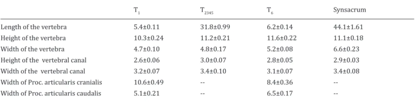
Table 2. Morphometric measurements of thoracal and lumbosacral vertebrae of rock partridges (mean±SE).
T1 T2345 T6 Synsacrum
Length of the vertebra 5.4±0.11 31.8±0.99 6.2±0.14 44.1±1.61
Height of the vertebra 10.3±0.24 11.2±0.21 11.6±0.22 11.1±0.18
Width of the vertebra 4.7±0.10 4.8±0.17 5.2±0.08 6.6±0.23
Height of the vertebral canal 2.6±0.06 3.0±0.07 2.8±0.05 2.9±0.03 Width of the vertebral canal 3.2±0.07 3.4±0.10 3.1±0.07 3.4±0.08 Width of Proc. articularis cranialis 10.6±0.49 -- 8.4±0.36 --Width of Proc. articularis caudalis 5.1±0.21 -- 6.5±0.17
Discussion
Cervical vertebra is S-shaped in the poultry and cervi-cal column is composed of 12 vertebrae in pigeons, 14 in chickens, 14 in ducks, 17-18 in geese and 23-25 in swans. Animals stick out their cervical verte-brae when they try to catch something with their beaks. They keep cervical vertebra S-shaped in or-der to prevent the brain from jounces when walking, jumping (Doğuer and Erençin 1964, King and McLel-land 1975, Bahadır et al 1993, Taşbaş 2001). In this study, number of cervical vertebrae was seen to be 13 in pheasants and rock partridges (Table 1-4). The smallest cervical vertebra, atlas, has been stated to be a ring shaped bone composed of upper and lower parts (Zweers et al 1987, Bahadır 2002).
Morphomet-ric measurements could not be done for atlas also in pheasants and rock partridges as it composed of ven-tral and dorsal parts.
Thoracic vertebrae have been reported to compose of 7 vertebrae in chickens and pigeons, 9-10 verte-brae in ducks and geese (Doğuer and Erençin 1964, Nickel et al 1977, Bahadır et al 1993, Bahadır 2002) and this part of spinal column has been reported to be short and immobile (Nickel et al 1977, Taşbaş 2001). Vertebrae thoracicae were detected to be 6 in pheas-ants and rock partridges in this study. Although 1. and 6. vertebrae are separate bones in both species, 2. - 5. thoracic vertebrae were detected to union and present as a single bone.
Synsacrum was detected to compose as the result of union of 14 lumbar and sacral vertebrae in rock par-tridges and pheasants and length was found to be 44.1-6.61mm in rock partridges and 62.6-1.73 mm in pheasants. Aforementioned vertebrae support the lit-erature reporting that union of these vertebrae form synsacrum also in other poultry.
Conclusions
When morphometric measurements of cervical ver-tebrae of pheasants and rock partridges were statis-tically evaluated together, the longest bone was de-tected to be C8, the highest C13, the widest C3, bones that have maximum canal height C8, C9, C10 and C12 and the bone that has the widest canal C13. C13 was the bone that has the widest proc. articularis cranialis in both species. Vertebrae that have the widest proc. articularis caudalis were observed to be C3 and C7 in greek partridges and C3 and C13 in pheasants.
Acknowledgement
The current study was supported by Scientific Re-search Projects Coordination of Selcuk University (09202054).
References
Bahadır A, 2002. Hareket sistemi, in; Evcil Kuşların Anato-misi, Ed; Dursun N, Medisan Yayınevi, Ankara, Türkiye, s: 1-29.
Bahadır A, Yıldız B, Serbest A, Yıldız B, Yılmaz O, 1993. Evcil Su kuşlarından yerli kaz, yerli ördek ve pekin ördeğinin iskeletleri üzerinde karşılaştırmalı makro-anatomik araştırmalar. Uludağ Ünv Vet Fak Der, 12, 1-12.
Baumel JJ, King AS, Breazile JE, Evans HE, Vanden Berge JC, 1993. Nomina anatomica avium puplished by the nuttall orniholgical clup, Cambridge, USA.
Çetin O, Kırıkçı K, 2000. Alternatif Kanatlı Yetiştiriciliği, Konya, S.Ü. Vak Yayın, Konya, Türkiye.
Demirkan AÇ, 2002. Ördekte İskelet Sistemi, A.Ü. Sağ Bil Ens Doktora Tezi, Ankara, Türkiye.
Doğuer S, Erençin Z, 1964. Evcil Kuşların Komparativ Anato-misi, Ankara Ünv Basımevi, Ankara, s: 2-23.
Dyce KM, Sack WO, Wensing CJG, 1987. Texbook of Veteri-nary Anatomy, Philadelphia, USA, pp: 775-779.
Getty R, 1975. Sisson and Grossman’s the Anatomy of the Domestic Animals, New York, USA, pp: 1790-1801. Howman K, 1993. Pheasants of the World, Their Breeding
and Management, Hancock House Publishers, Washing-ton, USA.
Table 4. Morphometric measurements of thoracal and lumbosacral vertebrae of the pheasants (mean±SE).
T1 T2345 T6 Synsacrum
Length of the vertebra 6.6±0.11 34.6±0.67 8.3±0.19 62.6±1.73
Height of the vertebra 11.0±0.17 15.0±0.45 14.7±0.32 7.1±0.19
Width of the vertebra 6.3±0.25 5.9±0.16 6.3±0.14 7.9±0.11
Height of the vertebral canal 3.1±0.08 2.9±0.06 2.9±0.03 3.1±0.07 Width of the vertebral canal 4.2±0.13 3.4±0.07 3.2±0.07 3.5±0.06 Width of Proc. articularis cranialis 13.60.19 -- 8.4±0.12 --Width of Proc. articularis caudalis 6.2±0.15 -- 7.4±0.26
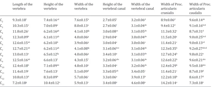
--Table 3. Morphometric measurements of cervical vertebrae of the pheasants (mean±SE).
Length of the
vertebra Height of the vertebra Width of the vertebra Height of the vertebral canal Width of the vertebral canal Width of Proc.articularis cranialis Width of Proc. articularis caudalis C2 9.3±0.18f 7.4±0.16cd 7.6±0.15b 2.7±0.05f 3.2±0.06d 8.9±0.06d 9.6±0.14ab C3 10.3±0.15e 7.0±0.09e 8.0±0.13a 2.7±0.06f 3.1±0.04de 9.4±0.12d 9.1±0.16bcd C4 11.8±0.26c 6.2±0.16gh 4.1±0.10fg 3.0±0.08ce 3.1±0.05de 11.3±0.32c 8.7±0.31d C5 12.3±0.09b 6.1±0.11gh 4.0±0.06g 2.9±0.04e 3.0±0.04de 11.5±0.20c 9.0±0.25bcd C6 12.6±0.15ab 6.2±0.10h 3.9±0.06g 3.0±0.04e 3.0±0.06e 11.4±0.21c 9.0±0.13cd C7 12.7±0.21ab 6.2±0.11gh 4.1±0.08fg 3.1±0.06bce 3.1±0.04de 12.3±0.35b 9.2±0.27abcd C8 13.0±0.13a 6.5±0.12fg 4.0±0.06g 3.4±0.10a 3.1±0.03de 12.7±0.24b 9.8±0.22a C9 12.5±0.16ab 6.6±0.13f 4.3±0.15f 3.2±0.06abc 3.1±0.06de 12.6±0.22b 9.6±0.21abc C10 12.4±0.18b 7.1±0.09de 4.8±0.10e 3.3±0.04a 3.2±0.06de 12.4±0.29b 9.5±0.18abc C11 11.4±0.19c 7.6±0.13c 5.1±0.09d 3.3±0.05ab 3.4±0.05c 11.4±0.21c 8.7±0.19d C12 10.8±0.13d 8.3±0.09b 5.7±0.06c 3.3±0.06a 3.9±0.13b 12.2±0.18b 8.6±0.17d C13 7.2±0.18g 10.4±0.12a 5.9±0.13c 3.4±0.08a 4.6±0.08a 14.2±0.14a 7.3±0.18e
King AS, McLelland J, 1975. Outlines of Avian Anatomy, Bail-liere Tindal, London, UK, pp: 1-22.
Nickel R, Schummer A, Seiferle E, 1977. Anatomy of the Do-mestic Birds, Verlag Paul Parey, Hamburg, Germany, pp: 3-25.
Roenigk WP, 1999. World poultry consumption symposi-um: Muscle growth and development, keynote address. Poultry Sci, 78, 722-728.
Taşbaş M, 2001. Veteriner Anatomi, Yorum Matbaası, An-kara, Türkiye, s: 223-232.
Wise DR, Jennings AR, 1973. The Development and mor-phology of the growth plates of two long bones of the turkey. Res Vet Sci, 14, 161-166.
Woodard AE, Abplanalp H, Phisenti JM, Snyder RL, 1983. Inbreeding effects on reproductive traits in the ring-necked pheasant. Poultry Sci, 62, 1725-1730.
Yılmaz A, 2004. Kuluçkalık keklik (Alectoris graeca) yumurtalarının depolanmasında süre, pozisyon ve ön ısıtmanın kuluçka sonuçlarına etkileri. S.Ü. Sağ Bil Ens Doktora Tezi, Konya, Türkiye.
Zweers GA, Vanden Berge JC, Koppendraier R, 1987. Avian cranio- cervical systems, Part I: Anatomy of the cervical column in the chicken. Acta Morphol, 25, 131-155.