1 Ceylanpınar State Hospital, Department of Biochemistry, Şanlıurfa, Turkey 2 Dicle University Faculty of Medicine, Department of Biochemistry, Diyarbakır, Turkey 3 Dicle University Faculty of Medicine, Dept Physical Medicine and Rehabilitation, Diyarbakır, Turkey
4 Batman State Hospital, Dept., Physical Medicine and Rehabilitation, Batman, Turkey 5 Dicle University Faculty of Medicine, Dept., Family Medicine, Diyarbakır, Turkey Yazışma Adresi /Correspondence: Dr. Mustafa Akif Sariyildiz, Dicle University Faculty of Medicine, Department of Physical Medicine and Rehabilitation, Diyarbakır, Turkey Email: [email protected]
Geliş Tarihi / Received: 21.09.2012, Kabul Tarihi / Accepted: 23.10.2012
Copyright © Dicle Tıp Dergisi 2012, Her hakkı saklıdır / All rights reserved ORIGINAL ARTICLE / ÖZGÜN ARAŞTIRMA
The relationship between bone mineral density and levels of RANKL,
osteoprotegerin and cathepsin-K in patients with rheumatoid arthritis
Romatoid artritli hastalarda kemik mineral yoğunluğu ile RANKL, osteoprotegerin ve
katepsin-K düzeyleri arasındaki ilişki
Gökhan Çakırca1, Nuriye Mete2, Ibrahim Batmaz3, Mustafa Akif Sarıyıldız3, Mehmet Ali Ulu4,
Levent Yazmalar3, Tahsin Celepkolu5, Remzi Çevik3
ÖZET
Amaç: Bu çalışmanın amacı Romatoid Artrit’li (RA) has-talarda Osteoprotegerin (OPG), Nükleer faktör kappa B reseptör aktivatörü ligandı (RANKL), katepsin-K düzeyle-rini ve ayrıca bu parametrelerin kemik mineral yoğunluğu (BMD) ile ilişkisini araştırmaktır.
Gereç ve yöntem: Çalışmaya postmenopozal sağlıklı (n=30), postmenopozal osteoporozlu (n=30) ve postme-nopoz RA’lı (n=30) toplam 90 olgu alındı. Serum RANKL, OPG ve katepsin-K ELİSA yöntemiyle çalışıldı.
Bulgular: Postmenopozal RA’ lı hastalarda serum RANKL ve OPG seviyeleri postmenopozal sağlıklı kontrollere kıyasla anlamlı yüksekken, serum OPG/RANKL oranı anlamlı düşük bulundu. Bununla birlikte postmenopozal RA’lı hastalarda, postmenopozal osteoporozlu hastalara göre serum OPG ve OPG/RANKL oranı anlamlı düşük iken, serum RANKL seviyesi anlamlı yüksekti. Postme-nopozal RA’lı hastalarda lomber spine (LS) ve femur neck (FN) kemik mineral dansiteleri (BMD) ile OPG/RANKL oranı arasında pozitif korelasyon saptanırken; RANKL düzeyleri ile negatif korelasyon bulundu.
Sonuç: RANKL/RANK/OPG sistemi osteoporoz ve RA patogenezinde rol alabilmektedir ve OPG/RANKL oranı kemik dansitesinin önemli bir belirleyicisi olabilir.
Anahtar kelimeler: Osteoporoz, romatoid artrit, RANKL, OPG, Katepsin-K
ABSTRACT
Objectives: The aim of this study was to evaluate the levels of osteoprotegerin (OPG), nuclear factor kappa B receptor activator ligand (RANKL), cathepsin K in patients with rheumatoid arthritis (RA) and the relation between these parameters and bone mineral density (BMD). Materials and methods: Totally 90 cases including 30 postmenopausal and healthy women, 30 with postmeno-pausal osteoporosis and 30 with postmenopause RA were enrolled in the study. The serum RANKL, OPG and cathepsin K were measured by ELISA method.
Results: The levels of serum RANKL and OPG in the pa-tients with postmenopausal RA were found significantly higher compared to the postmenopausal healthy women whereas the rate of serum OPG/RANKL was found sig-nificantly lower. In addition, the rate of OPG and OPG/ RANKL were significantly lower in patients with post-menopausal RA compared to the postpost-menopausal osteo-porosis, whereas the level of serum RANKL was signifi-cantly higher. Positive correlation was detected between bone densities of lumbar spine (LS), femur neck (FN) and the rate of OPG/RANKL in patients with postmenopausal RA. Also negative correlation was detected between LS and FN bone densities and RANKL levels.
Conclusions: The system of RANKL/RANK/OPG may have a role in osteoporosis and RA pathogenesis and the rate of OPG/RANKL might be a significant determiner of bone density.
Key words: Osteoporosis, rheumatoid arthritis, RANKL, OPG, cathepsin-K
INTRODUCTION
Osteoporosis (OP) is a disease of muscle skeleton system which is characterized by the increase of possibility of bone fragility and fracture as a result of low bone mass and deterioration of microarchi-tecture form of bone tissue.1 It is necessary to make
a multilateral evaluation on bone metabolism so as to diagnose, cure and observe OP. In addition to radiographic methods, biochemical determiners of bone circle are used nowadays in order to evaluate early diagnosis, clinic progress and feedback of the treatment.2
Osteoprotegerin (OPG) functions as a trap re-ceptor by attaching Nuclear Factor Kappa B Re-ceptor Activator Ligand (RANKL), which is a key mediator of a bone resorption and it prevents the at-tachment to Nuclear Factor Kappa B Receptor Acti-vator (RANK). As a result, it prevents RANKL from developing bone resorption by inhibiting osteoclast differentiation and activation.3 It is important to find
out new determiners which show bone cycles such as OPG/RANK/RANKL in order to cure bone dis-eases and detect new therapeutic agents.4,5,6
It was shown that OPG/RANK/RANKL have a role in not only focal and generalized bone lost but also joint inflammation pathophysiology in Rheu-matoid Arthritis.7,8 It was shown in the studies that
active T cells are important resources of RANKL in inflame synovium, synovial fibroblast and synovial tissue.9 Furthermore, the studies specify that the rate
of OPG/RANKL can be used as a biologic diagnosis way for non-malignant pathologies such as osteoly-sis and bone fractures in relation with osteoporoosteoly-sis, ankylosing spondylitis, rheumatoid arthritis, bone tumors and hip prosthesis.10
Cathepsin K is a cysteine protease which is necessary for the deterioration of bone matrix pro-tein component and for the osteoclast functions. It is produced with bone resorption macrophages and synovial fibroblasts. It has a role in the fragmenta-tion of osteonectin, type 1and type 2 of collagene proteins.11 In addition to that, it has a key role in
osteoporosis, osteolytic bone metastasis and bone remodeling and resorption in RA.12,13
We aimed to evaluate the levels of cathepsin K, OPG, RANKL and relation with bone mineral density (BMD) in patients with RA.
MATERIALS AND METHODS
The present study had a cross-sectional design ap-proved by the local ethics committee. All the re-cruited subjects were approved with an informed consent form before participating in the study. Ninety cases who applied to the Dicle Faculty of Medicine, Physical Medicine and Rehabilitation outpatient clinic between November 2010 and Sep-tember 2011; 30 recently diagnosed with postmeno-pausal osteoporosis and did not take antiresorptive treatment; 30 healthy female (employee in our hos-pital) postmenopausal individuals and 30 patients diagnosed with RA according to the 1987 American College of Rheumatology criteria 14 were included
in this study; (did not use biologic agents, 20 of them were with osteoporosis and 10 of them were osteophenic).
Patients with thyroid disease or parathyroid glands disease, other endocrine disorders, serious liver or kidney disease, radiological abnormali-ties (scoliosis, platyspondyly, and others) were ex-cluded from the study. Patients were also exex-cluded if they had concomitant use of estrogen, androgen, anticonvulsant or anticoagulant drugs. Other exclu-sion criteria were alcohol users, smokers, and HIV subjects.
Measurement of the clinical variables and laboratory tests in patients with RA
The demographic and clinical characteristics of the patients such as age, sex, height, weight, education-al level, the disease duration (which is defined as the duration since the onset of the first symptoms of RA), degree of morning stiffness, tender-swol-len joint count and the physician’s and patient’s global assessments were determined. The duration of the morning stiffness (minutes), mean pain and mean daytime fatigue [100 mm visual analogue scale (VAS)] were also noted. The disease activity (DAS28) scores were evaluated using the disease activity score calculator.15 The erythrocyte
sedimen-tation rate (ESR) was measured through the Wester-gren method (mm/h) and the serum C-reactive protein (CRP) level was determined with the help of nephelometry (mg/dl). The RF titres were also measured through the nephelometric immunoassay method (IU/ml).
Sample collection of OPG, RANKL and cathepsin K
Blood samples were obtained after an overnight fast. Serum was separated and frozen immediately after blood drawing. All the serum were stored at -80°C until analysis. Serum concentrations of OPG, RANKL and cathepsin K of all individuals were both measured by an ELISA. The optical density was measured at 450 nm using an automatic ELISA reader.
Measurement of BMD
BMD was determined by dual X-ray absorptiom-etry (DXA) at the lumbar spine (L1-L4; BMD-LS) and the femoral neck (BMD-FN). Patients with T scores higher than -1.0 at both sites (LS and FN) were considered to have normal BMD, those with T score of -1 to - 2.5 as patients with osteopenia and those with a T score lower than -2.5 at LS and/or FN were defined as patients with osteoporosis.
Statistical analysis
The calculations were performed using the Statisti-cal Package for Social Sciences for Windows soft-ware version 16.0. The Kolmogorov-Smirnov test was used to confirm that data within the ranges of normal distribution in both groups. A non-paramet-ric test was employed for the variables outside the normal distribution. The comparison of the data between reciprocal groups was carried out through the Mann-Whitney U-test. Variance analysis
(ANO-VA) was used for the comparison among multiple groups and Tukey HSD test was used so as to find the groups which cause the difference at the end of the test. Correlations between OPG, RANKL, OPG/ RANKL, BMD and the RA-related variables were investigated with the help of Spearman’s correla-tion. Statistical significance was based on a value of p<0.05 with a 95% confidence interval.
RESULTS
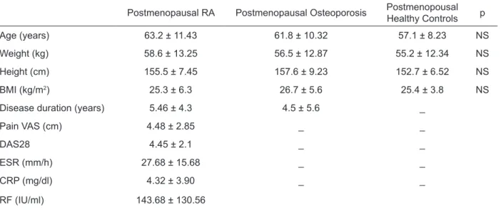
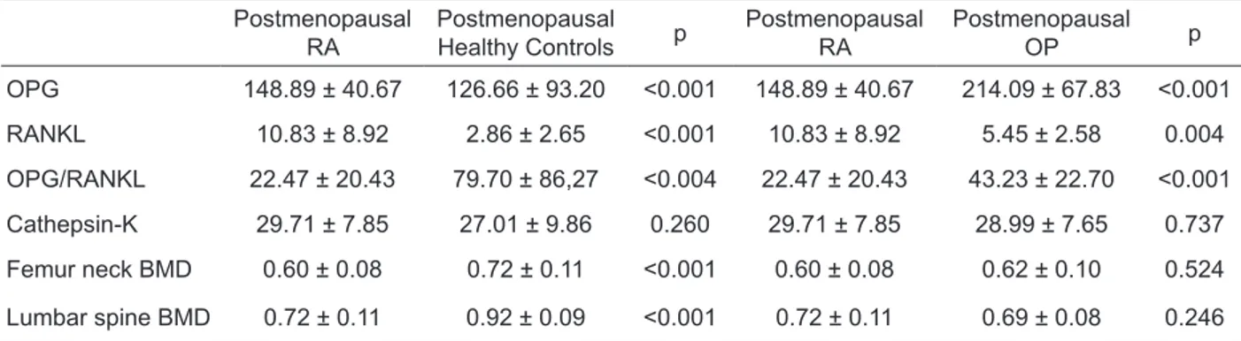
Totally 90 cases whose ages were between 42 and 65 (mean 50.8 ± 11.2) years, were included in this study. Demographic data of the all groups were shown in Table 1. Patients with postmenopausal RA showed obviously higher plasma levels of RANKL compared with other two groups, with a consequent significantly lower OPG/RANKL. Whereas OPG level was significantly higher in postmenopausal RA patients than the postmenopausal healthy group, OPG level in the postmenopausal RA patients was significantly lower than the ones with OP (table 2). Correlation analyses of serum OPG, OPG/RANKL, RANKL, Cathepsin K and clinical variables in pa-tients with postmenopausal RA was demonstrated in Table 3. Whereas there was a significantly negative correlation between levels of cathepsin K, RANKL and BMD, the here was also a significantly positive correlation between the rate of OPG/RANKL and BMD.
Table 1. Characteristics of patients and controls (mean ± SD)
Postmenopausal RA Postmenopausal Osteoporosis PostmenopousalHealthy Controls p
Age (years) 63.2 ± 11.43 61.8 ± 10.32 57.1 ± 8.23 NS
Weight (kg) 58.6 ± 13.25 56.5 ± 12.87 55.2 ± 12.34 NS
Height (cm) 155.5 ± 7.45 157.6 ± 9.23 152.7 ± 6.52 NS
BMI (kg/m2) 25.3 ± 6.3 26.7 ± 5.6 25.4 ± 3.8 NS
Disease duration (years) 5.46 ± 4.3 4.5 ± 5.6 _
Pain VAS (cm) 4.48 ± 2.85 _ _
DAS28 4.45 ± 2.1 _ _
ESR (mm/h) 27.68 ± 15.68 _ _
CRP (mg/dl) 4.32 ± 3.90 _ _
RF (IU/ml) 143.68 ± 130.56
VAS visual analog scale, ESR Erythrocyte sedimentation rate, CRP C-reactive protein, RF rheumatoid factor, DAS disease activity score, BMI Body mass index, NS Not significant
Table 2. Mean scores of the postmenopausal RA, postmenopausal healthy controls, and postmenopausal OP (mean ± SD) Postmenopausal
RA PostmenopausalHealthy Controls p PostmenopausalRA PostmenopausalOP p OPG 148.89 ± 40.67 126.66 ± 93.20 <0.001 148.89 ± 40.67 214.09 ± 67.83 <0.001 RANKL 10.83 ± 8.92 2.86 ± 2.65 <0.001 10.83 ± 8.92 5.45 ± 2.58 0.004 OPG/RANKL 22.47 ± 20.43 79.70 ± 86,27 <0.004 22.47 ± 20.43 43.23 ± 22.70 <0.001 Cathepsin-K 29.71 ± 7.85 27.01 ± 9.86 0.260 29.71 ± 7.85 28.99 ± 7.65 0.737 Femur neck BMD 0.60 ± 0.08 0.72 ± 0.11 <0.001 0.60 ± 0.08 0.62 ± 0.10 0.524 Lumbar spine BMD 0.72 ± 0.11 0.92 ± 0.09 <0.001 0.72 ± 0.11 0.69 ± 0.08 0.246 OPG Osteoprotegerin, RANKL receptor activator of nuclear factor kappa B ligand, BMD bone mineral density, RA rheumatoid arthritis
study, whereas the rate of serum OPG/RANKL was significantly lower and serum RANKL and the level of OPG were significantly higher in patients with postmenopausal RA in comparison to the post-menopausal healthy controls. Furthermore, the rate of serum OPG and OPG/RANKL were significantly lower and the level of serum RANKL was signifi-cantly higher in patients with postmenopausal RA compared to the patients with postmenopausal OP.
Patients with RA are at increased risk for os-teoporosis and bone fractures.8 Moreover, a positive
correlation was found out between LS, FN BMD and OPG/RANKL in the patients with
postmeno-Table 3. Coefficiens (r) of Spearman correlation analy-sis of serum OPG, RANKL, Cathepsin K levels and clini-cal characteristics in patients with postmenopausal RA
Characteristics OPG RANKL OPG/RANKL Cathepsin-K
Age 0.110 0.325** 0.255* 0.205
Disease duration -0.060 0.119 0.012 0.149
Tender joint count 0.079 0.250* 0.121 0.204
Duration of morning stiffness 0.181 0.106 0.139 0.166
Pain VAS 0.155 0.090 0.305** 0.285** Fatigue VAS -0.190 0.121 0.209* 0.351** ESR (mm/h) 0.235* 0.300** 0.124 0.177 CRP (mg/dl) 0.276** 0.256* 0.191 0.189 RF -0.193 -0.088 -0.043 0.015 DAS28 0.095 0.243** 0.301** Femur neck BMD 0.181 -0.369** 0.285** -0.289** Lomber spine BMD 0.155 -0.361** 0.406** -0.316**
VAS visual analog scale, ESR Erythrocyte sedimentation rate, CRP C-reactive protein, OPG Osteoprotegerin, RANKL receptor activator of nuclear factor kappa B ligand, RF rheumatoid factor, DAS disease activity score, RA rheumatoid arthritis, BMD bone mineral density
*p<0.05, **p<0.01 DISSCUSSION
The aim of our study was to investigate the rela-tionship between laboratory markers of bone me-tabolism and BMD in RA patients with a particular focus on serum levels of bone-resorbing cytokines such as RANKL and its’ physiological antagonist OPG. Especially in patients with RA the assessment of correlations between soluble parameters of bone metabolism and BMD is problematical because BMD is a result of bone loss during a relatively long period of time, whereas soluble parameters of bone metabolism can change in a relatively short time and are influenced by disease activity. In our
pausal RA whereas a negative correlation was found between the levels of RANKL. It has been suggested again that the ratio of OPG/RANKL may be more important than single OPG or RANKL in regulating osteoclast formation and regulating bone destruc-tion in RA.16,17 Haugeberg et al.18 reported, for the
first time, the overall frequency of osteoporosis was doubled in the female RA population. In accordance with results of Haugeberg, in fact, we also observed that bone metabolic status was worse in RA patients than postmenopausal healty controls. Changes of overall bone mass in this study were similar to other researches.19,20 Moreover, it was also found in
another study that serum concentrations of OPG, RANKL, and OPG/RANKL ratio were significantly different among groups of normal, osteopenia, and osteoporosis in RA. These differences in RA pa-tients were confirmed again by multiple linear re-gression analysis, serum RANKL levels negatively related to BMD, in contrast to RANKL, there were positive linear correlations between BMD and OPG/ RANKL ratio.21 Data in the literature, with regard to
such relationship between the OPG/RANKL system and BMD in RA, was only reported by Oelzner P et al. (20) who found only in RA patients older than 60 years old, serum RANKL levels showed a negative correlation with BMD at the LS and serum OPG/ RANKL ratio did not relate to BMD.
Xu et al. 21 found that age and CRP
concentra-tions were identified as the crucial risk determinants of osteoporosis in RA. Also in our study we found that ESR, CRP and age were positively correlated with serum RANKL levels in RA. Inflammation, reflecting the activity of disease which associ-ated with generalized bone loss in RA in previous reports.22 This information is likely to become
in-creasingly important as more bone-directed treat-ments become part of RA management paradigms. Thus, denosumab, a monoclonal antibody directed against RANKL, should suppress bone loss in RA and could be a potential target for novel therapeutic agents.23
Proinflammatory cytokines like interleukin-1 and tumor necrosis factor alpha induce an over expression of cathepsin K, which is responsible for increased bone resorption and skeletal compli-cations in RA. Bone resorption and formation is a well-balanced system and is mediated by osteo-clasts. Cathepsin K is essential for bone resorption,
which depends on the production of cathepsin K by osteoclasts and its secretion into the extracellular department.24 In our study, we found out a negative
correlation between cathepsin K and LS, FN BMD in the patients with RA. It may result from the fact that cathepsin K kas a role bone resorption.
The limitation of the present study was its cross-sectional design. Finally, although this study is somewhat limited in statistical power because of the small sample size and further studies with a greater number of patients are needed. Another limitation of the study was that the relation between the remedies used for RA (for instance DMARD, corticosteroid) and OPG, RANKL, cathepsin K could not be evaluated. It should not be forgotten that medicine such as methotrexate and corticoste-roid may cause decrease in BMD as well.
In conclusion, present study demonstrates that the OPG/RANKL system represents an important mediator of bone resorption in RA-induced osteo-porosis. Besides, we found out that the levels of ca-thepsin K and RANKL are related to BMD in the patients with RA. The close follow-up of the levels of OPG/RANKL, RANKL and cathepsin K in the early RA patients may be a messenger of a possible osteoporosis in these patients.
The authors reports no coflicts of interest REFERENCES
1. Armas LA, Recker RR. Pathophysiology of osteoporosis: new mechanistic insights. Endocrinol Metab Clin North Am 2012;41(3):475-86.
2. Delmas PD. The role of markers of bone turnover in the as-sesment of fracture risk in postmenopausal women. Osteo-poros Int 1998;8(S1):32-6.
3. Kurban S, Mehmetoğlu İ. Osteoprotegerin RANK and RANKL. Turk J Biochem 2007;32(3);178-84.
4. Kostenuik PJ, Shalhoub V. Osteoprotegerin: A Physiological and Pharmacological Inhibitor of Bone Resorption. Curr Pharm Des 2001;7(8):613-35.
5. Boyce BF, Xing L. Biology of RANK, RANKL, and osteo-protegerin. Arthritis Res Therap 2007;9(S1):1-7.
6. McClung M. Role of RANKL inhibition in osteoporosis. Ar-thritis Res Therap 2007;9 (S1):S3.
7. Vega D, Maalouf NM, Sakhaee K. The role of receptor ac-tivator of nuclear factor-kB (RANK)/RANK ligand/os-teoprotegerin: clinical implications. J Clinical Endocrinol Metab 2007;92(12):4514-21.
8. Romas E, Sims NA, Hards DK, et al. Osteoprotegerin re-duces osteoclast numbers and prevents bone erosion in col-lageninduced arthritis. Am J Pathol 2002;161(4):1419-27.
9. Gravallese EM, Manning C, Tsay A, et al. Synovial tissue in rheumatoid arthritis is a source of osteoclast differentiation factor. Arthritis Rheum 2000;43(2):250-8.
10. Baud’huin M, Lamoureuxa F, Duplomba L, et al. RANKL, RANK, osteoprotegerin: key partners of osteoimmünology and vascular diseases. Cell Mol Life Sci 2007;64(18):2334-50.
11. Hou WS, Li Z, Gordon RE, et al. Cathepsin K is a critical protease in synovial fibroblast-mediated collagen degrada-tion. Am J Pathol 2001;159(6):2167-77.
12. Goto T, Yamaza T, Tanaka T. Cathepsins in the osteoclast. J Electron Microsc. 2003;52(6):551-8.
13. Rieman DJ, McClung HA, Dodds RA, et al. Biosynthesis and processing of cathepsin K in cultured human osteo-clasts. Bone 2001;28(3):282-9.
14. Arnett FC, Edworthy SM, Bloch DA, et al. The Ameri-can Rheuma¬tism Association 1987 criteria for the classification of rheumatoid arthritis. Arthritis Rheum 1988;31(3):315-24.
15. Wong BR, Rho J, Arron J, et al. TRANCE is a novel li-gand of the tumor necrosis factor receptor family that ac-tivates c-Jun N-terminal kinase in T cells. J Biol Chem 1997;272(40):25190-4.
16. Haynes DR, Crotti TN, Capone M, et al. Osteoprote-gerin and receptor activator of nuclear factor kappaB li-gand (RANKL) regulate osteoclast formation by cells in the human rheumatoid arthritic joint. Rheumatology 2001;40(6):623-30.
17. Nosaka K, Miyamoto T, Sakai T, et al. Mechanism of hy-percalcemia in adult T-cell leukemia: overexpression of re-ceptor activator of nuclear factor jB ligand on adult T-cell leukemia cells. Blood 2002;99(2):634-40.
18. Haugeberg G, Uhlig T, Falch JA, et al. Bone mineral den-sity and frequency of osteoporosis in female patient with rheumatoid arthritis: result from 394 patients in the Olso county rheumatoid arthritis register. Arthritis Rheum 2000;43(3):522-30.
19. Urbanek R, Tlustochowicz W, Patola J, et al. Incidence of osteoporosis in patients with rheumatoid arthritis. Przegl Lek 2000;57(2):103-7.
20. Oelzner P, Franke S, Lehmann G, et al. Soluble recep-tor activarecep-tor of NFkappa B-ligand and osteoprotegerin in rheumatoid arthritis relationship with bone mineral den-sity, disease activity and bone turnover. Clin Rheumatol 2007;26(12):2127-35.
21. Xu S, Wang Y, Lu J and Xu J. Osteoprotegerin and RANKL in the pathogenesis of rheumatoid arthritis-induced osteo-porosis. Rheumatol Int 2011,DOI 10.1007/s00296-011-2175-5.
22. Lodder MC, De Jong Z, Kostense PJ, et al. Bone mineral density in patients with rheumatoid arthritis: relation be-tween disease severity and low bone mineral density. Ann Rheum Dis 2004;63(12):1576-80.
23. Cohen SB, Dore RK, Lane NE, et al. The Denosumab Rheu-matoid arthritis Study Group Denosumab treatment effects on structural damage, bone mineral density, and bone turn-over in rheumatoid arthritis: a twelvemonth, multicenter, randomized, double-blind, placebo-controlled, phase II clinical trial. Arthritis Rheum 2008;58(5):1299-309. 24. Skoumal M, Kolarz G, Woloszczuk W, et al. Serum
cathep-sin K levels of patients with destructive rheumatoid arthri-tis: correlation with radiological destruction. Arthritis Res Ther 2005;7(1):65-70.