Effect Of NaOH Concentration On The Degradation Properties Of
CHA/PCL Composites For Bone Replacement Applications
NaOH Konsantrasyonunun ve NaOH Bekletme Süresinin Kemik Greftlerinde Kullanılan CHA/PCL
Kompozitlerinin Biyolojik Yıkımına Etkisi
Duygu Ege
11 University of Cambridge Department of Materials Science &
Metallurgy Aim: To study the effect of NaOH concentration and duration of treatment on the % mass loss
of 19 wt% uncalcined carbonated hydroxyapatite (CHA)/Poly-ε -caprolactone (PCL)
To analyze the degradation behaviour of composites and to evaluate the possibility of bone replacement by the study of buffer saline solution (PBS) following NaOH application.
Material and Method: CHA was produced using a wet precipitation method. The CHA/PCL composites were prepared by twin screw extrusion followed by injection moulding. In order to accelerate the degradation rate of 19 wt% uncalcinated CHA/PCL, the samples were surface treated with various concentrations of NaOH (Sigma Aldrich, UK) for various durations. Biological degradation rates were analyzed in PBS
Results: The increase of the concentration of NaOH from 3 M to 5 M increased the % weight loss of the samples after NaOH treatment and subsequent PBS studies. However, increasing the durahon of surface treatment from one day to three days in 5 M NaOH did not affect the % weight change.
Conclusions: Biological degradation rate was accelerated with the increase of NaOH concentration. 19 wt% uncalcined CHA/PCL samples that were surface treated with 3 M NaOH for one day had a more controlled degradation during subsequent PBS studies
Key Words: Poly- ε -caprolactone, carbonated hydroxyapatite, NaOH treatment, biological degradation study, twin screw extrusion, injection moulding
Amaç: NaOH konsantrasyonu ve bekletme süresinin ağırlıkça % 19 kalsine edilmemiș karbonlanmıș hidroksiapatit (CHA)/Poly- ε -kaprolakton (PCL)’nin kütle kaybına etkisinin incelenmesi
NaOH uygulamasının ardından gerçekleștiren fosfatla tamponlanmıș salin (PBS) çalıșmasında, kompozitlerin biyolojik yıkımlarının analizi ve kemik grefti uygulanabilirliklerinin incelenmesi Gereç ve Yöntemler: CHA ıslak çökeltme yöntemi kullanılarak üretilmiștir. CHA/PCL kompozit, ikiz vidalı ekstrüzyon ve enjeksiyon kalıplama ile hazırlanmıștır. Ağırlıkça % 19 kalsine edilmemiș CHA/PCL’in biyolojik yıkım hızını arttırmak amacıyla, numuneler farklı sürelerde, çeșitli konsantrasyonlardaki NaOH solüsyonları (Sigma Aldrich, UK) ile muamele edilmiștir. Daha sonra biyolojik yıkım hızları PBS içinde analiz edilmiștir.
Bulgular: NaOH konsantrasyonunun 3 M‘dan 5 M’a artması, NaOH ve PBS’de muhafaza edildikten sonra numunelerin % ağırlık kaybını arttırmıștır. Ancak, 5 M NaOH’da numuneleri 1 ve 3 gün bulundurmak % ağırlık değișimini etkilememiștir.
Sonuç: NaOH konsantrasyonunun artması ile numunelerin biyolojik yıkımları hızlandırılmıștır. Ağırlıkça % 19 kalsine edilmemiș CHA/PCL’in 1 gün sureyle 3 M NaOH içinde muhafaza edilmesi, PBS çalıșmalarında daha kontrollü bir șekilde biyolojik yıkıma uğradıklarını göstermiștir.
Anahtar Sözcükler: Poli-ε -kaprolakton, karbonatlı hidroksiapatit, NaOH muamelesi, biyolojik yıkım çalıșması, çift vidalı ekstrüzyon, enjeksiyon dökümü
Bone grafting, the surgical procedure used to replace bone to aid the healing process, has been used to fill bone defects for the last
century, most commonly as autografts or allografts (1). From 1980’s, there have been attempts to replace bone using biomaterials
Received : Dec 05,2013 Accepted: Jan 25,.2014 Correspondig Author
Duygu Ege,
GSM : + 90 533 342 25 77 Phone : + 44 1223 33430 E-mail : [email protected] 27 Charles Babbage Road Cambridge
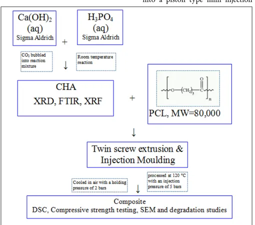
Figure 1: The schematic diagram of the CHA/PCL composite preparation
including, calcium phosphates, hydroxyapatite, glass–ceramics, bioactive glass, and bioresorbable polymers. There is growing attention to the use of bioresorbable polymers in synthetic biomaterial composites. Some of the commonly used bioresorbable composites are
poly-ε-caprolactone (PCL), polyglycolic acid (PGA), polylactic acid (PLA) and polylactide–co–glycolide (PLGA). Bioresorbable composites transfer load gradually to the surrounding tissue whilst guiding new bone formation (2). The optimisation of the degradation rate of the biomaterial support to facilitate new bone formation is vital to sustain healthy bone growth. PCL is hydrophobic in nature due to the linear chains of methylene groups (3,4). In vivo, the total degradation time of PCL goes up to 2–3 years (5-8). Ideally, degradable polymers for load bearing or non–major load bearing applications need to degrade in around 6 months in order to sustain healthy bone in-growth (9). The groups of Chandrasekaran (Ang et al., 2007) and Teoh (9-11) enhanced the degradation rate of porous PCL composites by surface treating it with NaOH (9, 10, 12, 13). The cleaved carboxyl and hydroxyl chains are then easily removed from the surface. This leaves a rough surface texture and increases the surface area between the polymer and the medium (9, 14). Incorporation of Bioglass®, HA or β–TCP filler further accelerated the degradation rate during the NaOH treatment (15-19). The aim of this paper is to study the effect of surface treatment on the degradation rate of CHA/PCL nanocomposites. 19 wt% uncalcined CHA/PCL was
surface–treated for various durations and for different concentrations of NaOH. In order to observe the effect of NaOH treatment on the composites, a
PBS study was carried out subsequent to the NaOH surface treatment.
1. Materials and Methods
1.1. Materials
PCL pellets with a molecular weight of 80,000 were purchased from Sigma Aldrich. CHA was produced using a wet precipitation method (20). The CHA/PCL composites were prepared by twin screw extrusion followed by injection moulding. The above process can be outlined in a schematic diagram as in Figure 1
1.2. Preparation of CHA
Carbonated hydroxyapatite was prepared in–house by a wet precipitation reaction (20).
1.3. Fabrication of the
composite
A mini 5 cm3, co–rotating twin screw
extruder (DSM, Netherlands) was used at Imperial College London to disperse the CHA powder in a PCL matrix. PCL pellets and CHA powder was fed into the extruder simultaneously without any premixing. Table 1 shows the wt% of CHA, CHA processing condition and PCL that were used for extruded samples.
Twin screw extrusion was used to draw material through a 1 mm gauge strand die to compound and disperse the filler particles in the matrix material at 120°C. Around 6 g material was fed into the extruder at a screw speed of 10 rpm. The material was mixed in the extruder for 30 minutes with a screw rotation speed of 20 rpm. The material was taken out from the extruder with a screw speed of 20 rpm.
After cutting the extruded rods into cylinders of approximately 10 cm in length, the samples were fed into a piston type mini injection
moulder (DSM, Xplore, Geleen, The Netherlands) with a capacity of 5 cm3. After injection moulding,
samples were obtained with a diameter of 6 mm and a length of 12 mm. The material was injection moulded with a flow temperature of 120 °C, a mould temperature of 23–25°C with an injection pressure
of 5 bar. It was cooled and hardened at a holding pressure of 2 bar and a holding time of 3 seconds which compensated for material shrinkage (21).
1.4. X–Ray Fluorescence
(XRF) and Leco carbon
analysis
XRF was carried out at London & Scandinavian Metallurgical Co Ltd (Fullerton Road, UK). 2 g of uncalcined CHA powder was analysed after wet ball milling the powder and sieving it with a 75 µm gauge sieve.
1.4.
X–RayDiffraction (XRD)
XRD was carried out for about 50 mgof uncalcined CHA with a Philips PW1730 diffractometer. The X– ray generator was operated at 40 kV and 40 mA using CuK
α
(λ
= 0.15404 nm). The data were collected over a 2θ
range of 20– 40ºC using a step size of 0.04º with a dwell time of 10 s. The slit sizes used were 1º divergence, 0.2º receiving and 1º antiscatter. Identification of phases was achieved by comparing the diffraction patterns of CHA with ICDD [Joint Committee ofPowder Diffraction Standards (JCPDS)] (HA: 09–432).
1.6 Scanning Electron
Microscopy (SEM)
Using a field emission gun scanning electron microscope (JEOL 6340F FEGSEM) operating at 5 kV, in
backscattering electron imaging mode, the morphology and the particle size distribution of uncalcined CHA powder were analysed. CHA powder was scattered onto an aluminium stub covered by double adhesive carbon tape and sputter–coated with a thin layer of platinum in an argon– purged chamber for 30 seconds.
1.7.NaOH surface treatment
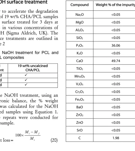
In order to accelerate the degradationrate of 19 wt% CHA/PCL samples were surface treated for 3 days at 37C in various concentrations of NaOH (Sigma Aldrich, UK). The surface treatments are outlined in Table 2
After the NaOH treatment, using an electronic balance, the % weight loss was calculated for the NaOH treated samples using Equation 1. three repeats were conducted for each sample. % weight loss= i f i M M M 100 (20)
2. Results
In Table 3, XRF analysis shows that CHA samples were all phase pure. The phase purity of CHA was
analyzed by XRD. Figure 2 shows the XRD patterns of uncalcined XRD patterns of CHA revealed the
presence of all the major HA peaks, such as (002), (211), (112) and (300) at 25.88°, 31.78°, 32.18° and 32.98° respectively at 2
θ
(22). All CHA peak positions and intensities matched to the ICDD standard reference sample for hydroxyapatrite (09-0432). The main calcium oxide peak at 37.2° (23) was not present in the XRD trace. No other secondary phases such as tri–calcium phosphate were detected, which suggests that the uncalcined and calcined CHA powder were phase pure.Table 3: XRF analysis of the
carbonated hydroxyapatite
Compound Weight % of the impurity
Na2O <0.05 MgO <0.05 Al2O3 <0.05 SiO2 <0.05 P2O5 36.06 K2O <0.05 CaO 49.74 TiO2 <0.05 Mn3O4 <0.05 V2O5 <0.05 Cr2O3 <0.05 Fe2O3 <0.05 BaO <0.05 ZrO2 <0.05 ZnO <0.05 SrO <0.05 C 1.98 Table 2: NaOH treatment for PCL and
CHA/PCL composites
NaOH
treatment 19 wt% uncalcined CHA/PCL
5 M, 3 d
5 M, 1 d
3 M, 1 d
Table 1: wt% of CHA, CHA processing conditions for extruded samples Sample list wt% of CHA Processing condition of CHA
1 0 –
2 10 Uncalcined
3 20 Uncalcined
In Figure 3, overall, CHA particles were observed to be dispersed quite well in the polymer matrix. Figure 4 shows the % weight change of 19 wt% uncalcined CHA/PCL after different surface treatments. The values were given in the range of the standard errors. It shows that for 19 wt% uncalcined CHA/PCL, the mass loss is 12±0.74 %, 31±5.96 % and 32±22.65 % for 1 day treatment in 3 M NaOH, 1 day treatment in 5 M NaOH and 3 days treatment in 5 M NaOH, respectively. By increasing the concentration of NaOH from 3 M to 5 M at 1 day, the % weight loss
of the samples increased. Increase of the time of surface treatment with 5 M NaOH did not affect the % weight change but the sample variability was observed to be much greater.
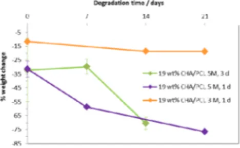
Figure 5 shows the % weight loss during the PBS study of 19 wt% uncalcined CHA/PCL after different NaOH surface treatments. When 19 wt% uncalcined CHA/PCL was surface–treated with 5 M NaOH for 1 day, during the subsequent PBS study, most of the material degraded within 21 days. On the other hand, surface treatment with 3 M NaOH for 1 day resulted in a more steady degradation rate than the other surface treatments.
3. Discussion
Prior work shows that all the composites have a minimal % weight loss over a 1 month period (7). It was found that the degradation rate also rose with the increase of NaOH concentration and the time of surface treatment. Similar results to this study were found by Lam et al (10). and Ang et al (11). who saw faster degradation rates for calcium phosphate/PCL than the pure PCL.
For 19 wt% uncalcined CHA/PCL after 3 days 5 M NaOH treatment, total mass loss was observed in PBS within a 14-day period. This was
possibly due to pass of a form of percolation threshold for NaOH treatment. NaOH treatment and water penetration to the internal surface between particles and matrix resulted in fast disintegration of the composites. As surface treatment of 19 wt%
uncalcined CHA/PCL with one day surface treatment with 3 M NaOH was observed to have a more controlled degradation rate, it could be beneficial to further study this system and similar systems.
4. Conclusions
As, PCL and CHA/PCL composites have a relatively slow degradation rate for bone replacement, CHA/PCL composites were surface treated with NaOH in order to accelerate their degradation rate. During the subsequent PBS studies, it was observed that for 19 wt% uncalcined CHA/PCL the degradation rate was accelerated significantly. This is possibly due to pass of a form of percolation threshold for NaOH treatment. Additionally, it was observed that the degradation rate was accelerated with the increase of NaOH concentration. 19 wt% uncalcined CHA/PCL samples that were surface treated with 3 M NaOH for 1 day were observed to have a more controlled degradation rate. In future studies, in vivo studies would be useful in order to examine the compatibility of these composites for bone replacement.
Figure 3: FEGSEM (BSE) micrographs of the fracture surface of 19 wt% uncalcined CHA/PCL Scale bar is 1 µm.
Figure 4: % weight change of 19 wt%
uncalcined CHA/PCL after different NaOH pre–treatments (Results mean ± standard error, n=3)
Figure 5: % weight loss after the PBS study of 19 wt% uncalcined CHA/PCL with various NaOH surface treatments (Results mean ± standard error, n=3)
Figure 2: X–ray diffraction pattern of CHA uncalcined CHA powder (* indicates the HA peaks)
REFERENCES
1. Aho, Heikkila. Clinical Applications
of Bone Allografts and Substitutes. Phillips GO, editor. Singapore: World Scientific Publishing Co. Ptc. Ltd.; 2005.
2. Hench LL, Paschall HA. Direct
chemical bond of bioactive glass-ceramic materials to bone and muscle. Journal of Biomedical Materials Research. 1973;7:25-42.
3. Tiaw KS, Goh SW, Hong M, Wang Z,
Lan B, Teoh SH. Laser surface modification of poly(epsilon-caprolactone) (PCL) membrane for tissue engineering applications. Biomaterials. 2005;26:763-769.
4. Chouzouri G, Xanthos M.
Degradation of aliphatic polyesters in the presence of inorganic fillers. Journal of Plastic Film & Sheeting. 2007;23:19-36.
5. Chen DR, Bei JZ, Wang SG.
Polycaprolactone microparticles and their biodegradation. Polymer Degradation and Stability. 2000;67:455-459.
6. Lowry KJ, Hamson KR, Bear L, Peng
YB, Calaluce R, Evans ML, et al. Polycaprolactone/glass bioabsorbable implant in a rabbit humerus fracture model. Journal of Biomedical Materials Research. 1997;36:536-41.
7. Chouzouri G, Xanthos M. In vitro
bioactivity and degradation of polycaprolactone composites containing silicate fillers. Acta Biomaterialia. 2007;3:745-756.
8. Labet M, Thielemans W. Synthesis of
polycaprolactone: a review. Chemical Society Reviews. 2009;38:3484-3504.
9. Yeo A, Sju E, Rai B, Teoh SH.
Customizing the Degradation and Load-Bearing Profile of 3D Polycaprolactone-Tricalcium
Phosphate Scaffolds Under Enzymatic and Hydrolytic Conditions. Journal of Biomedical Materials Research Part B-Applied Biomaterials. 2008;87B:562-569.
10. Lam CXF, Savalani MM, Teoh SH,
Hutmacher DW. Dynamics of in vitro polymer degradation of polycaprolactone-based scaffolds: accelerated versus simulated physiological conditions. Biomedical Materials. 2008;3(3).
11. Ang KC, Leong KF, Chua CK,
Chandrasekaran M. Compressive properties and degradability of
poly(epsilon-caprolatone)/hydroxyapatite
composites under accelerated hydrolytic degradation. Journal of Biomedical Materials Research Part A. 2007;80A:655-660.
12. Tsuji H, Ishida T. Poly(L-lactide). X.
Enhanced surface hydrophilicity and chain-scission mechanisms of poly(L-lactide) film in enzymatic, alkaline, and phosphate-buffered solutions. Journal of Applied Polymer Science. 2003;87:1628-1633.
13. Yeo A, Wong WJ, Khoo HH, Teoh
SH. Surface modification of PCL-TCP scaffolds improve interfacial mechanical interlock and enhance early bone formation: An in vitro and in vivo characterization. Journal of Biomedical Materials Research Part A. 2010;92A:311-321.
14. Htay M. Water vapour transmission
and degradation properties of biaxiallt stretched PCL films and cell-permeable membranes 2004.
15. Azevedo MC, Reis RL, Claase BM,
Grijpma DW, Feijen J. Development
and properties of polycaprolactone/hydroxyapatite
composite biomaterials. Journal of Materials Science-Materials in Medicine. 2003;14:103-107.
16. Tsuji H, Suzuyoshi K, Tezuka Y,
Ishida T. Environmental degradation of biodegradable polyesters: 3. Effects of alkali treatment on biodegradation of poly(epsilon-caprolactone) and poly (R)-3-hydroxybutyrate films in controlled soil. Journal of Polymers and the Environment. 2003;11:57-65.
17. Kikuchi M, Koyama Y, Yamada T,
Imamura Y, Okada T, Shirahama N, et al. Development of guided bone regeneration membrane composed of beta-tricalcium phosphate and poly (L-
lactide-co-glycolide-epsilon-caprolactone) composites. Biomaterials. 2004;25:5979-5986.
18. Rich J, Jaakkola T, Tirri T, Narhi T,
Yli-Urpo A, Seppala J. In vitro evaluation of poly(epsilon-caprolactone-co-DL-lactide)/bioactive glass composites. Biomaterials. 2002;23:2143-2150.
19. Rai B, Ho KH, Lei Y, Si-Hoe KM, Teo
CMJ, bin Yacob K, et al. Polycaprolactone-20% tricalcium phosphate scaffolds in combination with platelet-rich plasma for the treatment of critical-sized defects of the mandible: A pilot study. Journal of Oral and Maxillofacial Surgery. 2007;65:2195-2205.
20. Gibson IR, Bonfield W. Novel
synthesis and characterization of an AB-type carbonate-substituted hydroxyapatite. Journal of Biomedical Materials Research. 2002;59:697-708.
21. Wilberforce SIJ, Finlayson CE, Best
SM, Cameron RE. The influence of the compounding process and testing conditions on the compressive mechanical properties of poly(D,L-lactide-co-glycolide)/alpha-tricalcium phosphate nanocomposites. Journal of the Mechanical Behavior of Biomedical Materials. 2011;4:1081-1089.
22. Panda RN, Hsieh MF, Chung RJ,
Chin TS. FTIR, XRD, SEM and solid state NMR investigations of carbonate-containing hydroxyapatite nano-particles synthesized by hydroxide-gel technique. Journal of Physics and Chemistry of Solids. 2003;64:193-9.
23. Slosarczyk A, Paszkiewicz Z, Paluszkiewicz C. FTIR and XRD evaluation of carbonated hydroxyapatite powders synthesized by wet methods. Journal of Molecular Structure. 2005;744:657-661.