Ankara Üniv Vet Fak Derg, 62, 243-246, 2015 DOI:
Short Communication / Kısa Bilimsel Çalışma
Chylothorax associated with Ebstein’s anomaly in a Golden Retriever
dog
Zeki YILMAZ
1, Pınar LEVENT
1, Meriç KOCATURK
1, Hüseyin CIHAN
1, Elif PARS
2,
Saim SAĞ
31
Department of Internal Medicine, Veterinary Faculty, Uludag University, Bursa; 2Anatolia Animal Hospital, Suadiye, İstanbul;
3Department of Internal Medicine, Cardiology, Medical Faculty, Uludağ University, Bursa, Turkey.
Summary: 2-year-old, intact female, a Golden Retriever Dog was presented with the signs of respiratory distress, exercise intolerance, and muscular weakness. A high-grade systolic murmur was auscultated over the tricuspid valvular area. Thoracic radiographs showed an enlarged D-shape heart and free fluid (chylothorax) in the pleural space. ECG revealed small QRS complexes. Doppler echocardiography disclosed severe tricuspid insufficiency with moderate pulmonary hypertension, and tricuspid leaflets also were displaced down into the right ventricle, substantiating Ebstein’s anomaly. Five-weeks after the medical therapy, patient died at home. The owner of the patient refused the necropsy. This is the first case report of tricuspid dysplasia and Ebstein’s anomaly along with chylothorax in dogs.
Key words: Chylothorax, Ebstein’s anomaly, tricuspid valve dysplasia, dog
Golden Retriever ırkı bir köpekte Ebstein’s anomalisi ilişkili şilotoraks
Özet: 2 yaşında, kısırlaştırılmamış, dişi Golden Retriever köpek solunum güçlüğü, egzersiz intoleransı ve kas zaafiyeti bulguları ile kliniklerimize sunuldu. Triküspit kapak alanında yüksek derece sistolik kalp üfürümü oskülte edildi. Torasik radyografide; D-şeklinde kalp büyümesi ve plöyral alanda serbest sıvı (şilotoraks) belirlendi. EKG’de küçük QRS kompleksleri gözlendi. Doppler ekokardiyografide orta dereceli sistolik pulmoner hipertansiyon, ciddi triküspid kapak yetmezliği ve Ebstein anomalisi olarak tanımlanan triküspit kapağın sağ ventriküle doğru yer değiştirmesi saptandı. Beş hafta medikal tedaviden sonra hasta, evinde hayatını kaybetti. Hasta sahibi nekropsiyi reddetti. Bu rapor köpeklerde Ebstein anomalisi, triküspit displazi ve şilotoraksın birlikte seyrettiği ilk olgu rapordur.
Anahtar sözcükler: Şilotoraks, Ebstein anomalisi, triküspit kapak displazisi, köpek.
Tricuspid valve dysplasia (TVD) is a congenital malformation of the tricuspid valve (TV), chordae tendineae, or papillary muscles, resulting in TV insufficiency (29). The basal attachments of TV are ventrally displaced into the right ventricle (RV) resulting in a further malformation named Ebstein’s anomaly (14, 22).
Chylothorax a triglyceride (TG) and cholesterol (Chol) rich fluid is an infrequent cause of pleural effusion (10). There is limited information on chylothorax relating with heart diseases in human (1, 7) and animals (9). Although only two dogs with chylothorax associated with congenital cardiac disorders of TVD (9, 25) have been described, chylothorax due to Ebstein’s anomaly in dogs has not been reported yet. Thus, we reported here, for the first time, Ebstein’s anomaly as well as its possible mechanism(s) associated with chylothorax in a dog.
The dog (2-year-old, intact female, Golden Retriever, 24.5 kg) was presented to the private clinic with the signs of exercise intolerance and respiratory distress. The prevention of heartworm was complete. Despite the treatment, dog was worsened, and referred to the small animal clinic (Veterinary Faculty, Uludag University, Bursa) with the similar symptoms. Heart rate, capillary refill time and body temperature were within reference range. Respiratory rate increased (64/min.). TV insufficiency was suspected based on the high-grade regurgitant systolic murmur over the TV area. Since complete blood count and serum biochemistry panel were within reference ranges (data not shown), haematological problems were excluded in the differential diagnosis. After these clinical and laboratory observations, the dog was examined for cardiopulmonary disorders by thoracic radiography, ECG, and echocardiography (CarisPlus, Esoate).
Zeki Yılmaz - Pınar Levent - Meriç Kocaturk - Hüseyin Cihan - Elif Pars - Saim Sağ 244
Since thoracic radiography showed a severe right-sided cardiomegaly (D-shape) (Fig.1), the differential diagnosis included congenital heart anomalies, TVD, right heart failure, left to right shunts, pulmonary stenosis, and tetralogy of Fallot (2). Clinical (weak heart pulse) and ancillary findings (small QRS complexes and a decreased visualization of the heart) were strongly suggestive for a pleural effusion in this dog. A right atrial
(RA) enlargement in a dog with right apical systolic murmur was generally pathognomonic for severe TVD and TV insufficiency (14). But this is not sensitive, small QRS complexes in dogs with TVD were reported, as well (15).
TVD was confirmed by 2D and color Doppler echocardiography. The dog had a markedly enlarged RA (6.9 cm), a large TV annulus (6.5 cm), and malformation
Figure 1: A: Cardiomegaly, caudal vena cava distension, decreased visualization of the heart as a result of moderate pleural effusion, and dorsal displacement of the trachea. B: Right atrial and right ventricular enlargement (D-shape)
Şekil 1: A: Kardiyomegali, kaudal vena kavada genişleme, orta derece perikardiyal efüzyon nedeniyle görünebilirliği azalmış olan kalp ve tracheanın dorsal yer değiştirmesi. B: Sağ atrium ve sağ ventrikülde genişleme (D-şekli)
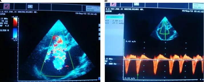
Figure 2: A: A high-grade tricuspidal regurgitation jet (TRJ) into a markedly enlarged right atrium. B: The pressure gradient (27.1 mmHg) and velocity (2.60 m/s) of TRJ on the left apical 4-chamber view.
Şekil 2: A: Belirgin büyümüş sağ atriyum içene yüksek yoğunluktaki trikuspidal geri akım (TRJ). B: Sol apikal 4 boşluk pozisyonunda TRJ’nin basınç gradienti (27.1 mmHg) ve velositesini (2.60 m/s) göstermektedir.
A
B
Ankara Üniv Vet Fak Derg, 62, 2015 245
of TV apparatus, resulting in TV insufficiency (Fig.2A). Also, in this case, 2D echo showed that a left ventricle (LV) diastolic D-shape at the level of papillary muscle suggestive for RA and RV overload, as described earlier (13-15, 21). A normal septal leaflet of TV attaches 0.5 - 1.0 cm more apical than the anterior mitral leaflet in the 4-chamber view. As a rule, if the tricuspid septal attachment lies more than 2 cm "beneath" (towards the apex) than mitral septal attachment, this is Ebstein’s anomaly (2). Thus, Ebstein’s anomaly was diagnosed, because of which tricuspid annulus was 2.5 cm lower than mitral annulus and TV was displaced toward RV, resulting in RV atrialization. Functional and geometric parameters of left heart were within reference ranges (data not shown), indicating preserved LV contractility.
Bubble contrast study was performed to check if right to left heart shunt is present (18), since atrial septal defect was reported in a dog with TVD (9) and Ebstein’s anomaly (6). Bubbles were seen only in the RA and RV just after the intravenous injection of normal saline-air mix solution. This result excluded to an additional congenital heart defect (ASD, VSD, tetralogy of Fallot etc.) for this case.
In the dog, to relieve the respiratory stress and allow further diagnostic investigation, thoracic fluid was drained with ultrasound guided procedure. Its physical appearance (blurry and semi-transparent) was not similar to typical chylous effusion (a white, odorless, and milky). Maldonado et al. (16) reported that out of 61 chylothorax, only 27 (44%) were milky, the rest being serous (26%), serosanguinous (26%) or bloody (3%). Strip analysis showed a high glucose and protein content with a specific gravity of 1.010, in consistence with modified transudate. Since fluid smear consisted of mostly lymphocytes (97%), chylothorax was suspected (1, 4). TG value greater than 110 mg/dl is highly suggestive of a chylous effusion (10, 23). For this case, fluid TG level (601 mg/dl) was higher than that of serum (120 mg/dl), and Chol/TG ratio was <1, indicative for chylothorax, as described earlier (4). In the dog, there was not a dramatically difference between fluid (172 mg/dl) and serum Chol levels (110 mg/dl).
Although the mechanism for chylothorax is unknown, possible mechanisms of chylothorax in this case may be related with four factors that govern fluid movement to or from the pleural space: hydrostatic pressure, colloid osmotic pressure, filtration coefficient, and lymphatic function. When any of these factors are altered, fluid accumulates within the pleural space (34). Congestive heart failure could evoke pleural effusions by altering normal fluid transport mechanisms (34). Also, as in this case, RA volume overload and moderate pulmonary hypertension (42 mmHg – Figure 2B) calculated using the modified Bernoulli equation (24) may be lead to increase in lymph production and
decrease in thoracic duct inflow that means lymphatic dilatation and hypertension, resulting in chylothorax. These explanations may be supported by other studies relating with chylothorax associated with right-sided congestive heart failure in 5 cats (11) and a dog (20).
In this dog, surgery was not selected because of the technical problems. Medical treatment included (12, 25); furosemide (2 mg/kg, po, q12h), enalapril plus hydrochlorothiazide (0.5 mg/kg, po, q12h,), and pimobendan (0.5 mg/kg/day) with salt-restriction in diet. Diuretics and an ACE inhibitor can improve the heart function by reducing cardiac workload, fluid volume in pleural space, and pulmonary hypertension (14, 25). Pimobendan was prescribed to improve LV and RV contraction and reduce venous congestion (19). Also, Rutin, a benzopyrine drug, was suggested to decrease lymph production and chyle leakage from thoracic duct (5). Five weeks after the treatment, the dog died at home, and owner refused the necropsy. Deprest et al. (8) reported that necropsy clearly showed the distal displacement of the malformed TV leaflets, chordae tendineae and papillary muscles in the RV in a dog, which is characteristic of Ebstein’s anomaly.
In conclusion, chylothorax should be diagnosed based on the analysis of TG and Chol levels in serum and pleural fluid, which is not mandatory to appear milky white. In differential diagnosis of dogs with right-sided cardiomegaly, Ebstein’s anomaly should also be included. Echocardiographic criteria for this anomaly is of a RA enlargement, TV regurgitation and an abnormally downward displaced TV. Owner should be informed that the prognosis is poor despite the medical therapy.
References
1. Agrawal V, Doelken P, Sahn SA (2008): Pleural fluid
analysis in chylous pleural effusion. Chest, 133,
1436-1441.
2. Attenhofer Jost CH, Connolly HM, Dearani JA, et al (2007): Ebstein's anomaly. Circulation, 115, 277-285. 3. Boon JA (2011): Veterinary Echocardiography, Second
Edition, Wiley Blackwell Publishing, USA.
4. Campbell SL, Forrester D, Johnston SA, et al (1995):
Chylothorax associated with constrictive pericarditis in a dog. J Am Vet Med Assoc, 206, 1561-1564.
5. Casley-Smith JR, Casley-Smith JR (1992): Modern
treatment of lymphoedema. II. The benzopyrones.
Australas J Dermatol, 33, 69-74.
6. Choi R, Lee SK, Moon HS, Park IC, Hyun C (2009):
Ebstein's anomaly with an atrial septal defect in a jindo dog. Can Vet J, 50, 405-410.
7. de Bruijn D, van Oort A, Kapusta L (2007): Ebstein's
anomaly with severe hypoplastic and stenotic pulmonary venous connections: an unusual cause of congenital chylothorax. Ultrasound Obstet Gynecol, 30, 910-912.
8. Deprest C, French A, Chiers K (2006): Congenital
Zeki Yılmaz - Pınar Levent - Meriç Kocaturk - Hüseyin Cihan - Elif Pars - Saim Sağ 246
Golden Retriever. Vlaams Diergeneeskundig Tijdschrift,
75, 380-384.
9. Diana A, Guglielmini C, Acocella F, et al (2009):
Chylothorax associated with tricuspid dysplasia and atrial septal defect in a bullmastiff. J Am Anim Hosp Assoc, 45,
78-83.
10. Fossum TW, Jacobs RM, Birchard SJ (1986):
Evaluation of cholesterol and triglyceride concentrations in differentiating chylous and nonchylous pleural effusions in dogs and cats. J Am Vet Med Assoc., 188, 49-51.
11. Fossum TW, Miller MW, Rogers KS, et al (1994):
Chylothorax associated with right-sided heart failure in five cats. J Am Vet Med Assoc., 204, 84-89.
12. Gutberlet M, Oellinger H, Ewert P, et al (2000): Pre-
and postoperative evaluation of ventricular function, muscle mass and valve morphology by magnetic resonance tomography in Ebstein's anomaly. Rofo, 172, 436-442.
13. Hirschklau MJ, Sahn DJ, Hagan AD, et al (1977):
Cross-sectional echocardiographic features of Ebstein's anomaly of the tricuspid valve. Am J Cardiol. 40, 400-404.
14. Kittleson MD, Kienle RD (1998): Small Animal
Cardiovascular Medicine. Mosby. Snt Louis, USA.
15. Kornreich BG, Moïse NS (1997): Right atrioventricular
valve malformation in dogs and cats: an electrocardiographic survey with emphasis on splintered QRS complexes. J Vet
Intern Med, 11, 226-230.
16. Maldonado F, Hawkins FJ, Daniels CE, et al (2009):
Pleural fluid characteristics of chylothorax. Mayo Clin
Proc, 84, 129-133.
17. Noone KE (1985): Pleural effusions and diseases of the
pleura. Vet Clin North Am Small Anim Pract., 15,
1069-1084.
18. Otto C (2004): Textbook of Clinical Echocardiography, Saunders, Phyledelphia.
19. Papich MG (2011): Saunders Handbook of Veterinary
Drugs- Small and Large Animal, Third Edition, Elsevier
Saunders, USA.
20. Salci H, Yilmaz Z, Bayram AS, et al (2009): Medical
and surgical treatment of chylothorax in a dog with right-sided heart failure. Turk J Vet & Anim Scien, 33, 165-170.
21. Skouras V, Kalomenidis I (2010): Chylothorax:
diagnostic appoach. Curr Opin Pulmon Med, 16, 387-393.
22. Sousa MG, Gerardi DG, Alves RO, Camacho AA (2006): Tricuspid valve dysplasia and Ebstein's anomaly in
dogs: case report. Arq Bras Med Vet Zootec, 58, 762-767.
23. Staats BA, Ellefson RD, Budahn LL, et al (1980): The
lipoprotein profile of chylous and nonchylous pleural effusions. Mayo Clin Proc., 55, 700-704.
24. Tai TC, Huang HP (2013): Echocardiographic assessment of right heart indices in dogs with elevated pulmonary artery pressure associated with chronic respiratory disorders, heartworm disease, and chronic degenerative mitral valvular disease. Veterinarni Medicina, 58, 613–620.
25. Takemura N, Machida N, Nakagawa K, et al (2003):
Ebstein's anomaly in a beagle dog. J Vet Med Sci., 65,
531-533.
Geliş tarihi: 00.00.2014/ Kabul tarihi: 00.00.2014
Adress for correspondance;
Prof. Dr. Zeki Yılmaz
Internal Medicine Department, Animal Teaching Hospital,
The Faculty of Veterinary Medicine, University of Uludağ, Bursa, Turkey. e-mail: [email protected]