Ankara Üniv Vet Fak Derg, 58, 141-144, 2011
Short Communication / Kısa Bilimsel Çalışma
Myxoid liposarcoma in a cat
Yonca Betil KABAK1, Cenk YARDIMCI2, Semra OKUR-GUMUSOVA3,
Mustafa Yavuz GULBAHAR1
Ondokuz Mayıs Üniversitesi, Veteriner Fakültesi, 1Patoloji, 2Cerrahi, 3Viroloji Anabilim Dalı, Samsun.
Summary: In the study, a myxoid liposarcoma in the proximal region of the left humerus of a 15-year-old male, shorthair cat was described. Left arm along with scapula was amputated. The cut section of tumor was tan to white with soft multilobulated mass which contained hemorrhagic and necrotic foci as well as mucuous composition. Microscopically, the tumor was composed of round-to-polygonal cells. The cells had numerous round, variable-sized, well-defined, intracytoplasmic vacuoles. The lipid droplets in the cytoplasmic vacuoles of the tumor cells was showed with Oil Red O staining. A myxoid extracellular substance remind of mucin was stained by Alcian blue staining (pH = 2,5). Tumor cells were strongly positive for vimentin, but negative for pancytokeratin and S-100. A polymerase chain reaction (PCR) assay from samples showed no evidence of feline leukemia virus (FeLV) / feline
sarcomavirus (FeSV) or feline immunodeficiency virus (FIV) deoksiribonucleic acid (DNA). This case describes a first report of
myxoid liposarcoma in a cat.
Key words: Cat, liposarcoma, myxoid, tumor.
Bir kedide miksoid liposarkom
Özet: Bu çalışmada, 15 yaşlı erkek bir kedinin sol humerusunun proksimal bölgesindeki miksoid liposarkom tanımlanmıştır. Skapula ile birlikte sol kol ampute edildi. Tümörün kesit yüzü krem- beyaz renkte, müköz yapı yanında hemorajik ve nekrotik odaklar içeren yumuşak loplu bir yapıdaydı. Mikroskobik olarak tümör, yuvarlak– polygonal hücreler içeriyordu. Hücreler çok sayıda yuvarlak, değişik büyüklüklerde, belirgin intrasitoplazmik vakullere sahipti. Oil Red O boyası ile tümör hücrelerinin sitoplazmik vakuollerindeki yağ damlacıkları gösterildi. Müsini akla getiren ekstrasellüler miksoid yapı Alcian blue boyası ile boyandı. Tümör hücreleri vimentin pozitif fakat pansitokeratin ve S-100 negatifti. Örneklerde polimeraz zincir reaksiyonu (PZR) ile
feline leukemia virus (FeLV), feline sarcomavirus (FeSV) veya feline immunodeficiency virus (FIV) deoksiribonükleik asit
(DNA)’leri görülemedi. Bu vaka, kedilerdeki miksoid liposarkomu tanımlayan ilk raporudur. Anahtar sözcükler: Kedi, liposarkom, miksoid, tümör.
Liposarcoma is a rare tumor in domestic animals and has been most frequently described in dogs (1,6). In cats, there are limited numbers of liposarcoma cases in the literature (8,9). A myxoid variant of liposarcoma is the least common for the cat, and has been mostly reported in dogs (1). According to the literature, myxoid liposarcoma has not been reported in cats. This study is suggested to be the first case of myxoid liposarcoma in a cat.
A 15-year-old male, shorthair cat showed a gradually increased swelling in the proximal region of the left humerus, by initial clinical examination. During radiographic examination, the complete encirclement of humerus with tumor and the destruction of bone were seen. After three months of the first examination, even though destruction of the humerus had not progressed, the swelling had increased (Fig. 1A). Therefore, the limb was amputated totally together with the humerus and
scapula. The cat was in good health for 10 months post-surgical period, there was no evidence of distant metastasis or regional recurrence of the tumor. The tumor samples were stained with hematoxylin-eosin, Oil Red O, Alcian Blue (pH = 2.5), and Masson’s trichrome stains in routine processes. Additional sections were immunostained by the avidin-biotin peroxidase complex method (ABPC) (Zymed Histostain Plus Kit, California, USA) with the chromogenic substrate AEC (3-Amino-9-Ethyl Carbazole) (Zymed AEC RED substrat kit, Kaliforniya, ABD) using mouse monoclonal anti-vimentin (Clone V9, Lab Vision Corporation, Fremont, USA), mouse anti-human pancytokeratin (AE1 / AE3, Dako Carpenteria, USA), and rabbit polyclonal anti-S-100 (Clone A1, Lab Vision Corporation, Fremont, USA).
The blood (leukocyte and serum) samples and parafin-embedded tissues were tested against feline leukemia virus (FeLV)/ feline sarcomavirus (FeSV) and
Yonca Betil Kabak - Cenk Yardımcı - Semra Okur-Gumusova - Mustafa Yavuz Gulbahar 142
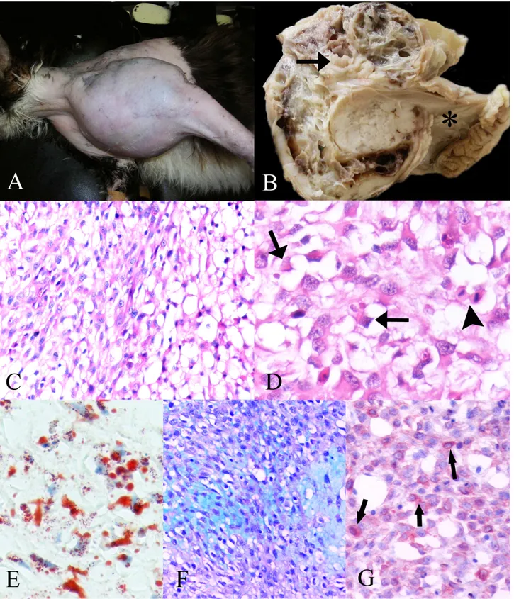
Figure 1. A) The proximal region of the left humerus of cat before the surgery. B) Tan to white, multilobulated mass which contains wide hemorrhage and necrotic foci in the cross section of the tumor with humerus (arrow), (* scapula). C) Round or oval atypical nuclei with one or more prominent nucleoli in the neoplastic cells, HE x240. D) Mitotic figure (arrowhead) and nuclear indentation (arrow), HE x320. E) Oil Red O-positive lipid droplets in cytoplasmic vacuoles of the tumor cells, frozen section, Oil Red O stain x320. F) Myxoid extracellular substance, Alcian blue stain (ph=2.5), x240. G) Vimentin- positive tumor cells (arrow). ABPC, AEC chromogen, hematoxylin counterstain, 240x.
Şekil 1. A) Ameliyat öncesi kedinin sol humerusunun proximal bölgesi. B) Krem -beyaz renkte hemorajik ve nekrotik odaklar içeren, loplu yapıda tümörün kesit yüzü ve humerus(ok), (* scapula). C) Neoplastik hücrelerdeki yuvarlak- oval atipik çekirdek ile bir veya daha fazla belirgin çekirdekcik, HE x240. D) Mitotik figür (okbaşı) ve nüklear çentiklenme (ok), HE x320. E) Tümör hücrelerinin sitoplazmik vakuollerinde Oil Red O-pozitif lipid damlacıkları, dondurulmuş kesit, Oil Red O x320. F) Miksoid ekstrasellüler madde, Alsian blue x240. G) Vimentin pozitif tümör hücreleri (ok), ABPC, AEC, hematoksilen karşıt boyaması, 240.
Ankara Üniv Vet Fak Derg, 58, 2011 143
feline immunodeficiency virus (FIV) by nested polymerase chain reaction (PCR) assay. Primers were designed according to the sequence data described previously (10). 5’TTACTCAAGTATGTTCCCATG3’ and 5’CTGGGGAGCCTGGAGACTGCT3’ primers were used for the first round PCR amplification of the 166-bp segment of FeLV. For the second round PCR, 5’GGTTAAGCACCTGGGCCCCTG3’ and 5’GCAGCG GCCTTGAAACTTCTG3’ primers amplified an 85-bp fragment of FeLV. Besides the semi-nested PCR amplification for FIV proviral DNA, the first round primers which were 5’CTACTGCTGCTGCAGCTGAA3’ and 5’CACTGCATCCTAGCTGGTGC3’ yielded a 485 bp product of the gag gene and the second round primers which were 5’CACTGCATCCTAGCTGGTGC3’ and 5’GGATGAAAGCTTAAAGCAAC3’ yielded a 159 bp product of FIV. This analysis detected FeLV and FeSV proviral DNA together. The DNA was isolated from the samples with a genomic DNA purification kit (Qiagen DNeasy Blood and Tissue Kit, Cat No. 69506, Hilden, Germany) according to the manufacturer’s instructions. PCR amplification was performed as described elsewhere (10) with minor modifications. The samples were subjected to the nested PCR to identify FeLV / FeSV and
FIV proviral DNA. The resulting DNA products
(amplicon) were analyzed on agarose gel (1.5%) after electrophoresis at 80 V for 30 minutes. The DNA bands were observed under ultraviolet light.
At the end of the PCR assay, all samples evaluated for the presence of FeLV, FeSV and FIV DNA and the results were appeared as negative.
Grossly, the tumor was a tan-to-white, soft, multilobulated mass, with mucous composition, including a wide hemorrhage and necrotic foci (Fig. 1B). Microscopically, the tumor was composed of round-to-polygonal cells arranged in solid or loose sheets. The cells had numerous clear, round, variable-sized, well-defined, intracytoplasmic vacuoles. The neoplastic cells had round or oval atypical nuclei with one or more prominent nucleoli (Fig. 1C). Some tumor cells showed nuclear indentation by lipid-laden cytoplasmic vacuoles, typical of liposarcoma. Frequently, mitosis was also detected (Fig. 1D). There were multiple foci of necrosis accompanied by inflammatory infiltration and hemorrhagic foci. The loose myxoid stroma in which the neoplastic cells were embedded was abundant in some areas. On frozen sections, Oil Red O demonstrated lipid droplets with varied sizes in the cytoplasmic vacuoles of the tumor cells (Fig. 1E). Alcian blue (pH = 2,5) showed mucins in the myxoid extracellular substance (Fig. 1F). Additionally, the tumor cells were strongly positive for vimentin (Fig. 1G), but negative for pancytokeratin and S-100.
The tumor was diagnosed as myxoid liposarcoma based on gross, histochemical and immunohistochemical
findings. The histopathology of the neoplasia are consistent with round, spindle or stellate cells, with vacuolated cytoplasm and nuclear indentation is achieved by the coalescence of vacuoles (7). For the differential diagnosis of liposarcoma from other mesenchimal neoplasmas, typical histologic features and Oil Red O staining may be adequate (6,7). In the frozen sections of the tumor, the cytoplasmic vacuoles in the tumor cells showed a typical staining of the red–orange color by Oil Red O, indicative of lipid droplets (6). The most common type of liposarcoma is myxoid variant in humans (7) and it is infrequent in the canine (1). The myxoid variant is identified by the presence of neoplastic cells embedded within the Alcian blue-positive, loose, mucoid stroma (6). The myxoid background was detected among the tumor cells with vacuolated cytoplasm by using Alcian blue at pH = 2,5, in this study. The microscopical features of tumor cells together with Oil Red O positive-vacuoles and Alcian blue positive-stroma verified the first case of the myxoid variant of liposarcoma in a cat. Vimentin and S-100 immunolabeling are suggestive of liposarcoma (11). Our case showed only vimentin immunolabeling, but was negative for S-100. In humans, liposarcomas have been located in the lower extremities, retroperitoneal, mesenteric, and shoulder areas (7). In dogs, liposarcoma has been located in the shoulder, thorax, axilla, tail base, or hip regions as subcutaneous masses and within the viscera (1). In the present case, it was located essentially in the left proximal humerus, extending to the scapular region to a lesser degree. The liposarcoma tends to recur locally and metastasizes rarely to the lung, liver, and bone (1,7). After surgery, follow-up information has shown no evidence of local recurrence and / or distant metastasis for 10 months post-surgical time and the cat is in health.
In veterinary literature, the etiology of liposarcoma has been determined by limited reports. In cats, the increased incidence of feline vaccine-associated sarcoma is a significant problem in veterinary medicine (2) and the risk factor of postvaccinal sarcomas is considered to be a fibrous connective tissue reaction to the aluminium within the vaccine and leads to tumors. Vaccine-associated sarcomas are fibrosarcomas, malignant fibrous histiocytomas, osteosarcomas, chondrosarcomas, rhabdomyosarcomas, myxosarcomas, and liposarcomas (2). The present case was localized on the proximal humeral region on the first clinical examination, and we suspected that this region was not the usual vaccination site in cats. Moreover, there was no information about the anatomical region where the vaccination(s) was applied by the hospital or veterinarian. Several studies have reported that feline leukemia virus, polyomavirus, and feline immunodeficiency virus do not have any direct involvement in the pathogenesis of vaccine-associated sarcomas of cats (3-5). However, another liposarcoma
Yonca Betil Kabak - Cenk Yardımcı - Semra Okur-Gumusova - Mustafa Yavuz Gulbahar 144
case has been determined in a cat having serological evidence of feline leukemia virus infection (8) and a tumor that includes type C virus particles that are transmitted to kittens (9). In the study showed no evidence of FeLV – FeSV and FIV DNA in the etiology of liposarcoma.
In conclusion, the present case is the first description of spontaneous myxoid liposarcoma in a cat and there is no association with FeLV – FeSV and FIV infections or the vaccination site.
Acknowledgements
A part of this study was presented as a poster at the 27th Annual Meeting of European Society of Veterinary Pathology, 9 –12 September 2009, Olsztyn – Kraków, Poland and published in J Comp Pathol, 141(4):288, (Meeting abstract 43), as abstract.
References
1. Goldschmidt MH, Hendrick MJ (2002): Liposarcoma. 97-99. In:Tumors in Domestic Animals, DJ Meuten (Ed). 4th ed., Iowa State Press, Ames, IA.
2. Hendrick MJ, Goldschmidt MH, Shofer FS, Wang YY, Somlyo AP (1992): Postvaccinal sarcomas in the cat:
epidemiology and electron probe microanalytical identification of aluminum. Cancer Res, 52, 5391-5394.
3. Kidney BA, Ellis JA, Haines DM, Jackson ML (2000):
Evaluation of formalin-fixed paraffin-embedded tissues obtained from vaccine site-associated sarcomas of cats for DNA of feline immunodeficiency virus. Am J Vet Res, 61,
1037-1041.
4. Kidney BA, Ellis JA, Haines DM, Jackson ML (2001):
Comparison of endogenous feline leukemia virus RNA content in feline vaccine and nonvaccine site-associated sarcomas. Am J Vet Res, 62, 1990-1994.
5. Kidney BA, Haines DM, Ellis JA, Burnham M, Jackson ML (2001): Evaluation of formalin-fixed
paraffin-embedded tissues from vaccine site-associated sarcomas of cats for polyomavirus DNA and antigen. Am J Vet Res,
62, 828-832.
6. Masserdotti C, Bonfanti U, De Lorenzi D, Ottolini N (2006): Use of Oil Red O stain in the cytologic diagnosis
of canine liposarcoma. Vet Clin Pathol, 35, 37-41.
7. Rosai J (2004): Soft Tissues. 2279-2285. In: Rosai and Ackerman's Surgical Pathology. J. Rosai. (Ed), 9th ed., Vol 2, Mosby, Inc., St Louis.
8. Stephens LC, Tsai CC, Raulston GL, Jardine JH, MacKenzie WF (1983): Virus-associated liposarcoma
and malignant lymphoma in a kitten. J Am Vet Med
Assoc, 183, 123-125.
9. Stephens LC, King GK, Jardine JH (1984): Attempted
transmission of a feline virus-associated liposarcoma to newborn kittens. Vet Pathol, 21, 614-616.
10. Stiles J, Bienzle D, Render JA, Buyukmihci C, Johnson EC (1999): Use of nested polymerase chain reaction
(PCR) for detection of retroviruses from formalin-fixed, parafin-embedded uveal melanomas in cats. Vet
Ophthalmol, 2, 113-116.
11. Wang FI, Liang SL, Eng HL, Jeng CR, Pang VF (2005): Disseminated liposarcoma in a dog. J Vet Diagn Invest, 17, 291-294.
Geliş tarihi: 25.03.2010 / Kabul tarihi: 26.07.2010
Corresponding author
Yonca Betil Kabak
University of Ondokuz Mayis, Faculty of Veterinary Medicine,
Department of Pathology, Kurupelit 55139 Samsun-Turkey Tel: +90 362 3121919 ext.3914
Fax: +90 362 4576922
E-mail: [email protected] or [email protected]