Short Communication / Kısa Bilimsel Çalışma
Histopathological and immunohistochemical evaluation of congenital
cutaneous melanomas in calves (3 cases)
Enver BEYTUT
1, Engin KILIÇ
2, Sadık YAYLA
21Kafkas University, Faculty of Veterinary Medicine, Department of Pathology, Kars; 2Kafkas University, Faculty of Veterinary
Medicine, Department of Surgery, Kars, Turkey.
Summary: Three male calves with a dark mass, hanging down on the skin at various parts of the body, were presented for
clinical examination. All masses were present at the birth of the calves and their size increased with time. The masses were extirpated by a routine surgical operation and presented for histopathological examination. Tumour cells having cytoplasmic melanin granules showed a round or spindle-shaped nuclei. The mean mitotic rate was 5-7 at a high power field. Neoplastic melanocytes displayed an evident cytoplasmic immunolabeling to Melan A, HMB-45, S-100, NSE, and Vimentin. Tumour cells showed a nuclear positivity for Ki-67. Nuclear pleomorphism and a high mitotic rate along with the positive reaction of neoplastic cells to the melanocytic markers were indicative of melanoma. Nevertheless, neoplasms originating from melanocytes always need advanced imaging techniques along with histopathological and immunohistochemical examination while assessing biological behavior of the tumour.
Keywords: Calves, congenital cutaneous melanoma, histopathology, immunohistochemistry.
Buzağılarda kongenital deri melanomlarının histopatolojik ve immunohistokimyasal olarak
değerlendirilmesi (3 vaka)
Özet: Vücutlarının farklı bölgelerindeki deri üzerinde siyah kitle bulunan 3 erkek buzağı klinik olarak muayene edildi. Kitlelerin
doğumla beraber görüldüğü ve zamanla büyüdükleri bildirildi. Tüm kitleler rutin cerrahi yolla alındı ve histopatolojik olarak değerlendirildi. Tümör kitlelerinin yuvarlak veya iğsi çekirdekli hücrelerden oluştuğu ve sitoplazmalarının melanin granülleri ile dolduğu görüldü. Mitoz oranı yüksek büyütmede ortalama 5-7 olarak belirlendi. Neoplastik melanositlerde Melan A, HMB-45, S-100, NSE ve vimentin için sitoplazmik reaksiyon görüldü. Yine tümör hücreleri Ki-67 nükleer pozitif reaksiyon gösterdi. Nükleer pleomorfizim ve yüksek mitotik oranla birlikte, tümör hücrelerinin melanositik markırlara karşı pozitif reaksiyonu kitlelerin melanom olduğunu göstermiştir. Bununla birlikte, melanosit kökenli tümörlerin biyolojik karakterinin belirlenmesinde ileri görüntüleme teknikleri ile birlikte histopatolojik ve immunohistokimyasal değerlendirmeler yapılmalıdır.
Anahtar sözcükler: Buzağı, histopatoloji, immunohistokimya, kongenital deri melanoma.
Melanomas are tumours arising from
neuroectodermal melanoblasts or melanocytes (5). Benign forms of the neoplasm are referred as melanocytoma in animals and nevus in humans and the malignant forms are called melanoma or malignant melanoma in both medical practices (4, 23, 24). These tumours often occur in large domestic animals including cattle (2, 4, 5, 11), but the neoplasms are most significant in gray or white horses (14). In cattle, melanomas are rare than dogs and horses and have been documented to represent less than 2 % of all bovine tumours (5, 14). Miller et al. (13) reported that even though melanocytic tumours occur in older bovines, most cases are seen in calves younger than 2 years old. This neoplasm is occasionally seen as a congenital lesion in Duroc pigs (14), goats (1) and in calves (15, 22).
Melanomas are firm, hyperpigmented, discrete and single growths in cattle, located dermo-epidermally or subcutaneously (5, 11, 14, 25), and found often in the skin of animals (3, 4, 22). Although excessive exposure to the direct sunlight is noted to be a risk factor in humans and angora goats, there is no evidence for this in domestic animals (2, 11).
Melanomas are classified as epithelioid, spindle cell, mixed and whorled or dendritic melanomas due to the predominant cell type (2, 11). Diagnosis of the neoplasm is based on the histological and cytological evaluation. The presence of melanin pigment is the most valuable feature for the identification of melanocyte tumours (11). Our literature reviews found no detailed record of immunohistochemical expression of melanoma markers in
the congenital cutaneous melanomas of calves. Thus, the present study was undertaken to investigate the immunohistochemical expression of Melan-A, HMB-45, S-100, Neuron Specific Enolase (NSE), Vimentin, and Ki-67 and to evaluate the histopathological results of congenital cutaneous melanomas in calves.
Case 1: A six-months-old Simmental male calf with a cutaneous mass on the tibiotarsal joint of the right hind limb was presented to the Department of Veterinary Surgery, University of Kafkas, for clinical examination.
The calf was of a fairly general condition at the clinical examination. The neoplastic mass appeared large, round, black and hanging on the skin (Figure 1A). The dimensions of the mass in the case were approximately 20x17x15 cm in diameter. The rectal temperature was 38.5C; the heart rate was 90 beats/min; and the respiratory rate was 50 breaths/min. Upon auscultation of the lung, the sounds were clear. Superficial lymph nodes were not seen to be enlarged. No mass was detected in thoracic or abdominal cavities on roentgen examination.
Figure 1. Tumour masses on the tibiotarsal joint (A, case 1) and perianal area of calves (B, case 2). Şekil 1. Buzağılarda tibiotarsal eklem (A, vaka 1) ve perianal bölgede tümöral kitle (B, vaka 2).
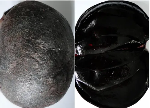
Figure 2. Hairless tumour mass and its cut surface with severe black appearance (case 3). Şekil 2. Kılsız tümöral kitle ile kesit yüzünde şiddetli siyahımsı görünüm (vaka 3).
Case 2: A Simmental male calf of five-months-old with a large mass on the skin of the perianal area was also admitted to the same Department, for a clinical examination. The mass with black in colour was ruptured and appeared to have haemorrhages (Figure 1B). The mass was measured as approximately 22x13x10 cm in diameters. The calf developed progressive weight loss in spite of normal feeding.
Case 3: A brown Swiss male calf of about one month of age with a black skin mass on the left shoulder was admitted for clinical examination. The mass dimensions in this case were smaller than others and were measured as approximately 10x8x7 cm in diameters (Figure 2).
Based on the information given by the owners, all masses were present at the birth and their sizes increased with the time. All routine clinical examinations of the calves were carried out and the masses were extirpated in a routine surgical operation. Subcutaneous, muscular and tendinous invasion by tumour masses were detected during the surgery.
All masses following surgical operation were submitted to the Department of Veterinary Pathology for histopathological evaluation. On the cross-section, the cut surface of the masses showed soft to moderately firm in consistency and severe black in colour. The masses were supposed to be a melanocytic tumour based on their appearance. Tissue samples taken from the masses were fixed in 10% buffered formalin, processed routinely, and the sections taken in 4 µm thickness were stained with haematoxylin and eosin (H&E). For bleaching, some heavily pigmented tumour sections were deparaffinized, hydrated, and incubated in 10% solution of hydrogen peroxide (H2O2) for 24 hours at 65oC until sections
appeared clear. The conventional mitotic rate in the cases was calculated in five areas of tumour with greatest mitotic activity on the bleached sections stained with H&E (40x objective).
Both bleached and unbleached sections were stained with avidin-biotin-peroxidase complex (ABC) technique (8) for Melan-A, HMB-45, S-100, NSE, vimentin, and Ki-67. Paraffin sections were dewaxed in xylene and hydrated through graded alcohols. Endogenous peroxidase activity was blocked with 3 % H2O2 in distilled water for 20 min.
The sections were subsequently put in citrate buffer saline (pH 6.0) in a microwave oven for 20 min for antigen retrieval. The sections were incubated with 5% normal rabbit (Melan A, HMB-45, Vimentin, NSE, Ki-67) or goat (S100) serum at room temperature (RT) for 60 min. The sections were then incubated with monoclonal mouse anti-human Melan A/ (Leica, PA0233), monoclonal mouse anti-human HMB-45 (Cell Marque, 282M-98), polyclonal rabbit-anti cow S100 (Novocastra, NCL-L-S100), monoclonal mouse anti-NSE (Genemed, GM021),
monoclonal mouse anti-porcine vimentin (Leica, PA0640) and monoclonal mouse anti-Ki-67 (Genemed, GM010) primary antibodies. The sections were incubated with biotinylated rabbit anti-mouse IgG (Melan A, HMB-45, NSE, Vimentin, Ki-67), and with biotinylated goat anti-rabbit IgG (S100) at a dilution of 1/100 in TBS for 60 min at RT, the secondary antibodies supplied by Dako, Carpinteria, USA. All sections were treated with
streptavidin-peroxidase complex (ABC; Dako,
Carpinteria, USA), at a dilution of 1/300 for 30 min at RT. The sections were rinsed three times with TBS for 5 min in each step. Immunostaining was obtained using 3-amino-9-ethylcarbazole (AEC) and 3.3 diaminobenzidine (DAB) as the chromogens. Harris’ hematoxylin was used as the counterstain. The sections taken from human skin were used as the positive control during all the immunostaining protocol of the primary antibodies. Primary antibodies were omitted from negative control sections and incubated with diluted normal serum.
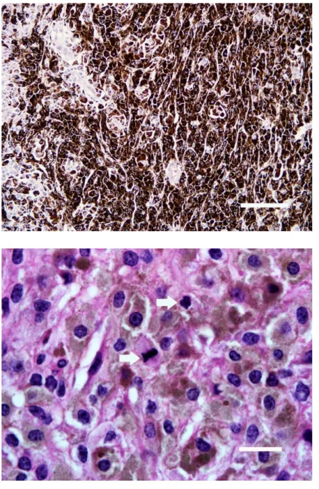
Microscopically, all tumour masses were composed of heavily pigmented epithelioid or spindle-shaped cells with a highly angular shape or stellate which were often arranged in sheets in a band-like pattern. Epithelioid type of melanocytes revealed commonly a moderate to large, round to oval nuclei containing 1-2 nucleoli with a delicate chromatin and abundant granular black pigment in their cytoplasm (Figure 3). Nuclear details of the neoplastic cells were mainly observed after bleaching of the sections. Neoplastic melanocytes exhibited nuclear pleomorphism and numerous giant cells with a heavily pigmented cytoplasm. Even though mitosis was difficult to observe in the H&E-stained sections, the bleached sections displayed about 5-7 mitotic figures at a high power field-HPF (Figure 4). Neoplastic cells were often arranged in a fusiform pattern with some parts arranged in small nests. In particular, most tumour cells with dense pigment particles darkened the cellular margin, and therefore, nuclear details of tumour cells could only be observed with difficulty. Melanocyte dendrites filled with the pigment particles and were noticed as thin and long extensions. Occasionally, a few macrophages with phagocytosed melanin granules were seen to be distributed throughout the tumour masses. Stromal collagen was minimal in the masses. Neoplastic cells were generally seen to be located within the collagenous stroma. The masses were infiltrated by numerous neutrophil leukocytes and a few lymphocytes, with the predominance in the areas next to the epidermal layer. Blood vessels often occluded with thromboses, and lymphatics around the blood vessels contained multiple tumour cells. Epidermis revealed distributed melanocytes within all layers and pigments desquamated from the corneal layer.
Figure 3. Tumour cells containing a high amount of melanin pigment (H&E, Bar=166 μm).
Şekil 3. Aşırı miktarda melanin pigmenti taşıyan tümör hücreleri (HE, Bar=166 μm).
Figure 4. Tumour cells showing many mitotic figures (arrows) following H2O2 bleaching
(H&E, Bar=11 μm).
Şekil 4. H2O2 ile beyazlatmadan sonra çok
sayıda mitoz (oklar) gösteren tümör hücreleri (HE, Bar=11 μm).
Figure 5. A strong immunolabeling in the cytoplasm of neoplastic melanocytes to Melan A (ABC, Bar=166 μm).
Şekil 5. Neoplastik melanositlerin sitoplazmasında Melan A için kuvvetli immunoreaktivite (ABC, Bar=166 μm).
Figure 6. Weak immunoreactivity for HMB-45 in the cytoplasm of neoplastic melanocytes following bleaching (ABC, Bar=51 μm). Şekil 6. Beyazlatmayı takiben neoplastik melanositlerin sitoplazmasında zayıf HMB-45 immun reaksiyon (ABC, Bar=51 μm).
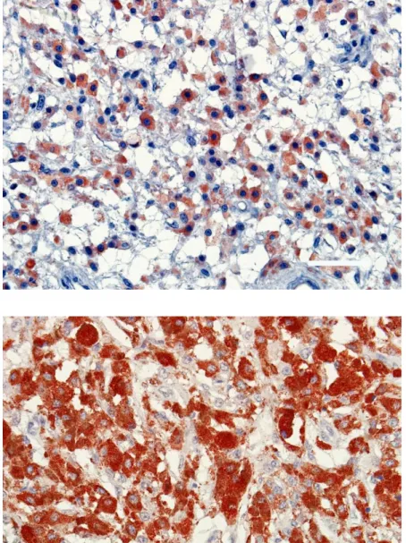
Figure 7. A strong S100 protein immunoreactivity in the cytoplasm of neoplastic melanocytes (ABC, Bar=51 μm). Şekil 7. Neoplastik melanositlerin sitoplazmasında S100 proteini için kuvvetli immun reaksiyon (ABC, Bar=51 μm).
Figure 8. Positive immunoreactivity of the neoplastic cells to vimentin (ABC, Bar=51 μm). Şekil 8. Neoplastik melanositlerde vimentine karşı pozitif immunreaksiyon (ABC, Bar=51 μm).
All neoplastic melanocytes displayed a diffuse positive cytoplasmic immunolabeling to Melan A in both bleached and unbleached sections (Figure 5). The tumour cells revealed an evident immunoreactivity to the marker. However, the intensity of staining for Melan A was strong in the cytoplasm of neoplastic cells from the unbleached sections compared to the bleached ones. Positive labeling to Melan A was higher in the center of the mass. Interestingly, a positive reaction to Melan A was strong around some cells having heavy cytoplasmic melanin pigmentation. Neoplastic melanocytes from all three masses showed a similar staining pattern to HMB-45 (Figure 6). Positive immunolabeling to HMB-45 occurred commonly in the cytoplasm of tumour cells. However, not all neoplastic cells displayed a positive labeling to the marker, and staining intensity varied among the tumour cells. Like Melan A, positive labeling to HMB-45 was clearly stronger in unbleached sections compared to the bleached sections treated by hydrogen peroxide solution. S-100 immunoreactivity was found to be very strong in all neoplastic melanocytes. Positive labeling to S-100 occurred in both the nuclei and cytoplasm of tumour cells and moreover, in some cells, strong immunoreactivity masked cellular margins and cytoplasmic details. The reaction products of S-100 were fine granular within the cytoplasm and distributed diffusely within all the cytoplasm (Figure 7). NSE immunostaining was detected in the cytoplasm of neoplastic melanocytes. But, not all neoplastic cells displayed a positive reaction to NSE. The
immunoreaction products showed fine granular
distribution in the cytoplasm of the neoplastic cells. Vimentin immunopositive reaction was detected in the cytoplasm of neoplastic melanocytes and stromal tissue (Figure 8). The intensity of staining to vimentin varied in different parts of the neoplastic tissue, with a strong reaction in dermal cells and in the deep area of the neoplasm. A staining in the wall of blood vessels and epidermis was not seen. In the unbleached sections, immunoreactivity to vimentin was openly stronger compared to bleached slides. Ki-67 immunolabeling was only found in the nuclei of tumour cells and the germinative layer of the epidermis.
Cutaneous melanomas are often congenital and occur in cattle younger than two years old (5, 9, 13, 24). Even though the site of the melanomas may vary in various species, the neoplasm can occur in various parts of the body where melanocytes are normally present (2-5, 25). The tumours reported in this paper appeared to have arisen from the cutaneous tissues of tibiotarsal joint, perianal area and left shoulder, with heavily pigmented which caused the black appearance facilitating clinical diagnosis of melanomas, consistent with the results of others (4, 6, 13, 21). It is well known that metastasis is an important feature for melanomas (11). Other criteria used
for distinguishing melanoma from melanocytomas are reported to be based on the cytological features (20, 24). In the present cases, metastase or swelling to the regional lymph nodes was not noticed at the clinical examination. However, histopathological examination suggested a malignant potential for the masses because of nuclear pleomorphism, high mitotic rate, giant cells and nests of the neoplastic cells within some lymphatics, as reported by others (21, 24). Neoplastic cells predominantly consisted of a mixture of epithelioid and spindloid types of melanoma and determined numerous mature melanocytes with an abundant cytoplasmic granular black pigment. Millanta et al. (12) reported that a histopathological examination of malignant skin and oral melanomas in dogs revealed predominantly epithelioid types of tumour cells and that proliferative activity of neoplastic melanocytes was a valuable criterion in the evaluation of prognosis and in the discrimination of benign and malignant melanomas. Consistent with the gross appearance of the tumours, the cytoplasm of the neoplastic cells showed a heavy deposition of brownish black melanin granules, and therefore, the morphological features of the nuclei could not be seen or were difficult to distinguish, as stated by others (3, 4, 11, 24). However, the bleaching of the sections was found to be very useful in the evaluation of details of neoplastic melanocytes, consistent with the results of Millanta et al. (12).
The present work mainly aimed to evaluate the expression of Melan A, HMB-45, S-100 and NSE with Vimentin and Ki-67 that might be useful in the diagnosis and prognosis of the congenital cutaneous melanomas in calves. Immunohistochemistry found an evident reaction in neoplastic melanocytes from both bleached and unbleached sections, with a stronger positive reaction in the unbleached sections compared to bleached ones. Nevertheless, hyperpigmented melanomas should be bleached to distinguish neoplastic cells showing cytoplasmic immunolabeling to the immune markers. Additionally, in such works instead of DAB, because of brownish colour of end product, AEC should be preferred as a chromogen.
Melan A is one of the most important melanocytic differentiation markers for both human and canine melanomas, and the marker can be particularly informative in the estimation of the biological behaviors of melanomas (10, 18, 19). In this study, Melan A immunoreactivity occurred in the cytoplasm of the neoplastic melanocytes, without any nuclear staining. Staining intensity for the marker was strong in the central areas of tumours, as reported by Aydoğan et al. (1). S100 has also been commonly used as a melanocytic marker with a high sensitivity for canine (18) and bovine (13) melanomas. This protein is also very beneficial in the diagnosis of neuronal and neuroectodermal neoplasms
(10). Our study revealed that, like Melan A, S100 immunoreactivity was stronger in unbleached sections. Even though both Melan A and S100 were found to be immunopositive in the neoplastic melanocytes, S100 reactivity was stronger than Melan A, consistent with the results reported by Koenig et al. (10). However, it should be mentioned here that even Melan A immunoreactivity was weak compared to the S100, Melan A is a more reliable indicator for malignant potential, justifying its use in an immunohistochemical panel for melanomas. Similar studies to further evaluate the sensitivity of Melan A and S100 in canine (10) and feline (16) melanomas reported that although Melan A is less sensitive than S100 protein, this marker is more specific for melanoma, and it is useful in differentiating feline cutaneous melanomas from the more common pigmented basal cell carcinomas. In this study, even though S100 protein immunoreactivity was more sensitive compared to Melan A in detecting of bovine melanocytic tumours, its specificity was low. Likewise, S100 protein is not strictly specific to melanocytes and the protein stains nonmelanocytic cells including glial cells, neurons, Schwann cells, and skin Langerhan’s cells, as stated by Ramos-Vara et al. (16). A weak HMB-45 immunopositive reaction was detected in both bleached and unbleached sections. It was reported that HMB-45 is also a useful marker for improved diagnosis of tumours originating from melanocytes in bovine and other species (9). However, the standard practice in the diagnosing of melanomas is to use a panel of the antibodies including HMB-45 and S100 (9). NSE is a glycolytic enzyme expressed by cells of neuronal or neuroectodermal origin and it was reported to be a useful marker in the diagnosis of neuronal neoplasms (10). A weak immunoreaction was seen in the cytoplasm of tumour cells for NSE. But, not all neoplastic cells showed a positive reaction against the marker, as reported in oral melanomas of dogs by others (7, 17). Vimentin is an intermediate filament expressed by mesenchymal and neuroectodermal cells. Vimentin immunopositivity was strong in the cytoplasm of neoplastic cells from congenital cutaneous melanomas. Consistent with our results, vimentin immunoreactivity was reported in the cytoplasm of neoplastic cells from melanomas in different animal species (7, 13, 17, 21). In the present cases, mitotic rate was 5-7/10 HPF in the bleached sections stained with H&E. Ki-67 immunostaining confirmed many mitoses in the neoplastic melanocytes. The mitotic index is used often for the prognostic criteria in melanomas of animals and is accepted generally >3/10 HPF in melanomas of dogs (14, 20).
In conclusion, histopathological findings including nuclear pleomorphism and high mitotic rate of the masses along with the positive immunolabeling of neoplastic cells to melanoma-sensitive markers were indicative of
malignancy. However, as the main feature for tumor malignancy is metastasis property, congenital cutaneous melanomas in calves need a further radiological, histopathological and immunohistochemical examination for uncertain biological behavior.
References
1. Aydoğan A, Haligür M, Özmen O (2013): Hepatic malignant melanoma in a goat, primary or metastatic? Isr J Vet Med, 68, 124-127.
2. Brito MF, Franca TN, Jabour FF, et al. (2009): Metastasizing oral melanoma in a cow. Ciência Rural, Santa Maria, 39, 1248-1252.
3. Canpolat İ, Yaman İ, Günay C (2007): A case of primary intraocular malignant iris melanoma in an Akkaraman sheep. Revue Méd Vét, 58, 171-173.
4. Chandrashekaraiah GB, Ballari SV, Manjunatha K, et al. (2014): Malignant melanoma in a Hallikar bullock. Int J Vet Sci, 3, 65-67.
5. Garma-Avina A, Valli VE, Lumsden JH (1981): Cutaneous melanomas in domestic animals. J Cutan Pathol, 8, 3-24.
6. Goldschmidt MH, Hendrick MJ (2002): Tumors of the skin and soft tissues. 45-117. In: DJ Meuten (ed), Tumors in Domestic Animals. 5th (edt), Iowa State Press, Ames, Iowa.
7. Gülbahar MY, Özak A, Kabak YB, et al. (2016): Oral metastatic melanoma with neurosarcomatous transformation in a dog. Ankara Univ Vet Fak Derg, 63, 89-92.
8. Hsu S, Raine L, Fanger H (1981): Use of avidin–biotin peroxidase complex (ABC) in immunoperoxidase techniques: a comparison between ABC and unlabeled antibody (PAP) procedures. J Histochem Cytochem, 29, 577-580.
9. Javanbakht J, Sasani F, Adibhashemi F, et al. (2014): Comparative histopathological diagnosis of cutaneous melanoma by H&E, special staining and immunohistochemical methods against cutaneous squamous cell carcinoma in horse and bovine. J Bioanal and Biomed, 6, 19-23.
10. Koenig A, Wojcieszyn J, Weeks BR, et al. (2001): Expression of S100a, Vimentin, NSE, and Melan A/MART-1 in seven canine melanoma cell lines and twenty-nine retrospective cases of canine melanoma. Vet Pathol, 38, 427-435.
11. Mesaric M, Zadnik T, Cerne M (2002): Malignant melanoma in a cow. Acta Vet-Beograd, 52, 59-64. 12. Millanta F, Fratini F, Corazza M, et al. (2002):
Proliferation activity in oral and cutaneous canine melanocytic tumours: correlation with histological parameters, location, and clinical behavior. Res Vet Sci, 73, 45-51.
13. Miller MA, Weaver AD, Stogsdill PL, et al. (1995): Cutaneous melanocytomas in 10 young cattle. Vet Pathol, 32, 479-484.
14. Moulton JE (1990): Tumors in Domestic Animals. University of California Press, Los Angeles, USA. 15. Pravettoni D, Ordobazari M, Beineke A (2003):
Congenital melanoma in a heifer. Dtsch Tierarztl Wochenschr, 110, 34-36.
16. Ramos-Vara JA, Miller MA, Johnson GC, et al. (2002): Melan A and S100 protein immunohistochemistry in feline melanomas: 48 Cases. Vet Pathol, 39, 127-132.
17. Ramos-Vara JA, Beissenherz ME, Miller MA, et al. (2000): Retrospective study of 338 canine oral melanomas with clinical, histologic, and immunohistochemical review of 129 cases. Vet Pathol, 37, 597-608.
18. Ramos-Vara JA, Miller MA (2011): Immunohistochemical identification of canine melanocytic neoplasms with antibodies to melanocytic antigen PNL2 and tyrosinase: comparison with melan A. Vet Pathol, 48, 443-450.
19. Rezaie A, Golshahi H, Hajikolaie MRH (2013): Epibulbar melanocytoma in a goat. Turk J Vet Anim Sci, 37, 479-481.
20. Sakai H, Yanai T, Kawakami S, et al. (2001): Malignant melanoma of the palpebral conjunctiva in a captive fallow deer. J Wildlife Dis, 37, 816-819.
21. Scandrett B, Wobeser G (2004): Malignant melanoma in a captive red deer (Cervus elaphus elaphus). J Wildlife Dis, 40, 808-810.
22. Schuh JCL (1989): Congenital intraocular melanoma in a calf. J Comp Pathol, 101, 113-116.
23. Smith SH, Goldschmidt MH, Mcmanus PM (2002): A comparative review of melanocytic neoplasms. Vet Pathol, 39, 651-678.
24. Vadalia JV, Fefar DT, Patel PB (2016): Surgical management of malignant melanoma in Kankrej cow. Intas Polivet, 17, 98-99.
25. Yager JA, Scott DW (1993): The skin and appendages. 531-738. In: Jubb KVF, Kennedy PC, Palmer N, (Eds), Pathology of Domestic Animals. 4th ed. vol. 1, Academic
press, San Diego, CA.
Geliş tarihi: 11.08.2017 / Kabul tarihi: 15.01.2018 Address for correspondence:
Prof. Dr. Enver BEYTUT
Kafkas University, Faculty of Veterinary Medicine, Department of Pathology, Kars, Turkey