Ankara Üniv Vet Fak Derg, 55, 173-176, 2008
Trypanosoma melophagium in blood cell culture
Serpil NALBANTOĞLU, Zafer KARAER
Department of Parasitology, Faculty of Veterinary Medicine, University of Ankara.
Summary: Trypanosoma melophagium is a nonpathogenic trypanosome in sheep. The parasite has a widespread distribution
and could be found everywhere along with the keds. Trypanosoma melophagium is a stercorarian species and is transmitted by the sheep ked, Melophagus ovinus. This is the first record of Trypanosoma from sheep PBL (peripheral blood lymphocytes) and blood culture in Turkey. It was identified these sheep Trypanosomes as Trypanosoma melophagium. In the present study, T.melophagium was detected in 7 (7.77%) of the 90 sheep cultures.
Key words: Cultivation, sheep, Trypanosoma melophagium
Kan hücre kültüründe Trypanosoma melophagium
Özet: Trypanosoma melophagium koyunlarda nonpatojen bir trypanosomadır. Parazit koyun biti veya bit sineğinin yaygın
olduğu her yerde bulunur. Trypanosoma melophagium Melophagus ovinus ile taşınır ve stercorarian bir türdür. Bu çalışma Türkiye’de koyunlarda PKL (perifer kan lenfositleri) ve kan kültürlerinde Trypanosoma melophagium’un ilk tespitidir. Çalışmada 90 koyun kültüründen 7’sinde (%7.77) T.melophagium saptanmıştır.
Anahtar sözcükler: Koyun, kültür, Trypanosoma melophagium.
Introduction
Trypanosoma melophagium is a nonpathogenic trypanosome in sheep. The parasite has a widespread distribution and could be found everywhere along with the keds (4, 10, 12). Trypanosoma melophagium is a stercorarian species and is transmitted by the sheep ked, Melophagus ovinus (8, 10). Melophagus ovinus is a pupiparous bloodsucking insect. The life cycle takes place on the host’s skin. They are transmitted by body contact within herd (9, 13, 14). The blood-sucking activity of the sheep keds causes skin infection, and allergic reaction that decreases meat production and wool quality in sheep. In addition, sheep keds may transmit parasites such as trypanosomes (8-10, 14, 17). The prevalence of T.melophagium in sheep is reported to reach a level of 80% in regions where control measures are not implemented for infestation with the indicated ectoparasite (12).
Transmission of T.melophagium occurs by contamination of the skin and if a ked is eaten by the sheep, the metacycle stages penetrate the buccal mucosa (4, 8, 9, 17). The trypomastigotes, which are the bloodstream forms of Trypanosoma melophagium, are 50-60 µm in length. The posterior end of trypomastigates is long and pointed. The undulating membrane is prominent, and a free flagellum is present. The kinetoplast is located near the posterior end, and posterior to the nucleus (10). In general, parasitemia is
very low therefore the probability of the detection of trypanosomes in blood smears is lower than in blood culture. Therapeutic approaches are usually not necessary (8, 10, 12).
The crithidia (epimastigote) forms of T.melophagium have been recovered from the gut of Melophagus ovinus (16), which is reported to be widespread in sheep and goats in Turkey (5, 15). However information does not exist on the detection of the parasite in blood smears and cultures prepared from sheep blood.
The present study was aimed for the detection of Trypanosoma melophagium in lymphocyte and blood cultures prepared from sheep blood, for the first time in Turkey.
Materials and Methods
The blood was collected in volumes of 10 ml by jugular venapuncture into heparinized tubes for the preparation of lymphocyte and blood cultures from a total of 90 sheep, comprising 52 sheep from Ankara, 31 sheep from Çankırı, 4 sheep from Mersin, 2 sheep from Bingöl and 1 sheep from Kayseri. Of the 52 sheep from Ankara, from which blood samples were collected, 29 were used for the preparation of lymphocyte cultures and 23 for blood cultures, whereas of the 31 sheep of Çankırı origin, blood samples collected from 27 animals were used for the preparation of lymphocyte cultures, and 4 were used for blood cultures. On the other hand, all the
Serpil Nalbantoğlu - Zafer Karaer 174
blood samples taken from the 7 sheep pertaining to the other provinces (Mersin, Bingöl and Kayseri) were used for the preparation of blood cultures, and 1 was also utilised for lymphocyte culture. The animals, from which blood samples were collected for the preparation of lymphocyte and blood cultures, were also used for the preparation of thin blood smears from peripheral blood.
For the preparation of lymphocyte and blood cultures, complete RPMI-1640 medium, containing 20% foetal calf serum, L-glutamine and penicillin + streptomycin, was used. The cultures, prepared in accordance with the method described by Brown (1) were incubated at 37 °C in an incubator containing 5% CO2, and were examined under a phase contrast inverted microscope for population growth, at an interval of 3 days, at which the medium was also renewed. Furthermore, the smears prepared from each culture, after being stained with Giemsa in a cell centrifuge (cytospin centrifuge, shadon, Cytospin 2) were examined for T.melophagium under a research microscope and 100x objective. The mean body length, the distances between posterior end and kinetoplast, posterior end and nucleus, kinetoplast and nucleus, nucleus and anterior end as well as the mean nuclear width, the length of the undulating membrane, the length of the free flagellum, and the body length including the free flagellum were measured in 50 of the trypomastigote forms of T.melophagium detected in the smears, using a micrometric ocular, for differential diagnosis. Based on these measurements, the nuclear index (=PN/NA) and kinetoplastic index (=PN/KN) were calculated.
Following the staining of the thin blood smears prepared from sheep blood, in accordance with the Giemsa method, these were examined for T.melophagium under a research microscope and 100x objective. The photographs of T.melophagium, detected in the slides, were taken under a Leica microscope and 40x objective.
Results
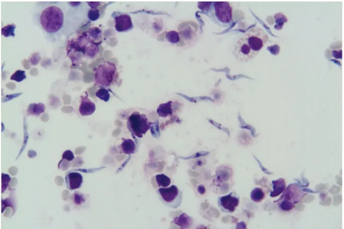
The development stages of T.melophagium in cell cultures are shown in Figure 1. According to measurements performed in the trypomastigote forms, the mean body length including the free flagellum, the mean body length excluding the free flagellum, the mean distances between posterior end and kinetoplast, posterior end and nucleus, kinetoplast and nucleus, nucleus and anterior end, the mean nuclear width, the mean length of the undulating membrane, and the mean length of the free flagellum were determined as 31.1 (25.5-40.5) µm, 23.6 (16.5-33) µm, 9.4 (6-13.5) µm, 10.9 (6-15.7) µm, 1.9 (0.1-3.7) µm, 11.5 (6-15.7) µm, 2 (1.5-3) µm, 11.3 (9-13.5) µm, and 6.8 (4.5-10.5) µm, respectively. Furthermore, the kinetoplastic and nuclear indices were calculated as 4.2 (2.5-6.6) µm and 0.9 (0.5-1.6) µm, respectively.
Figure 1. The development stages of T.melophagium in cell cultures
Şekil 1. Hücre kültüründe T.melophagium’un gelişim safhaları
The positivity of T.melophagium of lymphocyte and blood cell cultures prepared from ovine blood samples collected from Ankara, Çankırı, Mersin, Bingöl and Kayseri are indicated in Table 1.
Table 1. The positivity of T .melophagium of lymphocyte and blood cell cultures prepared from ovine blood
Tablo 1. Koyun kanlarından hazırlanan kan ve lenfosit hücre kültüründe T .melophagium pozitifliği
Location Lymphocyte culture (x/n) ٭ Blood culture (x/n) Ankara 0/29 2/23 Çankırı 5/27 0/4
Other provinces (Mersin,
Bingöl and Kayseri) 0/1 0/7
Total 5/57 2/34
٭x/n : (x= Number of infected animals; n= Number of animals examined)
٭x/n : (x= Enfekte hayvan sayısı; n= Muayene edilen hayvan sayısı)
Accordingly, T.melophagium was detected in 5 (8.77%) of the 57 lymphocyte cultures prepared from sheep, and 2 (5.88%) of the 34 blood cultures. Trypanosoma melophagium was not detected in the Giemsa stained smears prepared from the peripheral blood of the animals.
Discussion and Conclusion
Trypanosomes (genus Trypanosoma) are widespread parasites found in the blood of all classes of vertebrates. Although nonpathogenic stercorarian trypanosomes are easily detected in cultures, depending on the infected host, pathogenic salivarian trypanosomes are observed to grow at very low levels in cultures (12). Similarly, T.rangeli, T.theileri and T.melophagium, which generally cause either latent infections or infections characterised with low parasitemia in humans, and wild and domestic animals, are reported to be
Ankara Üniv Vet Fak Derg, 55, 2008 175
detected easier in blood cultures, when compared to other methods. Herbert (6), having examined the fine structure of the cytoplasmic inclusions, organelles and their function, and the cell chemistry of T.theileri and T.melophagium grown in in vitro cultures, electronmicroscopically, has reported these organelles to be viewed fully and quite evidently at the beginning of cultivation. However, this researcher has also reported these organelles to decrease gradually in subsequent days, and to disappear by the 10th day. In the present study, upon the passage of the cultivation, the intermediate forms of T.melophagium were determined to increase following the 6th passage, and degeneration was demonstrated to occur in further passages.
To date, two stercorarian species have been recorded in introduced vertebrates in Turkey (3, 7). Of these, Trypanosoma theileri was detected in cattle blood culture (7), and T. lewisi was detected in peripheral blood smears prepared from mole rats (Spalax leucodon) (3).
Trypanosoma melophagium, which is a stercorarian species, has been recorded in the gut of the sheep ked (Melophagus ovinus), which is the biological vector of the parasite, in Turkey (5, 15, 16). However the present study is the first record of T. melaphagim in blood cell cultures prepared from sheep, in Turkey.
Monophasic and diphasic blood agar cultures (e.g. NNN) and semisolid cultures and defined liquid media have been used for the growth of apathogenic stercorarian Trypanosoma species (T.theileri, T. lewisi, T. rangeli) (12). Lu (11) has reported T. melophagium to exhibit better population growth at 30°C and pH 7.25 in Modified Monophasic Medium for Trypanosomes (MMMT), in comparison to Medium 199 (supplemented with 5, 10, and 15% haemolysed defibrinated rabbit blood and 10% foetal calf serum), and has demonstrated particularly temperature to be the critical factor for the differentiation of epimastigotes to trypomastigotes. In the present study, isolation and continuance of the culture was maintained using the RPMI 1640 medium, and T. melophagium was determined to grow in the trypomastigote form at 37°C in both blood and lymphocyte cultures.
Upon the comparison of measurements reported by Büscher and Friedhoff (2) for the bloodstream trypomastigote forms of experimentally infected sheep (body length including the free flagellum 45.3 µm, the distance between posterior end and kinetoplast 14.7 µm, the distance between posterior end and nucleus 19.8 µm, the distance from the kinetoplast to the centre of the nucleus 5.1 µm, the distance between the nucleus and the anterior end 19.5 µm, the length of the free flagellum 6µm) with measurements of the trypomastigote forms detected to grow in cultures in the present study (the mean body length including the free flagellum 31.1 µm,
the mean distance between posterior end and kinetoplast 9.4 µm, the mean distance between posterior end and nucleus 10.9 µm, the mean distance between kinetoplast and nucleus 1.9 µm, the mean distance between nucleus and anterior end 11.5 µm, the mean length of the free flagellum 6.8 µm), similar to the report of Lu (11) the trypomastigote forms found in blood were observed to be larger in size when compared to the culture forms.
In conclusion, in the present study, for the first time in Turkey and the world, T.melophagium was detected in both peripheral blood lymphocyte cell culture and blood culture prepared from sheep blood using RPMI 1640 medium.
References
1. Brown CGD (1987): Theileriidae. 230-253. In: AER Taylor, JR Baker (Eds), In Vitro Methods for Parasite Cultivation. Academic Press, London.
2. Büscher G, Friedhoff KT (1984): The morphology of
ovine Trypanosoma melophagium (zoomastigophorea: kinetoplastida). J Protozool, 31, 98-101.
3. Candan S, Eren H ve Babür C (1994): Ankara yöresinde
yakalanan kör farelerde (Spalax leucodon) ilk Trypanosoma lewisi (Kent, 1880) olgusu. Turk Hij Biyol
Derg, 51, 145 – 147.
4. Eckert J, Friedhoff KT, Zahner H, Deplazes P (2005):
Lehrbuch der Parasitologie für die Tiermedizin. Enke
Verlag, Stuttgart.
5. Güralp N, Oğuz T (1967): Yurdumuz tiftik keçilerinde
görülen parazit türleri ve bunların yayılış oranı. Ankara
Üniv Vet Fak Derg, 14, 55-64.
6. Herbert IV (1965): Cytoplasmic inclusions and organelles
of in vitro cultured Trypanosoma theileri and Trypanosoma melophagium and some speculations on their function. Exp Parasitol, 17, 24-40.
7. İnci A, Sayın F (1995): Türkiye’de sığırlardan
Trypanosoma theileri (Laveran, 1902)’nin izolasyonu ve kültivasyonu. Ankara Üniv Vet Fak Derg, 42, 97-103.
8. Kaufmann J (1996): Parasitic Infections of Domestic
Animals. A Diagnostic Manual. Birkhäuser Verlag, Basel,
Boston, Berlin.
9. Kutzer E (2000): Arthropodenbefall bei Wiederkauern. 296-337. In: M Rommel, J Eckert, E Kutzer, W Körtig, T Schneider (Eds), Veterinärmedizinische Parasitologie. 5. Auflage, Parey Buchverlag Berlin.
10. Levine ND (1985): Veterinary Protozoology. Iowa State University Press, Ames.
11. Lu SC (1975): Culture and morphological observations on
Trypanosoma melophagium. Zhonghua Min Guo Wei
Sheng Wu Xue Za Zhi, 8, 20-35.
12. Mansfield JM (1977): Nonpathogenic trypanosomes of
mammals. 297-327. JP Kreier (Ed), Parasitic Protozoa, Vol
I, Taxonomy, Kinetoplastids, and Flagellates of Fish. Academic Press, New York.
13. Mehlhorn H, Düwel D, Raether W (1993): Diagnose und
Therapie der Parasitosen von Haus-, Nutz- und Heimtieren. 2nd edn. Fischer, Stuttgart.
14. Mehlhorn H, D’Haese J, Mencke N, Hansen O (2001):
Serpil Nalbantoğlu - Zafer Karaer 176
(Melophagus ovinus): a light and electron microscopic study. Parasitol Res, 87, 331-336.
15. Mimioğlu M (1973): Veteriner ve Tıbbi Artropodoloji. Ankara Üniversitesi Veteriner Fakültesi Yayınları No: 295, Ankara Üniversitesi Basımevi. Ankara
16. Mimioğlu M, Göksu K, Sayın F (1968): Veteriner ve
Tıbbi Protozooloji I. Ankara Üniversitesi Veteriner
Fakültesi Yayınları No: 232. Ankara Üniversitesi Basımevi, Ankara.
17. Rommel M (2000): Protozoeninfektionen der Wiederkäuer. 121-191. In: M Rommel, J Eckert, E Kutzer, W Körtig, T Schneider (Eds), Veterinärmedizinische Parasitologie. 5., Vollständing neubearbeitete Auflage, Blackwell Wissenschafts-Verlag, Berlin.
Geliş tarihi: 15.04.2008 / Kabul tarihi: 24.04.2008
Address for correspondance
Serpil Nalbantoğlu, DVM, PhD Associate Professor
Department of Parasitology Faculty of Veterinary Medicine
University of Ankara, 06110, Ankara, Turkey E-mail: [email protected]