Complications Following Endoscopic
Retrograde Cholangiopancreatography:
Minimal Invasive Surgical
Recommendations
Bora Koc1, Huseyin Yuce Bircan2, Gokhan Adas1, Ozgur Kemik3*, Adem Akcakaya1, Alpaslan Yavuz4, Servet Karahan1
1. Department of Surgery, Okmeydani Training and Research Hospital, Istanbul, Turkey, 2. Department of Surgery, Baskent University Faculty of Medicine, Istanbul Research Hospital, Istanbul, Turkey, 3. Department of Surgery, Yuzuncu Yil University Faculty of Medicine, Van, Turkey, 4. Department of Radiology, Yuzuncu Yil University Faculty of Medicine, Van, Turkey
Abstract
Background: ERCP has a complication rate ranging between 4% and 16% such as post-ERCP pancreatitis, hemorrhage, cholangitis and perforation. Perforation rate was reported as 0.08% to 1% and mortality rate up to 1.5%. Besides, injury related death rate is 16% to 18%. In this study we aimed to present a retrospective review of our experience with post ERCP-related perforations, reveal the type of injuries and management recommendations with the minimally invasive
approaches.
Methods: Medical records of 28 patients treated for ERCP-related perforations in Okmeydani Training and Research Hospital between March 2007 and March 2013 were reviewed retrospectively. Patient age, gender, comorbidities, ERCP indication, ERCP findings and details were analyzed. All previous and current clinical history, laboratory and radiological findings were used to assess the evaluation of
perforations.
Results: Between March 2007 and March 2013, 2972 ERCPs were performed, 28 (0.94%) of which resulted in ERCP-related perforations. 10 of them were men (35.8%) and 18 women (64.2%). Mean age was 53.36¡14.12 years with a range of 28 to 78 years. 14 (50%) patients were managed conservatively, while 14 (50%) were managed surgically. In 6 patients, laparoscopic exploration was performed due to the failure of non-surgical management. In 6 of the patients that ERCP-related perforation was suspected during or within 2 hours after ERCP, underwent to surgery primarily. There were two mortalities. The mean length of hospitalization stay was 10.46¡2.83 days. The overall mortality rate was 7.1%.
OPEN ACCESS
Citation: Koc B, Bircan HY, Adas G, Kemik O, Akcakaya A, et al. (2014) Complications Following Endoscopic Retrograde
Cholangiopancreatography: Minimal Invasive Surgical Recommendations. PLoS ONE 9(11): e113073. doi:10.1371/journal.pone.0113073 Editor: Tan To Cheung, The University of Hong Kong, Hong Kong
Received: June 19, 2014 Accepted: October 23, 2014 Published: November 26, 2014
Copyright: ß 2014 Koc et al. This is an open-access article distributed under the terms of the
Creative Commons Attribution License, which permits unrestricted use, distribution, and repro-duction in any medium, provided the original author and source are credited.
Data Availability: The authors confirm that all data underlying the findings are fully available without restriction. All relevant data are within the paper. Funding: The authors have no support or funding to report.
Competing Interests: The authors have declared that no competing interests exist.
Conclusion: Successful management of ERCP-related perforation requires immediate diagnosis and early decision to decide whether to manage
conservatively or surgically. Although traditionally conventional surgical approaches have been suggested for the treatment of perforations, laparoscopic techniques may be used in well-chosen cases especially in type II, III and IV perforations.
Introduction
Endoscopic Retrograde Cholangiopancreatography (ERCP) which is an important diagnostic and therapeutic modality for disorders of biliary tree and pancreas, has evolved over the decades, since first introduced in 1968 by McCune et al [1]. ERCP has a complication rate ranging between 4% and 16% such as post-ERCP pancreatitis, hemorrhage, cholangitis and perforation. Perforation rate was reported as 0.08% to 1% and mortality rate up to 1.5% [2–6]. Besides, injury related death rate is 16% to 18% [7,8].
Since the first endoscopic pancreatogram was obtained in 1968 and biliary sphincterotomy was first described in 1974, papillotomy for the management of choledocholithiasis have been widely used and in subsequent years, numerous endoscopic techniques evolved to address pancreaticobiliary disease [1,9]. As the indications for ERCP have increased, a greater focus on recognizing and
preventing complications has emerged [10]. ERCP has a complication rate ranging from 4% to 16% including asymptomatic hyperamylasemia, cardiopul-monary depression, hypoxia, aspiration, intestinal perforation, bleeding, cholan-gitis, adverse medication reactions, sepsis, acute pancreatitis and death. ERCP-related perforation is a rare but serious complication. The incidences of
perforation reported by recent series were ranged from 0.3% to 1.3% [4,6,11–14]. The most important point in the management of ERCP-related perforations is the definition of the injury type. However, the unusual and unexpected
complications are difficult to manage. The treatment of perforations varies from conservative management to urgent surgery according to the injury type and time of diagnosis. Majority of cases are retroperitoneal duodenal perforations usually due to papillotomy, whereas intraperitoneal perforations are less common and caused by the endoscope itself [15]. There has not been a consensus on management guidelines of ERCP related perforations, because of its low rate. There has been few case series in the literature that recommend different therapeutic modalities for ERCP-related perforation. Extensive drainage, repair with omental patch, pyloric exclusion, gastrojejunostomi, T-tube with or without cholecystectomy are surgical interventions that are used for the treatment of ERCP-related perforations [16–18]. Percutaneous drainage technique are generally used in the patients who managed conservatively.
Most recent studies indicate that, carefully selected patients may recover uneventfully with conservative management alone, while in the past, many
authors advocated early surgical management for ERCP-related perforations [11,19]. Many treatment guidelines have been proposed, but unfortunately there is still no consensus on it. Advances in laparoscopy and endoscopy led up to treat these unfortunate patients with minimal invasive techniques. In this study we want to present a retrospective review of our experience with post ERCP-related perforations, reveal the type of injuries and management recommendations with the minimally invasive approaches.
Materials and Methods
Medical records of 28 patients treated for ERCP-related perforations in
Okmeydani Training and Research Hospital between March 2007 and March 2013 were reviewed retrospectively. This study was approved by the institutional review board at our institution (Ethic Committee of Okmeydani Training and Research Hospital, Istanbul, Turkey) and informed written consent was obtained from all of the reviewed subjects for their clinical records to be used in this study.
Patient age, gender, comorbidities, ERCP indication, ERCP findings and details were analyzed. All previous and current clinical history, laboratory and
radiological findings were used to assess the evaluation of perforations.
Computerized tomography was planned on the onset of symptoms and repeated according the severity of the symptoms. Time between diagnosis of perforation and surgery (when used), the type of the operative intervention, the length of hospital stay, the complication rate and the ultimate patient outcome were also studied. The perforations were classified according to the site of perforation using the classification previously defined by Stapfer [11] (Table 1). Institutional ethic committee approved the evaluation of human subjects and the reporting of this study.
According to the management policy of our institution for ERCP-related perforations; extensive contrast extravasation on ERCP/CT, extraperitoneal or intraperitoneal fluid collection on CT with unsolved problem and severe peritonitis, duodenum lateral wall or jejunal injury and problem remaining unsolved with endoscopic procedure (retained hardware or biliary stone failed to be removed during ERCP) are candidates for urgent surgical repair. Patients without any of these conditions were managed conservatively. Conservative management consisted of close monitorisation with physical examination, nasobiliary drainage, antibiotic administration and parenteral nutritional support. All patients were monitored with white blood cell count and C-reactive protein (daily). Surgery was planned immediately when there is hypotension (systolic blood pressure #90 mmHg), tachycardia (heart rate >120/min), fever (axillary temperature >38
˚
C), worsening of abdominal symptoms and signs (signs of peritonitis).Results
Between March 2007 and March 2013, 2972 ERCPs were performed, 28 (0.94%) of which resulted in ERCP-related perforations. 10 of them were men (35.8%) and 18 women (64.2%). Mean age was 53.36¡14.12 years with a range of 28 to 78 years. ERCP was performed for treatment of bile duct stones in 20 patients with additional cholangitis in 7 patients, 4 patients for cholangitis, for benign biliary stricture in 2 patients and for pancreas head cancer in 2 patients. A complete ERCP procedure includes cannulation, sphincterotomy, and basket-balloon instrumentation for stone removal or relieving the bile duct passage.
ERCP-related perforation during the intervention was suspected in 23 patients, only 10 (35.7%) of the perforations were diagnosed during ERCP whereas the remaining 18 were (64.3%) diagnosed by physical examination, trans-abdominal ultrasound, computerized tomography and abdominal radiography.
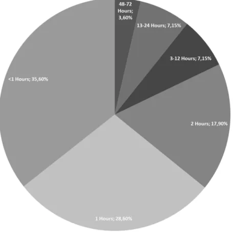
Demonstration of a perforation during ERCP was accomplished by a limited contrast study through the endoscope. Severe post-procedural abdominal pain with/without pancreatitis, signs of peritonitis, fever and increased levels of CRP and white blood cells were accepted as suspected perforation. The mean time of diagnosis after ERCP procedure was 5.57 hours, ranged between 1 and 72 hours (Figure 1).
Conservative management was successful in 14 (50%) patients (Table 2), while 14 (50%) were managed surgically. In 6 patients, laparoscopic exploration was performed due to the failure of non-surgical management. Laparoscopic cholecyctectomy+Laparoscopic common bile duct exploration (LCBDE)+T-tube+drainage was performed in 5 of these patients. Although the remaining patient (63 year-old-male) underwent laparoscopic exploration on the 48th hour because of the progression of physical and laboratory findings, no sign of perforation (intra-and retroperitoneal fluid) was found. Laparoscopic
cholecyctectomy+LCBDE+Stone extraction+Trans-cystic drain were successfully performed and the patient was discharged at the 12th post-operative day (Table 3). In 6 of the patients that ERCP-related perforation was suspected during or within 2 hours after ERCP, underwent to surgery primarily because of the extensive contrast extravasation on ERCP/CT and extraperitoneal or
intraperitoneal fluid collection on CT (also retained biliary stone failed to be removed during ERCP). LCBDE+T-Tube+Drainage were performed; additionally laparoscopic cholecystectomy was added to the surgery in non-cholecystecto-mized three patients (Table 4). In the remaining two patients, although minimal
Table 1. Classification of ERCP-Related Perforations [19].
Type Definition
1 Lateral or medial duodenal wall perforation (endoscope related) 2 Periampullary perforations (sphincterotomy related)
3 Ductal and duodenal perforations due to endoscopic instruments (not guide-wire) 4 Presence of retroperitoneal air due to guide-wire
invasive surgical approach was performed intra-abdominal abscess developed and required reoperation (Table 5). One of them was 63 year-old-female patient who underwent ERCP for CBD stone. The injury was near the ampulla consequent to precut sphincterotomy, showed contrast leakage. Laparoscopic
cholecyctectomy+LCBDE+Stone extraction+T-Tube+intra-and retroperitoneal drainage were performed two hours after perforation. 72 hours after ERCP, signs of peritonitis, fever, and white blood cell counts were increased. That’s why the surgical team decided to re-operate the patient. Intra-abdominal abscess was seen and pyloric exclusion+T-tube revision+gastrojejunostomi was performed. She was well discharged on the 14th day. The second patient was 68 years old female with the diagnosis of pancreas head cancer and liver metastasis underwent ERCP for biliary drainage. Type III injury in the distal common bile duct secondary to wire manipulation was suspected. LCBDE+T-Tube with intra- and retroperitoneal drainage was initially performed after two hours from ERCP. She was re-operated because of the intra-abdominal abscess on the 36th hour. There were two mortalities. The first one who was in failed non-surgical treatment group and died as a result of acute myocardial infarction on the 3rd day. Other patient with the
Figure 1. The figure shows the mean time of diagnosis after ERCP procedure (hours).
diagnosis pancreas head cancer who underwent surgery for ERCP related perforation died because of sepsis at the 10th day. The mean length of
hospitalization stay was 10.46¡2.83 days. The overall mortality rate was 7.1%.
Discussion
Although many patients with ERCP-related perforations can be managed expectantly, there is a dilemma for whom urgent operative intervention is necessary. In previous years some authors have suggested early operation for all endoscopic sphincterotomy perforations. However, with increasing experience with this rare but potentially lethal complication, there is increasing evidence that most perforations may be managed without surgery [20–23]. Early diagnosis of post-ERCP perforations is critical for successful management. Besides, the timing of operation is also important. The initial management is determined by the type and mechanism of injury. Progression of the symptoms and laboratory tests should be warning the surgeon for immediate surgical management. The key
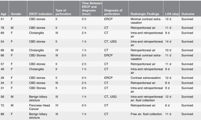
Table 2. Successful Nonsurgical Management of ERCP-Related Perforations.
Age Gender ERCP indication
Type of perforation Time Between ERCP and diagnosis (hour) Diagnosis of
perforation Radiologic Findings LOS (day) Outcome
51 F CBD stones II 0 h ERCP Minimal contrast
extra-vasation
12 d Survived
78 M CBD stones II 1 h CT Retroperitoneal air 11 d Survived
69 F Cholangitis III 2 h CT Intra-and retroperitoneal
air
9 d Survived
54 F CBD stones II 1 h CT, USG Intra-and retroperitoneal
air
14 d Survived
68 M Cholangitis IV 1 h CT Retroperitoneal air 10 d Survived
66 F CBD Stones III 0 h ERCP Minimal contrast
extra-vasation
11 d Survived
57 F CBD stones II 2 h CT Retroperitoneal air 11 d Survived
49 F Cholangitis II 1 h CT Intra-and retroperitoneal
air
9 d Survived
28 F CBD stones II 0 h ERCP Contrast extravasation 12 d Survived
34 F CBD stones III 2 h CT Retroperitoneal air 8 d Survived
31 F CBD Stones II 0 h CT Intra-and retroperitoneal
air
9 d Survived
58 M Benign biliary stricture
III 1 h CT, USG Intra-and retroperitoneal air, fluid collection
12 d Survived
72 M Pancreas Head Cancer
IV 0 h CT Retroperitoneal air 6 d Survived
68 F Benign biliary stricture
III 1 h CT Free air, fluid collection 11 d Survived
Footnotes: ERCP: Endoscopic Retrograde Cholangiopancreatography, CT: Computer Tomography, USG: Ultrasonography LOS: Length of Stay.
T able 3. Failed Non-S urgical Manag ement of ERCP -Re lated Perfora tions. Age Gende r ERCP indica tion T ype of perfo ration s Ti me between ERCP and diagnos is (hour) T ime betwee n ERCP and Oper ation (hour) T ype of Operat ion LOS (day) Outc ome 54 F Chol angit is + CB D ston e II 0 h 24 h Lapa roscop ic cholec yctec tomy + LCBDE + T-Tu be + draina ge 18 d Sur vived 45 M CBD ston es II 12 h 3 6 h Lapa roscop ic chol ecyct ectom y + LCBD E + Stone extrac tion + T-Tu be + draina ge 3 d Ex (AMI) 54 F Chol angit is + CB D ston e II 72 h 108 h Lapa roscop ic cholec yctec tomy + LCBDE + draina ge 10 d Sur vived 57 F CBD ston es III 24 h 9 6 h Lapa roscop ic cholec yctec tomy + Stone extr action + LCBD E + T-Tube 11 d Sur vived 71 F CBD ston es II 18 h 4 8 h Lapa roscop ic cholec yctec tomy + LCB DE + T-Tu be + dra inage 12 d Sur vived 63 M Chol angit is + CB D ston e III 12 h 4 8 h Lapa roscop ic cholec yctec tomy + LCB DE + Trans-Cystic drain 12 d Sur vived Foot notes: ERCP : En doscop ic R etrograde Chol angiopa ncreato graphy , LOS : Length of Stay , A MI: Ac ute Myoca rdia Infarcts, Ex :Exitus, LCB DE: Laparo scopic comm on bile duct explor ation. doi:10.1371/journal.pone.0113073.t003
point is to decide who can be conservatively managed and who should be promptly operated.
Although several researches have classified ERCP-related perforations according to the location or mechanism of injury and have recommended various
treatments, the most popular of these classifications was presented by Stapfer et al [11,13,14]. Stapfer et al classified perforations into four types according to the location and the mechanism of injury (Table 1) [11]. Type I perforations occur on the medial or lateral wall far from the ampulla and are caused by the
endoscope itself or by the stent. Type II perforations are generally retroperitoneal, are classified as peri-vaterian, occur during sphincterotomy. Type III perforations
Table 5. Failed primary minimal invasive surgical management of ERCP-related perforations.
Age Gender ERCP indication Type of Perforation Time Between ERCP and diagnosis (hour) Time between ERCP and Operation
(hour) Type of Operation
LOS (day) Outcome 63 F CBD stones II 0 h 1-) 2 h 2-) 72 h Laparoscopic cholecyctectomy+LCBDE+Stone extraction+T-Tube+drainage Re-operation: Pyloric exclusion+T-Tube revision+gastro-jejunostomy 14 d Survived 68 F Pancreas head cancer+liver metastasis III 1 h 1-) 2 h 2-) 36 h LCBDE+T-Tube+drainage Re-operation: explorative laparotomy+intra-abdominal abscess+drainage 10 d Ex (Sepsis)
Footnotes: ERCP: Endoscopic Retrograde Cholangiopancreatography, LOS: Length of Stay, LCBDE: Laparoscopic common bile duct exploration.
doi:10.1371/journal.pone.0113073.t005
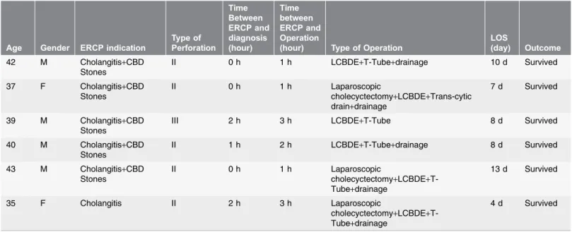
Table 4. Primary Minimal Invasive Surgical Management of ERCP-Related Perforations.
Age Gender ERCP indication
Type of Perforation Time Between ERCP and diagnosis (hour) Time between ERCP and Operation
(hour) Type of Operation
LOS (day) Outcome 42 M Cholangitis+CBD Stones II 0 h 1 h LCBDE+T-Tube+drainage 10 d Survived 37 F Cholangitis+CBD Stones II 0 h 1 h Laparoscopic cholecyctectomy+LCBDE+Trans-cytic drain+drainage 7 d Survived 39 M Cholangitis+CBD Stones
III 2 h 3 h LCBDE+T-Tube 8 d Survived
40 M Cholangitis+CBD Stones II 1 h 2 h LCBDE+T-Tube+drainage 8 d Survived 43 M Cholangitis+CBD Stones II 0 h 1 h Laparoscopic cholecyctectomy+LCBDE+T-Tube+drainage 13 d Survived 35 F Cholangitis II 2 h 3 h Laparoscopic cholecyctectomy+LCBDE+T-Tube+drainage 4 d Survived
Footnotes: ERCP: Endoscopic Retrograde Cholangiopancreatography, LOS: Length of Stay, LCBDE: Laparoscopic common bile duct exploration.
are due to wire manipulation or basket instrumentation during stone retrieval and occur in the distal common bile duct. Type IV perforations are tiny
retroperitoneal perforations caused by the use of compressed air during
endoscopy. Another classification was suggested by Howard et al. includes three groups; group I: guidewire perforations, group II: periampullary perforations, group III: duodenal perforations [13]. Another frequently used classification was presented by Enns et al; group I: esophageal, gastric and duodenal perforations, group II: sphincterotomy-related perforations, group III: guidewire-related perforations [14]. There were 17 type-II perforations, 9 type-III perforations and 2 type-IV perforations in our case series according to the classification system of Stapfer et al. No type-I injury was observed.
The initial clinical presentation of patients with ERCP-related perforation is non-specific. The classic presentation of perforation, with severe epigastric pain, vomiting and epigastric tenderness progressing to generalized rigidity is only seen in the minority of cases. Moreover, the diagnosis is likely to be delayed if the patient has elevated amylase levels and the clinical presentation is attributed to post-ERCP pancreatitis. The most accurate diagnose can be made when the rupture is seen during the procedure. When there is a suspected duodenal perforation, an ultrasound or CT scan is a sensitive method to judge the existence of peritoneal, retroperitoneal emphysema or fluid collection [24,25]. Genzlinger et al suggested that with routine post-ERCP computerized tomography in 13% to 33% of patients small amounts of retroperitoneal air may be detected, probably as a result of a post-procedural but non-significant micro-perforations [26]. Leukocytosis and fever that usually occur in the early phase are useful parameters for determining the management approach. Retroperitoneal nature of the injuries may mask severity; therefore, negative abdominal findings should not exclude surgery. Additionally, Mao et al suggested that subcutaneous emphysema is a sensitive physical sign that can be regarded as an effective parameter for an early diagnosis of perforation besides other radiologic examinations [16]. In our case series, perforation was suspected during the procedure in 23 (82%) patients and only in 8 (28.5%) of them it was diagnosed. Computerized tomography was performed within initial hours in 15 patients with suspected perforation to verify the diagnosis. These ratios are similar with other studies [3,11,13].
Most authors suggested to determine the type and mechanism of the
perforation before selecting the optimal treatment method. Many studies reported that around 70% of patients with ERCP-related perforation could be managed conservatively [11,17,18]. Although, Stapfer et al reported that Type I injuries required prompt surgical interventions, recent studies recommended successful endoscopic treatments with endoscopic clippings, endo-loop applications and endoscopic closure devices [27]. Additionally, Stapfer suggested conservative treatment strategy for type II and III injuries (periampullary and bile duct injuries). In the presence of significant peritoneal findings, type II and III perforations should be treated by surgery. Furthermore, type IV (retroperitoneal air alone) perforations are not regarded as real perforations and should be treated conservatively. The rate of conservative management may vary depending on the
management policies of the institutions. Our conservative treatment rate was 50% which is low compared to the other series. This is because these patients
underwent surgery not only for the injury but also for the underlying disease, which could not be treated by ERCP.
The extent of surgery was proportional to the degree of injury, and the intraabdominal contamination. The basic principles of surgical therapy are repair of the leakage with diversion of the gastric contents and control for the source of the sepsis by means of external drainage [28]. Generally, surgical interventions that are used for the treatment of ERCP-related perforations are as follows; extensive drainage, repair with omental patch, pyloric exclusion, gastrojejunos-tomi, T-tube with or without cholecystectomy [16–18]. Sarli et al reported a wide range of operative procedures for the treatment of ERCP-related perforations, including simple retroperitoneal drainage, duodenal repair around a T-tube inserted into the perforation, common bile duct exploration+T-tube placement, duodenal diversion by antrectomy+gastrojejunostomy or gastrojejunostomy with pyloric exclusion and pancreaticoduodenectomy [29]. No article was found in the literature about the laparoscopic management of ERCP-related perforations. In a study about the comparison of LCBDE and ERCP for the treatment of common bile duct stones, it was pointed that in experienced hands LCBDE is a safe and feasible option with the advantages of minimal access [30].
To summarize, we performed LC+LCBDE+T-tube+intra-or/and retroperito-neal drainage in 6 patients due to the failure of non-surgical management and LCBDE+with or without LC+T-tube+intra-or/and retroperitoneal drainage for 8 patients as a primary management. This approach failed in 2 (14%) patients, and our surgical mortality rate was 7.1%. These rates were similar to literature (Table 6). In this technique we can solve the injury and also the underlying disease (extraction of bile duct stones) in the same intervention. The most
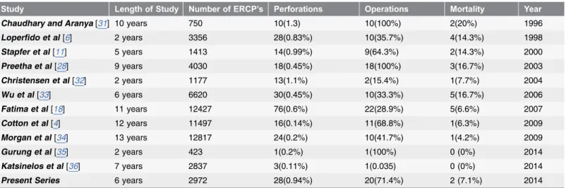
Table 6. Reported perforation rates with ERCP.
Study Length of Study Number of ERCP’s Perforations Operations Mortality Year
Chaudhary and Aranya[31] 10 years 750 10(1.3) 10(100%) 2(20%) 1996
Loperfido et al[6] 2 years 3356 28(0.83%) 10(35.7%) 4(14.3%) 1998 Stapfer et al[11] 5 years 1413 14(0.99%) 9(64.3%) 2(14.3%) 2000 Preetha et al[28] 9 years 4030 18(0.45%) 18(100%) 3(16.7%) 2003 Christensen et al[32] 2 years 1177 13(1.1%) 2(15.4%) 1(7.7%) 2004 Wu et al[33] 6 years 6620 30(0.45%) 10(33.3%) 5(16.7%) 2006 Fatima et al[18] 11 years 12427 76(0.6%) 22(28.9%) 5(6.6%) 2007 Cotton et al[4] 12 years 11497 16(0.14%) 11(68.8%) 1(6.3%) 2009 Morgan et al[34] 13 years 12817 24(0.2%) 10(41.7%) 1(4.2%) 2009 Gurung et al[35] 2 years 423 1(0.2%) 1(100%) 0 (0%) 2014 Katsinelos et al[36] 7 years 2837 3(0.11%) 1(0.035) 0 (0%) 2014
Present Series 6 years 2972 28(0.94%) 20(71.4%) 2 (7.1%) 2014
Footnotes: ERCP: Endoscopic Retrograde Cholangiopancreatography.
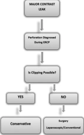
important limitation of our study was lack of type I injuries. It should be kept in mind that minimally invasive management (endoclipping) could be attempted if the perforation is diagnosed during ERCP. We suggest that, in patients with cholelitiasis and choledocholithiasis if type II and III perforation occurs during ERCP, CBDE+LC+T-tube+drainage with nasogastric suction can be performed as primary treatment or when conservative treatment fails. Based on our findings, we propose a simple management algorithm which can be readily and easily used (Figure 2). To design a prospective study about laparoscopic approach is not possible due to major ethical issues. That’s why the evaluation of this approach can only be made by retrospective case series. The source of severe sepsis and peritonitis may not be revealed objectively by laparoscopy laparoscopy which is a serious problem that needs to be resolved.
Conclusions
Successful management of ERCP-related perforation requires immediate diagnosis and early decision to decide whether to manage conservatively or surgically. While patients with type I perforation would invariably require
Figure 2. A simple management algorithm for the ERCP-related perforations.
immediate surgical intervention, those with type II or III, IV may often be managed conservatively. Otherwise, these types of injuries with retained stones and unrelieved bile obstruction should be explored. Although traditionally conventional surgical approaches have been suggested for the treatment of perforations, laparoscopic techniques may be used in well-chosen cases especially in type II, III and IV perforations.
Author Contributions
Conceived and designed the experiments: BK HYB AY OK. Performed the experiments: BK OK GA AA. Analyzed the data: GA OK AY SK. Contributed reagents/materials/analysis tools: BK AA SK. Wrote the paper: BK HYB GA AY OK.
References
1. McCune WS, Shorb PE, Moscovitz H (1968) Endoscopic cannulation of the ampulla of vater: a preliminary report. Ann Surg 167: 752–756.
2. Freeman ML (2003) Adverse outcomes of endoscopic retrograde cholangiopancreatography: avoidance and management. Gastrointest Endosc Clin N Am. 13: 775–798, xi.
3. Lai CH, Lau WY (2008) Management of endoscopic retrograde cholangiopancreatography-related perforation. Surgeon 6: 45–48.
4. Cotton PB, Garrow DA, Gallagher J, Romagnuolo J (2009) Risk factors for complications after ERCP: a multivariate analysis of 11,497 procedures over 12 years. Gastrointest Endosc 70: 80–88.
5. Silviera ML, Seamon MJ, Porshinsky B, Prosciak MP, Doraiswamy VA, et al. (2009) Complications related to endoscopic retrograde cholangiopancreatography: a comprehensive clinical review. J Gastrointestin Liver Dis 18: 73–82.
6. Loperfido S, Angelini G, Benedetti G, Chilovi F, Costan F, et al. (1998) Major early complications from diagnostic and therapeutic ERCP: a prospective multicenter study. Gastrointest Endosc 48: 1–10. 7. Cotton PB, Lehman G, Vennes J, Geenen JE, Russell RC, et al. (1991) Endoscopic sphincterotomy
complications and their management: an attempt at consensus. Gastrointest Endosc 37: 383–393. 8. Elder JB (1988) Surgical treatment of duodenal ulcer. Postgrad Med J 64 Suppl 1:: 54–59.
9. Peel AL, Hermon-Taylor J, Ritchie HD (1974) Technique of transduodenal exploration of the common bile duct. Duodenoscopic appearances after biliary sphincterotomy. Ann R Coll Surg Engl 55: 236–244. 10. Woods KE, Willingham FF (2010) Endoscopic retrograde cholangiopancreatography associated
pancreatitis: A 15-year review. World J Gastrointest Endosc 2: 165–178.
11. Stapfer M, Selby RR, Stain SC, Katkhouda N, Parekh D, et al. (2000) Management of duodenal perforation after endoscopic retrograde cholangiopancreatography and sphincterotomy. Ann Surg 232: 191–198.
12. Masci E, Toti G, Mariani A, Curioni S, Lomazzi A, et al. (2001) Complications of diagnostic and therapeutic ERCP: a prospective multicenter study. Am J Gastroenterol 96: 417–423.
13. Howard TJ, Tan T, Lehman GA, Sherman S, Madura JA, et al. (1999) Classification and management of perforations complicating endoscopic sphincterotomy. Surgery 126: 658–663; discussion 664–655. 14. Enns R, Eloubeidi MA, Mergener K, Jowell PS, Branch MS, et al. (2002) ERCP-related perforations:
risk factors and management. Endoscopy 34: 293–298.
15. Martin DF, Tweedle DE (1990) Retroperitoneal perforation during ERCP and endoscopic sphincterotomy: causes, clinical features and management. Endoscopy 22: 174–175.
16. Mao Z, Zhu Q, Wu W, Wang M, Li J, et al. (2008) Duodenal perforations after endoscopic retrograde cholangiopancreatography: experience and management. J Laparoendosc Adv Surg Tech A 18: 691– 695.
17. Knudson K, Raeburn CD, McIntyre RC Jr, Shah RJ, Chen YK, et al. (2008) Management of duodenal and pancreaticobiliary perforations associated with periampullary endoscopic procedures. Am J Surg 196: 975–981; discussion 981-972.
18. Fatima J, Baron TH, Topazian MD, Houghton SG, Iqbal CW, et al. (2007) Pancreaticobiliary and duodenal perforations after periampullary endoscopic procedures: diagnosis and management. Arch Surg 142: 448–454; discussion 454-445.
19. Avgerinos DV, Llaguna OH, Lo AY, Voli J, Leitman IM (2009) Management of endoscopic retrograde cholangiopancreatography: related duodenal perforations. Surg Endosc 23: 833–838.
20. Scarlett PY, Falk GL (1994) The management of perforation of the duodenum following endoscopic sphincterotomy: a proposal for selective therapy. Aust N Z J Surg 64: 843–846.
21. Sarr MG, Fishman EK, Milligan FD, Siegelman SS, Cameron JL (1986) Pancreatitis or duodenal perforation after peri-Vaterian therapeutic endoscopic procedures: diagnosis, differentiation, and management. Surgery 100: 461–466.
22. Dunham F, Bourgeois N, Gelin M, Jeanmart J, Toussaint J, et al. (1982) Retroperitoneal perforations following endoscopic sphincterotomy; clinical course and management. Endoscopy 14: 92–96. 23. Chung RS, Sivak MV, Ferguson DR (1993) Surgical decisions in the management of duodenal
perforation complicating endoscopic sphincterotomy. Am J Surg 165: 700–703.
24. Sezgin O, Ulker A, Temucin G (2000) Retroperitoneal duodenal perforation during endoscopic sphincterotomy: sonographic findings. J Clin Ultrasound 28: 303–306.
25. Zissin R, Shapiro-Feinberg M, Oscadchy A, Pomeranz I, Leichtmann G, et al. (2000) Retroperitoneal perforation during endoscopic sphincterotomy: imaging findings. Abdom Imaging 25: 279–282. 26. Genzlinger JL, McPhee MS, Fisher JK, Jacob KM, Helzberg JH (1999) Significance of retroperitoneal
air after endoscopic retrograde cholangiopancreatography with sphincterotomy. Am J Gastroenterol 94: 1267–1270.
27. Baron TH, Gostout CJ, Herman L (2000) Hemoclip repair of a sphincterotomy-induced duodenal perforation. Gastrointest Endosc 52: 566–568.
28. Preetha M, Chung YF, Chan WH, Ong HS, Chow PK, et al. (2003) Surgical management of endoscopic retrograde cholangiopancreatography-related perforations. ANZ J Surg 73: 1011–1014. 29. Sarli L, Porrini C, Costi R, Regina G, Violi V, et al. (2007) Operative treatment of periampullary
retroperitoneal perforation complicating endoscopic sphincterotomy. Surgery 142: 26–32.
30. Koc B, Karahan S, Adas G, Tutal F, Guven H, et al. (2013) Comparison of laparoscopic common bile duct exploration and endoscopic retrograde cholangiopancreatography plus laparoscopic
cholecystectomy for choledocholithiasis: a prospective randomized study. Am J Surg 206: 457–463. 31. Chaudhary A, Aranya RC (1996) Surgery in perforation after endoscopic sphincterotomy: sooner, later
or not at all? Ann R Coll Surg Engl 78: 206–208.
32. Christensen M, Matzen P, Schulze S, Rosenberg J (2004) Complications of ERCP: a prospective study. Gastrointest Endosc 60: 721–731.
33. Wu HM, Dixon E, May GR, Sutherland FR (2006) Management of perforation after endoscopic retrograde cholangiopancreatography (ERCP): a population-based review. HPB (Oxford) 8: 393–399. 34. Morgan KA, Fontenot BB, Ruddy JM, Mickey S, Adams DB (2009) Endoscopic retrograde
cholangiopancreatography gut perforations: when to wait! When to operate! Am Surg. 75: 477–483; discussion 483-474.
35. Gurung RB, Purbey B, Koju R, Bedi TR (2014) Endoscopic Retrograde Cholangiopancreatography at Dhulikhel hospital: Outcome Analysis. Kathmandu Univ Med J (KUMJ) 12: 55–59.
36. Katsinelos P, Lazaraki G, Chatzimavroudis G, Gkagkalis S, Vasiliadis I, et al. (2014) Risk factors for therapeutic ERCP-related complications: an analysis of 2,715 cases performed by a single endoscopist. Ann Gastroenterol 27: 65–72.
![Table 1. Classification of ERCP-Related Perforations [ 19 ].](https://thumb-eu.123doks.com/thumbv2/9libnet/3944873.50910/4.918.294.864.134.237/table-classification-ercp-related-perforations.webp)