Cukurova Medical Journal
Cukurova Med J 2016;41(1):187-190ÇUKUROVA ÜNİVERSİTESİ TIP FAKÜLTESİ DERGİSİ DOI: 10.17826/cutf.169024
Yazışma Adresi/Address for Correspondence: Dr. Merve Özen, Başkent University School of Medicine, Departmen of Radiology, Adana, Turkey. E-mail: [email protected]
Geliş tarihi/Received: 14.09.2015 Kabul tarihi/Accepted: 30.10.2015
OLGU SUNUMU/CASE REPORT
Granulocytic sarcoma of finger: a case report
Parmakta granülositik sarkom: olgu sunumu
Ayşin Purbager
1, Merve Özen
1, Hülya Aslan
1, Sermin Tok
1, Emrah Koçer
21Baskent University School of Medicine, Department of Radiology, 2Department of Pathology, Adana/Turkey Cukurova Medical Journal 2016;41(1):187-190.
Abstract Öz
Granulocytic sarcoma is an extramedullary tumor of immature myeloid cells. Such tumors may be located in any part of the body. We report a case of isolated granulocytic sarcoma of finger; which, to our knowledge, is the first report of that localization. A thirty-year-old woman with acute myeloid leukemia (AML) presented with progressive swelling and onychoptosis at her right fourth finger. The distal phalanx was excised and the tumor turned out to consisted of atypical myeloid cells, infiltrating bone trabeculae. Despite its rarity, considering myeloid sarcoma of the finger as a possible diagnosis may prevent inappropriate treatment in leukemia patients.
Granülositik sarkom, immatür myeloid hücrelerin oluşturduğu ekstramedüller bir tümördür. Vücudun herhangi bir yerinde görülebilir. Bu yazıda 30 yaşında, akut myeloid lösemi (AML) tanılı, kadın olguda el parmağında izlenen granülositik sarkom sunulmuştur. Literatürde bugüne kadar el parmağı lokalizasyonunda granülositik sarkom vakası bildirilmemiştir. Fizik muayene ve radyolojik incelemelerde distal falankstan köken alan yumuşak doku lezyonu izlenmiş ve lezyon eksize edilmiştir. Histolojisinde tümörün kemik yapıyı infiltre eden atipik myeloid hücrelerden oluştuğu gözlenmiştir. Nadir bir tümör olan granülositik sarkomun el parmağındaki görülebildiği, lösemi hastalarında en uygun tedaviyi gerçekleştirmek açısından akılda tutulmalıdır.
Key words: AML, granulocytic sarcoma Anahtar kelimeler: AML, granülositik sarkom
INTRODUCTION
Granulocytic sarcoma, also known as myeloid sarcoma or chloroma, is a proliferation of immature myeloid cells in the soft tissue or bone producing a clinically evident tumor. The lesion may precede or occur concurrently with acute or chronic myeloid leukemia or with other types of myeloproliferative disorders or myelodysplastic syndromes1.The incidence of myeloid sarcoma in the course of acute myeloid leukemia has been reported to range from 3–4.7%. The occurrence of extra-medullary lesions before the onset of overt disease is rare2.
Although acute myeloid leukemia is a marrow-based disease, bone involvement with erosion is very rare; most lesions are seen in solid organs or soft tissues3. Granulocytic sarcomas have been reported in the
temporal bone, shoulder bones, and tibia4-6, and most cases occur in patients with known leukemia, or those who eventually develop the disease1. To our knowledge, we report the first case of a chloroma localized to the finger.
CASE
A 30-year-old female patient with acute myeloid leukemia had been in post-bone-marrow-transplant remission for 4 years. She presented with onychoptosis and a 4-month history of progressive swelling of the fourth finger of her right hand starting from the nail bed, and erythema of the volar and dorsal sides of the phalanx. She had no recent history of trauma, fever, or weight loss.
Examination of her right hand revealed a vascular, soft tissue mass elevating the nail plate. The
Cilt/Volume 41 Yıl/Year 2016 Granulocytic sarcoma of the finger
188
maximum width of the mass was 3 cm. She reported no subjective paresthesia at the tip of the finger. Laboratory results were within normal ranges. The patient showed no other skeletal or soft tissue abnormalities and there was no family history of bone or hematologic cancers.
The orthopedist scheduled radiography and magnetic resonance imaging (MRI) of the hand for further evaluation. An antero-posterior (AP) radiograph showed medullary bone sclerosis and soft tissue swelling (Figure 1a). MRI demonstrated a
heterogenous, rounded, soft tissue mass measuring 20 × 15 mm surrounding the volar and dorsal aspects of the right fourth distal phalanx. The lesion demonstrated hyperintense signal on T2-weighted and fat-saturated T2-weighted images (Figure 1b, c). Contrast-enhanced fat-saturated T1-weighted images showed significant contrast enhancement (Figure1d).The bone cortex was not destroyed, but the medullary bone diameter was reduced compared with the other fingers. Based on the MRI findings, we made a presumptive diagnosis of a sarcomatous lesion.
Figure 1 (a) AP radiograph. Medullary bone sclerosis, soft tissue swelling of distal phalanx of fourth finger. (b) Sagittal T2-weighted MR image.The medullary bone and soft tissue mass are hyperintense. (c) Coronal fat-saturated T2-weighted image also shows the lesion as hyperintense. (d) Contrast enhanced fat saturated T1 weighted axial plane image shows contrast enhancement of the soft tissue component of the lesion (asterisks).
The distal phalanx of the fourth finger was surgically resected. Histopathological evaluation revealed a neoplastic mass of atypical myeloid cells, morphologically immature hematopoietic cells indicative of relapsed acute myeloblastic leukemia,
infiltrating the bone trabeculae, subcutaneous tissue, and epidermis. Immunohistochemical evaluations were positive for LCA, CD43, TdT, Bcl-2, CD34, and lysozyme. The histopathologic diagnosis was chloroma (Figure 2).
Purbager et al. Cukurova Medical Journal
189
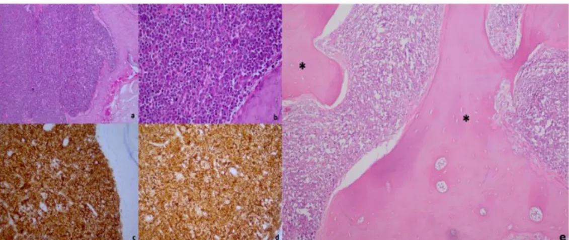
Figure 2. Histopathology. The set of microphotopgraph as reveals diffuse cutaneousin filtration (a) composed of medium sized cells some with round to oval nuclei that have finely dispersed chromatinand small nucleolei (b).The neoplastic cells were diffuse strong positive with CD43 (c) and CD34 (d). ( a: HE X100, b: HE X200, c: CD43 X200, d: CD34 X200). (e) HE X100, bone trabecula (asterisks). Diffuse infiltration of neoplastic myeloid cells between the bone trabecula
DISCUSSION
Myeloid sarcoma can develop at any age but is most common among young adults7. Musculoskeletal involvement is usually seen in symptomatic patients. Commonly involved sites include the orbit, sacrum, sinuses, spine, sternum, and ribs6. The tumor is thought to grow in the bone marrow and has a tendency for ligamentous and periosteal involvement, which most likely occurs by migration through the Haversian canals8. In advanced phases, osseous infiltration presents mainly as a focal radiolucent zone or sclerosis6. In this case, an AP radiograph showed no definitive bone changes, apart from minimal medullary bone sclerosis. However, medullary bone involvement was evident on MRI.
Local therapy (e.g., surgery or radiation therapy) can cure focal relapse, and early diagnosis is thus essential2. However, localization of the tumor on the hand is an important prognostic factor, regardless of the treatment method, given that the survival rate is much higher when the distal parts of the extremities are affected9.
Granulocytic sarcoma lesions are usually multiple and solid, recurring at different sites in nearly 50% of patients3. Granulocytic sarcoma lesions tend to be multicentered and solid, and recurrence is very common. Although these important clues can help to make a diagnosis, isolated lesions of granulocytic
sarcoma may be difficult to differentiate from other focal malignancies, such as lymphoma or carcinoma3. In the present case, there was no other soft tissue or bone lesion suggestive of granulocytic sarcoma.
The optimal therapeutic management of myeloid sarcoma remains uncertain. The options include chemotherapy, hematopoietic stem cell transplantation, radiotherapy, and surgery. Surgical resection was preferred by our orthopedicians because of the aggressive pattern of the tumor. To the best of our knowledge this is the first published report of myeloid sarcoma of the finger. Despite its apparent rarity, considering myeloid sarcoma of the finger as a possible diagnosis and carrying out the appropriate diagnostic tests (especially in leukemia patients) may prevent unnecessary or inappropriate treatment. Importantly, aspiration biopsy should be included in the diagnostic algorithm for any kind of soft tissue swelling or mass in a patient with acute myeloblastic lymphoma. Although AP radiographs are essential in the diagnostic work-up of musculoskeletal disorders, the absence of further imaging can lead to an incorrect diagnosis. MRI is necessary to reveal bone involvement and is an essential diagnostic step in leukemia patients presenting with bone or soft tissue lesions.
Cilt/Volume 41 Yıl/Year 2016 Granulocytic sarcoma of the finger
190
REFERENCES
1. Puranen MH, Ropponen KM, Kellokoski JK. Myeloid sarcoma: Case report with an unusual presentation in radicular cyst capsule. Oral Oncol Extra. 2006;42:190–3.
2. Breccia M, Mandelli F, Petti MC, D’Andrea M, Pescarmona E, Pileri SA et al. Clinico-pathological characteristics of myeloid sarcoma at diagnosis and during follow-up: report of 12 cases from a single institution. Leuk Res. 2004;28:1165–9.
3. Ooi GC, Chim CS, Khong PL, Au WY, Lie AKW, Tsang KWT et al. Radiologic manifestations of granulocytic sarcoma in adult leukemia. AJR Am J Roentgenol. 2001;176:1427–31.
4. Chang KH, Kim DK, Jun BC, Park YS. Temporal bone myeloid sarcoma. Clin Exp Otorhinolaryngol. 2009;2:198–202.
5. Gómez N, Ocón E, Friera A, Peñarrubia MJ, Acevedo A. Magnetic resonance imaging features of chloroma of the shoulder. Skeletal Radiol. 1997;26:70–2.
6. Fritz J, Vogel W, Bares R, Horger M. Radiologic spectrum of extramedullary relapse of myelogenous leukemia in adults. AJR Am J Roentgenol. 2007;189:209–18.
7. Zago LBR, Ladeia AAL, Etchebehere RM, de Oliveira LR. Testicular myeloid sarcoma: case report. Rev Bras Hematol Hemoter. 2013;35:68–70. 8. Haygood TM, Marom EM, Weber DM. Myeloma
and Leukemia. In: Silverman PM. Oncologic Imaging: A Multidisciplinary Approach. Elsevier Health Sciences. 2012;509–29.
9. Gokalp MA, Kaplanoglu V, Unsal SS, Erten R. Ewing’s sarcoma of the finger. J Clin Imaging Sci. 2014;4:57.