case report
Ann SAudi Med 2016 MARCH-APRiL www.AnnSAudiMed.net
148
V
aricocele is the most important identifiable pathological cause of male infertility. Although presenting on the left side in 85% to 90% of all cases, varicocele has rarely been observed on the right side in isolated cases.1 in cases of right-sided varico-cele, the presence of retroperitoneal masses should be considered and further studies be conducted for its detection. Although rarely observed, these retroperito-neal masses that are detected grow rapidly due to their location, and may be very large at the time of diagno-sis. Soft tissue sarcomas are the most common cause of retroperitoneal masses, while paragangliomas are rare causes.2 In this study, we present the first case of retroperitoneal paraganglioma as isolated right-sided varicocele that has been described in the published reports.Case
A 41-year-old man was admitted to our outpatient clinic suffering from severe right-sided scrotal pain. Physical examination revealed grade 3 isolated right-sided varicocele (Figure 1), while abdominal examination
revealed no palpable masses. The review of his
medi-Retroperitoneal paraganglioma presenting
as right-sided varicocele: case report
nihat Ates,
aMustafa Yüksel,
bSerdar Yılmaz,
cMani Habibi,
dtumay ipekci
eFrom the adepartment of urology, Antalya Ataturk State Hospital, Antalya, turkey; bdepartment of urology, Konya training and Research
Hospital, Antalya, turkey; cdepartment of urology, Konya training and Research Hospital, Antalya, turkey; ddepartment of General Surgery,
Esenler Maternity and Child Health hospital, Istanbul, Turkey; edepartment of urology, Baskent university Medical Faculty, Alanya Practice
and Research Center, Alanya, turkey
Correspondence: Dr. Mani Habibi · Department of General Surgery, Esenler Maternity and Child Health Hospital, Istanbul, Turkey, Havaalanı Mh., Taşocağı Cd. No:19 Esenler, Istanbul · T: +90 212 440 39 00 F: +90 212 629 13 03 · [email protected]
Ann Saudi Med 2016; 36(2): 148-151 dOi: 10.5144/0256-4947.2016.21.3.1135
Varicocele, the most important identifiable pathological cause of male infertility, is rarely observed on the right side. in isolated cases of right-sided varicocele, the presence of retroperitoneal masses compressing the inferior vena cava should be considered by further investigation. The retroperitoneal masses that are detected tend to grow rapidly due to their location and may be very large at the time of diagnosis. Soft tissue sarcomas are the most common cause of retroperitoneal masses, while paragangliomas are rare causes. Retroperitoneal paragangliomas should be diagnosed via detection of symptoms caused by hormonal activity, observation of the compression effect of the mass, or inci-dentally during imaging studies.
sIMILaR Cases PUBLIsHeD: this study presents the first case of retroperitoneal paraganglioma in
a 41-year old man as isolated right-sided varicocele that has been described in the published reports.
cal history and records revealed no personal or family history of variococele or related diseases. Right-sided varicocele showing reflux flow via the Valsalva maneu-ver was confirmed by scrotal Doppler ultrasonography.
case report
retroperitoneal paragangliomaann Saudi med 2016 marCH-april www.annSaudimed.net
149
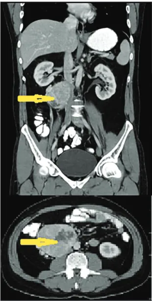
the parameters of the spermiogram test were normal. Contrast-enhanced Ct detected an irregularly shaped mass measuring 92×87 mm2 in the precaval and aor-tocaval areas at an infrarenal level contiguous with the duodenum (Figure 2). Although the mass was
com-pressing the inferior vena cava (iVC), no invasion was detected.
with an initial diagnosis of retroperitoneal mass, laparotomy was performed. during the removal of the retroperitoneal mass contiguous with the iVC and duo-denum, no symptoms of catecholamine discharge, such as hypertension or abnormalities in electrocardiogra-phy measurements, were observed. The pathological report described the detection of a capsulated,
well-Figure 2. Ct image of a 92×87 mm2, irregularly shaped,
contrast-enhanced mass detected in the precaval and aortocaval areas at an infrarenal level contiguous with the duodenum.
Figure 3. Positive staining with chromogranin in immunohistochemical examination.
grained tumor with cells containing pleomorphic–hy-perchromatic nuclei and wide cytoplasm and displaying intercellular mitosis. Histochemical staining revealed the sustentacular cells to be S100 protein positive and the tumor cells to be synaptophysin positive and chromogranin positive. Based on these histochemical findings, paraganglioma was diagnosed (Figure 3).
Postoperative urinary metanephrine and normeta-nephrine levels were normal, and the patient was dis-charged without any complications on postoperative day 4. Metaiodobenzylguanidine (MIBG) scintigraphy performed on the second postoperative week showed no metastasis or local enhancement.
DIsCUssIon
Defined as the dilatation of pampiniform plexus veins, varicocele affects 10% to 15% of the male population and is an important cause of male infertility.3,4 the most common type of varicocele is isolated left-sided disease due to the entrance angle of the left testicular vein into the left renal vein. Bilateral varicocele is observed in 10% of cases, while right-sided varicocele is rarely alone.1 As the right gonadal vein enters directly into the iVC, the presence of retroperitoneal masses compressing the IVC should be considered in cases of isolated right-sided varicocele. Although the most common causes of these retroperitoneal masses are renal masses and sar-comas, adrenocortical carcinoma,5,6 Burkitt lymphoma,7 wilms tumor,8 and IVC malformation caused by situs in-versus9 appear in the published reports. Review of the MEDLINE database indicates that the case reported here is the first case of retroperitoneal paraganglioma presenting as isolated right-sided varicocele, as de-scribed in the published reports.
case report
retroperitoneal paragangliomaAnn SAudi Med 2016 MARCH-APRiL www.AnnSAudiMed.net
150
tumors originating from extra-adrenal chromaf-fin cells are dechromaf-fined as paragangliomas and those from adrenal chromaffin cells as pheochromocytomas. Paraganglioma has an incidence of 2 to 8 per million cases per year10 and is primarily diagnosed between the third and fifth decades of life.11 Most (approximate-ly two thirds) of paragangliomas are seen in the head and neck. Approximately 75% of paragangliomas oc-cupying the sympathetic nervous chain originate from the abdomen.12 Paragangliomas might occur sporadi-cally or as symptoms of several hereditary syndromes. Approximately 25% of cases of pheochromocytoma and paraganglioma are associated with classical heredi-tary syndromes.10 Multiple endocrine neoplasia type 2 (Ret mutations), von Hippel Lindau disease (VHL muta-tions), hereditary paraganglioma/pheochromocytoma syndrome (succinate dehydrogenase [SdHx] tions), and, rarely, neurofibromatosis type 1 (NF1 muta-tions) are some of these syndromes.13 Retroperitoneal paragangliomas should be diagnosed via detection of symptoms caused by hormonal activity, observation of the compression effect of the mass, or incidentally dur-ing imagdur-ing studies.
Patients with hormonally active tumors might pres-ent with headache, sweating, and hypertension associ-ated with severe catecholamine release, symptoms that also present in pheochromocytoma. to date, no con-sensus regarding the histological criteria for determin-ing malignancy in paraganglioma has been reached. Nevertheless, the most applicable criterion is believed to be metastasis. The most preferred local imaging methods for detecting and examining paraganglioma
are currently Ct and MRi. MiBG scintigraphy is the pre-ferred imaging method due to its high specificity and sensitivity for examining residual tissues and detecting metastasis. Because of its cost-effectiveness, especial-ly in examining paragangliomas, which are related to SDHx mutations, fluorodeoxyglucose–positron emission tomography (FdG-Pet) is another option for imaging.14 in some series, the 18F-dOPA Pet scan was reported superior to MiBG scintigraphy.15 urinary metanephrine and normetanephrine levels are more significant in de-termining hormonal activity than plasma catecholamin levels because of their high specificities.
the gold standard treatment for paraganglioma is the aggressive surgical removal of the primer mass and metastatic masses. in hormonally active tumors, the administration of preoperative alpha-blockers is manda-tory for preventing intraoperative arrhythmia and hyper-tension. the use of this treatment option, together with embolization,17 radiotherapy,18 and chemotheraphy19 as adjuvant treatment options, has resulted in an intraoper-ative mortality rate for paraganglioma of less than 1%.16 in conclusion, isolated right-sided varicocele is a rare pathologic condition that may be accompanied by the compression of the IVC by retroperitoneal masses. Although this pathologic condition is mostly caused by the growth of renal masses and sarcomas, other rare causes, such as paragangliomas, should be considered. While the patient described here was diagnosed with hormonally inactive paraganglioma, preoperative hor-monal studies in cases of retroperitoneal masses should be considered due to the risk of intraoperative cardiac complications.
case report
retroperitoneal paragangliomaann Saudi med 2016 marCH-april www.annSaudimed.net
151
1. Skoog SJ, Roberts KP, Goldstein M, Pryor
JL. the adolescent varicocele: what’s new with an old problem in young patients? Pe-diatrics 1997;100:112–22.
2. Rajiah P, Sinha R, Cuevas C, Dubinsky
tJ, Bush wH, Jr, Kolokythas O. imaging of uncommon retroperitoneal masses. Radio-graphics. 2011;31:949-976.
3. Niedzielski J, Paduch D, Raczynski P.
As-sessment of adolescent varicocele. Pediatr Surg int 1997;12:410-3.
4. Meacham RB, townsend RR,
Rademach-er d, et al. the incidence of varicoceles in the general population when evaluated by physical examination, gray scale sonogra-phy and color doppler sonograsonogra-phy. J urol 1994;151:1535-8.
5. Brand tC, Morgan tO, Chatham JR, et
al. Adrenal cortical carcinoma presenting as right varicocele. J urol 2001;165:503.
6. Cheungpasitporn w, Horne JM, Howarth
CB. Adrenocortical carcinoma presenting as varicocele and renal vein thrombosis: a case report. J Med Case Rep 2011;5:337.
7. Roy CR, wilson t, Raife M, et al.
Varico-cele as the presenting sign of an abdominal
mass. J urol 1989;141:597-9.
8. Monroe K1, Navoy J, Odrezin GT,
Nich-ols MH, Polhill R. Varicocele as a present-ing feature of wilms’ tumor. Pediatr emerg Care. 1995 Oct;11(5):300-1.
9. Preziosi P, Miano R, Bitelli M, et al. Right
varicocele associated with inferior vena cava malformation in situs inversus: percu-taneous treatment with retrograde sclero-therapy. J endourol 2001;15:1001-3.
10. Stenstrom G, Svardsudd K.
Pheochro-mocytoma in Sweden 1958-1981. An anal-ysis of the national Cancer Registry data. Acta Med Scand 1986;220:225-32.
11. neumann HP, Bausch B, Mcwhinney
SR, et al. Germ-line mutations in nonsyn-dromic pheochromocytoma. n engl J Med 2002;346:1459-66.
12. Lee JA, duh, QY. Sporadic
paraganglio-ma. world J Surg 2008;32:683-7.
13. neumann HP. Harrison’s principles of
internal medicine. 17th ed. new York and London: McGraw-Hill Medical; 2008.
14. Van Berkel A, Rao Ju, Kusters B, et al.
Correlation between in vivo 18F-FDG PET and immunohistochemical markers of
glu-cose uptake and metabolism in pheochro-mocytoma and paraganglioma. J nucl Med 2014;55:1253-9.
15. Hoegerle S, Nitzsche E, Altehoefer C,
et al. Phaeochromocytomas: detection with 18F DOPA whole body PET––initial results. Radiology 2002;222:507-12.
16. niemann u, Hiller w, Behrend M. 25
years’ experience of the surgical treat-ment of phaeochromocytoma. eur J Surg 2002;168:716-9.
17. Carlsen CS, Godballe C, Krogdahl AS,
et al. Malignant vagal paraganglioma: re-port of a case treated with embolization and surgery. Auris nasus Larynx 2003;30:443-6.
18. Vogel J, Atanacio AS, Prodanov t, et al.
External beam radiation therapy in treat-ment of malignant pheochromocytoma and paraganglioma. Front Oncol 2014;4:166.
19. Niemeijer ND, Alblas G, van
Hul-steijn Lt, et al. Chemotherapy with cyclo-phosphamide, vincristine and dacarba-zine for malignant paraganglioma and pheochromocytoma: systematic review and meta-analysis. Clin endocrinol (Oxf) 2014;81:642-51.