MICRORNAS IN THE PERICARDIAL FLUID OF CORONARY
ARTERY PATIENTS
Reşat DİKME (Corresponding Author)
(Harran University Vocational School of Health Services, Perfusion Techniques Program, Harran University, 6300 Şanlıurfa-Turkey, 905354587844, Fax: 04143183209, e mail:
Name of other Authors Ataman Gönel
(Harran University Faculty of Medicine, Medical Biochemistry Department, Şanlıurfa, Turkey, 905075669870, [email protected])
İsmail Koyuncu
(Harran University Faculty of Medicine, Medical BiochemistryDepartment, Şanlıurfa, Turkey, 905317939878, [email protected])
Feridun Akkafa
(Harran University Faculty of Medicine, Medical BiologyDepartment, Şanlıurfa, Turkey, 905427125473, [email protected])
Mustafa Göz
(Harran University Faculty of Medicine, Cardiovascular SurgeryDepartment, Şanlıurfa, Turkey, 905322348245, [email protected])
Mehmet Salih Aydın
(Harran University Faculty of Medicine, Cardiovascular SurgeryDepartment, Şanlıurfa, Turkey, 905054850158, [email protected])
Ebru Temiz
(Harran University Faculty of Medicine, Medical BiochemistryDepartment, Şanlıurfa, Turkey, 905438904954, [email protected])
ABSTRACT
Background: microRNAs (miRNAs) that play important roles in organisms are associated
with usual function of cells, development of living, biological processes as well as many diseases involving cardiovascular disease. miRNAs are effective in endothelium disfunction, atherosclerosis, inflammation, apoptosis and angiogenesis related with coronary artery disease (CAD). miRNAs that are explained differently in cells, exist extracellularly, in circulation and body fluids. Many biomarkers related with CAD can get through circulation and pericard fluid (PF), the closest place to heart. That is why in etiopathogenesis evaluation of CAD miRNAs can be used diagnostically in PF and circulation. Since PF has a potential of
Purpose: The main purpose of this study is; to determine the amount of miRNA’s in the PF
and blood plasma of patients with coronary artery disease (CAD) and to contribute to the understanding of the pathophysiology of CAD.
Methods: 40 patients having coronary artery bypass surgery were involved to our study and
expression levels of miRNAs (miR-125b-5p, miR-320b, miR-34a-5p, miR-16-5p, miR-320a and miR-15a-5p) that their association with cardiovascular diseases was obtained, were studied in PF and blood plasma with qRT-PCR (Quantitative Real Time PCR) method.
Results: In gene expression of miRNAs as gradual change in plasma than PF was found for
miR16-5p 7.3539 and for miR320b 4.0715, as a result of this miR16-5p p=0.00001 value and miR320b p=0.00814 value were found statistically significant (p<0.05).
Conclusion: The reason of gene expression level of miR 16-5p and miR 320b were found
higher in plasma which is expected to be found high in PF may be extramyocardial sources. Obtaining of miR 16-5p and miR 320b in PF that have negative effect in terms of cardiovascular diseases strengthens the potential usage of these microRNAs as biomarker in cardiovascular disease. Although in previous studies it was defined as miRNA special to PF, obtaining of miR320b also in plasma shows that it is not only special to PF.
Keywords: Coronary artery disease, CAD, Pericardial fluid, PF, microRNA, miRNA.
INTRODUCTION
Despite of developments in treatment of cardiovascular diseases (CVD), these diseases are still at the first rank as cause of death (1). With studies done up to today it is estimated that mortality depending on CVD will rise to 36.3% from 28.9% until 2020 (2). World Health Organization predicts that in next couple of twenty years deaths depending on CAD will increase 120% in women and 137% in men. In the USA since CAD forms ¼ of treatment costs of all patients with 71.2 billion dollars, in order to treat and prevent disease it is necessary to understand molecular pathogenesis, find new biomarkers as well as define and use substances preventing disease (3). In defining of new biomarkers in CVD besides blood pericardial fluid (PF) can also be used. Pericardial fluid (PF) in two-walled pericardial sac surrounding the heart completely, shows dynamic changes. As pericard has immunologic, vasomotor and fibrinolytic activities, it affects myocyte structure, function and gene expression by synthesizing substances such as eicosanoids and prostaglandins (4).
LITERATURE REVIEW
Since pericard is the closest region to heart tissue PF analysis provides to understand many pathophysiological mechanisms in various pericardial and cardiovascular diseases. For example in a study done previously, finding atrial natriuretic factor and brain natriuretic factor in cardiac insufficiency approximately 12 times more in PF makes think that these factors play pathophysiological role as an autocrine or paracrine factor in cardiac insufficiency (5, 6). In unstable angina high levels of adhesion molecules such as ICAM-I, VCAM-I, e-selectine and proinflammatory cytokines such as interleukin 6 and 8 in PF supports the relation between coronary artery diseases and inflammation. miRNAs as a key molecule that play important roles in organisms are associated with usual function of cells, development of living, biological processes as well as many diseases involving cardiovascular disease (7). miRNAs that are defined among tissues in different ways exist in cell, out of cell, circulatory and in body fluids and each tissue and fluid can have specific miRNAs (8). They are carried by microvesicles, Argonaut 2, HDL lipoprotein and nucleophosmin and protect themselves against ribonuclease activity so by being carried to far regions, they affect gene expression in that region. Accordingly carried miRNAs play intercellular role (9). These miRNAs make progress being a biomarker in molecular diagnosis and treatment (7). In developments specific to tissue, they take role and make regulation in target genes. In cardiovascular diseases as they are at different level, they can give more precise datas compared to important cardiac biomarkers such as cTnI (9). In a study done by Feng Wang et. al. in CAD as diagnosis truth value for miRNA 133a was found as 0,918, it was found for cTnI as 0,741(9). However these miRNA are not only defined in heart but also skeleton muscle, this situation affects specificity of miRNAs negatively. In cardiovascular system as critical role of miRNAs is determined by inactivation of Dicer enzyme that processes miRNA, this situation causes angiogenesis, vascularization and defects in cardiac development (10). Approximately more than 60% of mammal transcript is under the control of miRNAs (11, 12). For diagnosis of diseases with miRNAs, biological fluids and tissues such as serum, cerebrospinal fluid, tear, milk, bile juice, peritoneum fluid, ovarian follicle fluid, urine, whole blood, nasal secretion, saliva, pleural effusion and feces, are used (11, 13-15). In a study done by Suvi M. Kuosmanen et. al. 742 miRNA were scanned in PF and in each sample approximately 256
found as miR 21-5p, miR 451a, miR 125b-5p, miR 7b-5p and miR 16-5p were identified most in all patients, miR-26b-5p, miR 29b, miR 125, miR 320, miR 34, miR 497 were found common in all fluids. Although these results were obtained, the meaning of these obtained miRNAs could not be obtained in terms of cardiac pathology. It was just predicted that these obtained miRNAs play role in relation between cardiac cells (7, 16). Towards treatment of these diseases new methods were tried to be developed by synthesizing miRNA mimics and antagomir. In many studies it was proved that artificial mimic miRNA and miRNA antagomir show effect just like main miRNAs and connect to target mRNA, inhibit or break up mRNA (17, 18).
The main aim of this study is to obtain gene expression of miRNAs (miR-125b-5p, miR-320b, miR-34a-5p, miR-16-5p, miR-320a and miR-15a-5p) that have previously relation with cardiovascular diseases in PF and plasma as well as identify their relation with coronary artery disease and with this contribute understanding of pathophysiology of disease.
Reasons of selecting miRNAs used in the study
In previous studies miR-125b-5p, miR-320b, miR-34a-5p that are found specific to PF, were associated with heart failure (HF) and other cardiovascular conditions before (7). miR-125b, miR-320b and miR34a-5p that were obtained to be related with acute myocard infarction (AMI) previously, affect formation of disease, pathogenesis and mortality risk (19-22). However in another study miR-125b, miR-320b were associated with atherosclerosis and CAD development (19, 23, 24). miR-34a-5p that was obtained in high quantities in PF samples, was defined as a factor contributing decrease regarding aging in cardiac function (25, 26). In the same research in addition to this miR-320a that was obtained in high quantity in all pericard fluid samples was defined as biomarker in cardiac failure (27, 28).
Again in another study in 96% of patients that had been taken to open heart surgery due to cardiac failure, miR-320b that was obtained as microRNA specific to PF, became the second most found microRNA with 3.5 Average Cp value (7). In the same study in 100% of patients miR-34a-5p was obtained as microRNA specific to PF and became the third most found microRNA with 2.3 Average Cp value, in addition miR-34a-5p took the 11th rank among 50 most found microRNAs. In another study about miR-34a-5p according to miR-34a-5p mirtarbase data base it was found to be associated with CAD (29). miR-125b-5p that was obtained as microRNA specific to PF, became the first microRNA at 98% with 5.1 Average
Cp value as well as it was among the most found five microRNA and was obtained to be associated with cardiovascular diseases (7).
In a study done by Suvi M. Kuosmanen et. al. miR-16-5p was obtained in all PF samples. It was among the most found five microRNA, became the fifth with 3.5 Average Cp among 50 miRNA and was found to be associated with cardiovascular diseases. In the same study miR-320a was obtained in PF of all patients. This miRNA became the second with 3.8 Average Cp value among 50 miRNA and was defined as biomarker of cardiac failure (7). In a study done about miR-15a-5p, it was obtained in pericardial fluid of all patients taken to open heart surgery due to cardiac failure and this miRNA became the seventh with 3.3 Average Cp value among 50 miRNA (7).
METHODOLOGY
40 patients that had coronary artery bypass surgery with cardiopulmonary bypass (CPB) method in surgery room of Harran University Medical Faculty Hospital Cardiovascular Surgery Department, were involved to the study. Before study an approval from Harran University Ethical Committee with date 05.01.2017 and number 17/01/23 was taken. PF and plasma samples were obtained from patients. Unsuitable 13 hemolyze sample was taken out from study and the rest 27 patient sample were taken to the scope of study.
Obtainment of Pericardial Fluid
During routine process done towards health problem of patient that his surgery was planned, PF taken routinely was used. In patients taken to coronary bypass surgery by CPB method after doing standard CPB procedures with median sternotomy, pericardium was opened and PF was aspirated by sterile injector. Then aspirated PF (2-25 ml) was taken to sterile non-anticoagulant tubes and the tubes were immediately put into an ice-filled container. PF in sterile tube was purified from cells by centrifuging at 5000 rpm for 5 minutes at 4°C. After centrifuge stage, supernatant part was centrifuged two times at 10000 rpm for 10 minutes at 4°C to seperate it from cell debrises and other microparticles. Then supernatant part was taken to RNase-free tube (ependorf tube) and kept at -80 °C to be studied. After samples were collected from sufficient patients, qRT-PCR study was done.
Obtainment of Blood Plasma
In patients taken to coronary bypass surgery by CPB method, blood was taken before cardiopulmonary bypass and put into sterile anticoagulant (heparinized) tubes. The tube was immediately put into an ice-filled container and sent to laboratory. Later sterile tube was centrifuged at 5000 rpm for 5 minutes. After centrifuge stage supernatant part was taken to RNase-free tube (ependorf tube) and kept at -80 °C to be studied. After collecting samples from sufficient patients qRT-PCR study was done.
Studying Stages of miRNA Expression in PF and Plasma
In all samples of patients taken to study, expression level of miRNAs was obtained.
microRNA isolation in PF and plasma
For microRNA isolation in PF and plasma, the processes were done according to working procedure of producing company. The isolation kit named Qiagen (Qiagen GmbH, Hilden, Germany) miRNeasy Serum/Plasma Kit (Cat No./ID: 217184) was used.
cDNA obtainment from miRNA
For cDNA obtainment from miRNA by using Qiagen miScript Reverse Transcription (RT) Kit II (Hilden, Germany, Cat No./ID: 218161), the study was done according to written procedure.
Quality and quantity identification from cDNAs
In order to determine quality and quantity of cDNA samples obtained before expression analysis experiments and put all samples to isodense reaction, Thermo Scientific Multiskan Spectrophotometer System (Waltham, Massachusetts, USA) equipment was used. Before expression analysis experiments, cDNA quantities obtained from samples were declined to 50 ng/μl.
qRT-PCR (quantitative Real Time PCR) study
Before qRT-PCR miRNA- cDNA preamplification (preAmp) was done in PF and plasma and then routine PCR protocole was applied. Reaction compounds for qRT-PCR were prepared by using Qiagen miScript SYBR Green PCR (Cat No./ID: 218073) kit. For concurrent PCR reaction Rotor Gene 6000 Real-Time PCR Machine (Qiagen GmbH, Hilden, Germany) equipment was used.
Primary selection of miRNA
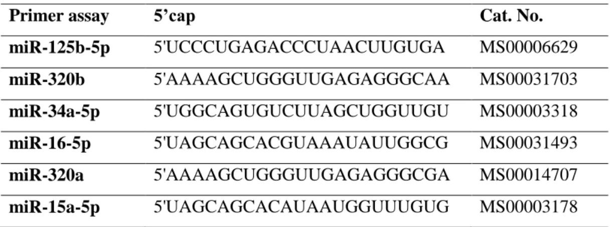
In the study 6 pieces of hsa miScript Primer Assays (miR-125b-5p, miR-320b, miR-34a-5p, miR-16-5p, miR-320a and miR-15a-5p) were used. hsa miScript Primer Assays was comercially taken from Qiagen (Qiagen GmbH, Hilden, Germany, product code: 218300) company. The ends of used hsa miScript Primer Assays 5’ and their commercial catalogue numbers are shown in Table 1.
Table 1. hsa miScript Primer Assays 5’ cap and their commercial catalogue numbers
Primer assay 5’cap Cat. No.
miR-125b-5p 5'UCCCUGAGACCCUAACUUGUGA MS00006629 miR-320b 5'AAAAGCUGGGUUGAGAGGGCAA MS00031703 miR-34a-5p 5'UGGCAGUGUCUUAGCUGGUUGU MS00003318 miR-16-5p 5'UAGCAGCACGUAAAUAUUGGCG MS00031493 miR-320a 5'AAAAGCUGGGUUGAGAGGGCGA MS00014707 miR-15a-5p 5'UAGCAGCACAUAAUGGUUUGUG MS00003178 Statistical Analysis
For evaluation and statistical analysis of obtained datas the programme in Qıagen web page (https://www.qiagen.com/us/shop/genes-and-pathways/data-analysis-center-overview-page/) was used. ‘’The Gene Globe Data Analysis Center’’ part in related web page is a web source for researchers analysing qRT PCR or NGS datas. qRT PCR modules in related web page transform threshold cycle-CT values to calcuated results for gene and miRNA expression. As values were entered to system in related web page, analysis was done by marking preamplification option. So more clear results were tried to be taken than other statistical programmes.
RESULTS
Demographic properties of 27 participant patients (9 female +18 male) were summarized in Table 2.
Table 2. Demographic properties of participant patients
Variable Patients (n = 27)
Gender (Female / Male)% 9/18 (% 33.3 / % 66.7)
Age (years) 61 ± 9,78
Length (cm) 166,96 ± 8,19
Weight (kg) 77,70 ± 20,10
BSA (Body surface area) (m2) 1,83 ± 0,15
miRNA Gene Expression Results in PF and Plasma
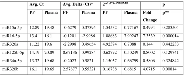
In normalization of miRNA expression, diligent values were not required in Qiagen web page, instead global values were used. In Web page PF was chosen as control group whereas plasma was chosen as studying group. As values were entered to system in related web page, analysis was done by marking preamplification option. All statistical results regarding Average (AVG) Ct, AVG Delta Ct, 2^(-Avg.(Delta(Ct)), Fold Change and p values obtained after entering miR15a-5p, miR16-5p, miR320a, miR125b-5p, miR34a-5p and miR320b gene expression datas to the system in related web site, were shown in Table 3. Fold change and p values are values obtained according to the comparison of plasma with PF.
Table 3. Statistical results of gene expression for 6 miRNAs in plasma and PF
Avg. Ct Avg. Delta (Ct)* 2^(-Avg.(Delta(Ct)) p
PF Plasma PF Plasma PF Plasma Fold
Change p** miR15a-5p 12.89 19.48 -0.6279 0.37395 1.54532 0.77167 0.4994 0.283504 miR16-5p 13.4 16.1 -0.1201 -2.9986 1.08683 7.99247 7.3539 0.000014 miR320a 11.22 19.6 -2.2998 0.49654 4.92374 0.7088 0.144 0.442233 miR125b-5p 14.19 20.09 0.67136 0.99284 0.62792 0.50249 0.8002 0.129741 miR34a-5p 13.32 19.68 -0.2023 0.5821 1.15057 0.66799 0.5806 0.324842 miR320b 16.1 19.65 2.57877 0.55321 0.16738 0.6815 4.0715 0.00814
*AVG Delta (Ct)=(Ct (GOI) - Avg Ct (HKG)), GOI: Gene of Interest, HKG: Housekeeping Genes
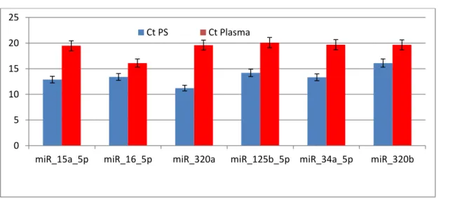
Average Ct values of miRNAs whose expression level was identified in PF and plasma, were given in Figure 1.
Figure 1. Average Ct values of miRNAs whose expression level was identified in PF and plasma
2^ (-Avg.(Delta(Ct)) values of miRNAs whose expression level was identified in PF and plasma,
were given in Figure 2.
Figure 2. 2^ (-Avg.(Delta(Ct)) values of miRNAs whose expression level was identified in PF and plasma
As a result of comparison of plasma with PF obtained fold change for miRNAs in plasma were calculated for miR16-5p as 7.3539, for miR320a as 0.144, for miR125b-5p as 0.8002, for miR34a-5p as 0.5806 and for miR320b as4.0715 (Figure 3).
0 5 10 15 20 25
miR_15a_5p miR_16_5p miR_320a miR_125b_5p miR_34a_5p miR_320b
Ct PS Ct Plasma 0 1 2 3 4 5 6 7 8 9
miR_15a_5p miR_16_5p miR_320a miR_125b_5p miR_34a_5p miR_320b 2^(-Avg.(Delta(Ct)) PF
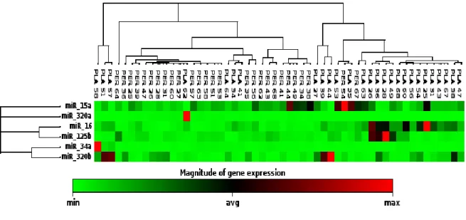
Figure 3. As a result of comparison of plasma with PF obtained fold change for miRNAs in plasma. As a result of calculation of replicate 2^(- Delta Ct) values for each miRNA gene expression in PF and plasma by using Stutent's t-test; In plasma compared to PF it was found for miR15a-5p p=0.283504, for miR16-5p p=0.000014, for miR320a p=0.442233, for miR125b-5p p=0.129741, for miR34a-5p p=0.324842 and for miR320b p=0.00814. As a result of this, miR16-5p and miR320b p<0.005 value was found statistically significant. Heatmap graphic that is formed by marking of gene expression of miRNAs obtained in PF and plasma one on one over a two-dimensional graphic with different colours, was shown in Figure 4.
Figure 4. Heatmap graphic for miRNAs in PF and Plasma (Green colored samples are expressed at minimum
level, red colored samples were expressed at maximum level) 0 1 2 3 4 5 6 7 8 9
DISCUSSION
In CAD pathological findings can show up in time and there occurs some changes in cells, out of cells, intercellular gap or in various factors such as substances, molecules, proteins etc. in body fluids before diseases shows up itself. Among these changes miRNAs take attention recently and these can be used as biomarker. As its role and predictive of its level in CAD have been recently discovered, its role in development and progress in CAD is not well known. However the role of this factor related with metabolism in myocard tissue is open to be discovered. Since the studies done in CAD about miRNA were not standardized completely, different results can come out. That is why the number of studies should be increased and a standard protocole should be prepared. Although miRNAs are in the way of being a good biomarker, there are question marks due to some deficiencies. For example defining some miRNAs related with CAD both from heart muscle and skeleton muscle causes complication. In order to annihilate this problem, it is important to make studies in region specific to tissue, on a cell basis and in tissue fluids. For example the changes in many substances, molecules, protein, hormone etc. in PF that is a specific tissue fluid of heart, give clear datas about the situation in heart tissue.
The studies done recently have made pericard an application area of new treatment methods. Making treatments that provide application of miRNA derivatives (mimic or antagomir) to pericardial gap just as giving intrapericardial basic fibroblast growth factor to stimulate collateral vein development to patients having no chance of percutan and surgical revascularization, can save patients from fatal situations. Accordingly invitro studies that will be done towards this aim, will provide development of new treatment methods applicable to clinic.
In CVD for diagnosis and course of disease without being dependent to only a kind of material existence, this study contributes development and maintenance of diagnostic and therapeutical processes of disease in systematic and healthy way. In this respect apart from usage of blood traditionally, it is important to use PF also. In cardiovascular diseases handling and conducting disease according to diagnosis and treatment processes by using two different
In our study as a result of comparison of plasma with PF, two of six miRNAs of plasma that are miR16-5p (p=0.000014) and miR320b (p=0.00814) were found statistically significant. In a previous study one of six microRNAs specific to PF that is miR-320b was associated with cardiovascular conditions such as HF and AMI (7), and was found to affect pathogenesis and mortality of cardiovascular disease (19-22). In many studies done recently miR-320b that is associated with development of atherosclerosis and coronary artery disease (19, 23, 24), was obtained in PF of patients taken to open heart surgery as 96% and became the second most found microRNA with 3.5 Average Cp- Assay Cp quantity value (7). In a study of Wang et. al. it was obtained that miR-320 shows an anti-angiogenetic effect to endethelium cells via exosomal transfer in type 2 diabetic rats (30). In studies done with animal models miR 320 was obtained as a factor that causes myocardial ischemia as well as stimulates cardiomyocyte apoptosis (31).
In our study as miR 320b was obtained in all patient samples, it became the most expressed miRNA in PF with 16.1 value in terms of average Ct. In plasma the most third expressed miRNa in terms of average CT with 19.65 value is miR 320 b showed change 4 times more in plasma that PF and this situation was found statistically significant. In previous studies finding miR 320b that has negative effect in terms of cardiovascular diseases, at high levels strengthens this situation more and intensifies its potential to be used as biomarker. As a result of our study, it was assumed that high expression levels of miR-320 can make negative effect on feeding of myocard by preventing development and bifurcation of coronary arteries with its anti-angiogenetic effect. Again due to myocardial ischemia and apoptosis caused by miR 320, there might occur a negative pressure on heart. That is why, high expression of miR 320 in patients that had coronary artery bypass, supports datas in the past.
In a study done by E.L. Vegter et. al. miR-16-5p was found in positive relation with CRP that is a negative biomarker in terms of cardiac indicator (32). In our study miR16-5p that was obtained in all samples in PF and plasma became the third most expressed miRNA in PF in terms of average Ct with 13.4 value whereas in plasma it became the least expressed miRNA with 16.1 value. As a result of comparison of PF with plasma, miR16-5p showed change 7.3 times more in PF than plasma and this situation was found statistically significant. In previous studies finding miR 16-5p that has negative effect in terms of cardiovascular diseases, at high levels points out its potential to be used as biomarker.
CONCLUSIONS
Finally as a result of evaluation of obtained datas and statistics in gene expression of miRNAs miR16-5p and miR320b was defined significantly more in plasma than PF. Accordingly, a result was found opposite to the hypothesis of “these parameters should be higher in PF that is close to heart tissue” that was presented pre-study. The main reason of finding gene expression of miR320b and 16-5p higher in PF than plasma may be the production of these parameters also by extramyocardial sources and are sent to plasma. Also obtaining miRNA 320b in plasma that was defined as a miRNA only specific to PF in previous studies, showed that this microRNA is not only specific to PF.
ACKNOWLEDGEMENTS
This work was supported by the Research Fund of Harran University (HUBAK Projects No. 17010) Sanliurfa, Turkey.
REFERENCES
1. Onat A, Keleş I, Çetinkaya A. On yıllık Tekharf çalışması verilerine göre Türk erişkinlerinde koroner kökenli ölüm ve olayların plevalansı. Türk Kardiyol Dern Arş- Arch Turk Soc Cardiology. 2001;29:8-19.
2. Zipes Douglas P, Libby P, Robert Bonow O, Eugene B (Ed). Braunwald’s Heart Disease, Textbook of cardiovascular medicine. 7th edition. Editör: Zipes Douglas P. 2012; p1243-1281.
3. Özkan Arat A. Akut koroner sendromlar: Epidemiyoloji, Türk Kardiyol Dern Arş- Arch Turk Soc Cardiology. 2013;41 Suppl 1:1-3.
4. Spodick DH, Microphysiology of the pericardium: substrate forintra pericardial therapeutics Herz. 2000;25:720-723.
5. Troughton RW, Asher CR, Klein AL. Pericarditis. Lancet 2004; 363:717-27.
6. Fujita M, Komeda M, Hasegawa K, Kihara Y, Nohara R, Sasayama S. Pericardial fluid as a new material for clinical heart research. Int J Cardiol 2001; 77:113-8.
7. M. Kuosmanen S, Hartikainen J, Hippeläinen M, Kokki H, Liisa Levonen A, Tavi P. MicroRNA Profiling of Pericardial Fluid Samples from Patients with Heart Failure.
8. Guo-Hua D, Pei-Ze M, Xian-Bo S, Ning L, Tong Z, Bo W. MicroRNA-223-3p Inhibits the Angiogenesis of Ischemic Cardiac Microvascular Endothelial Cells via Affecting RPS6KB1/hif-1a Signal Pathway. Plos One. October 2014. Volume 9, Issue 10, e108468.
9. Wang F, Chen C, Wang D. Circulating microRNAs in cardiovascular diseases: from biomarkers to therapeutic targets. Front. Med., 2014. 8(4): 404–418.
10. Kalozoumi G, Yacoub M, Sanoudou D. MicroRNAs in heart failure: Small molecules with major impact. Global Cardiology Science and Practice.2014;30.
11. Bissels U, Bosio A, Wagner W. MicroRNAs are shaping the hematopoietic landscape. Haematologica. 2012;97:160-7.
12. Großhans H. Regulation of microRNAs. Springer Science Business Media, Eds. New York. 2010.
13. Natarajan SK, Smith MA, Wehrkamp CJ, Mohr AM, Mott JL. MicroRNA function in human diseases. Med Epigenet. 2013;1:106-15.
14. Xiao YF, Yong X, Fan YH, Lü MH, Yang SM, Hu CJ. MicroRNA detection in feces, sputum, pleural effusion and urine: novel tools for cancer screening. Oncol Rep. 2013;30:535-44.
15. Park NJ, Zhou H, Elashoff D, Henson BS, Kastratovic DA, Abemayor E. Salivary microRNA: discovery, characterization, and clinical utility for oral cancer detection. Clin Cancer Res. 2009;15:5473-77.
16. Demyanets S, Kaun C, Pentz R, Krychtiuk KA, Rauscher S, Pfaffenberger S. Components of the interleukin-33/ST2 system are differentially expressed and regulated in human cardiac cells and in cells of the cardiac vasculature. J Mol Cell Cardiol. 2012;60:16–26.
17. Van Rooij E, Purcell AL, Levin AA. Developing microRNA therapeutics. Circ Res. 2012;110:496-507.
18. Broderick JA, Zamore PD. MicroRNA therapeutics. Gene Therapy. 2011; 18:1104-10. 19. Huang S, Chen M, Li L, He M, Hu D, Zhang X. Circulating MicroRNAs and the Occurrence of Acute Myocardial Infarction in Chinese Populations. Circ Cardiovasc Genet. 2014; 7: 189–98.
20. Gidlöf O, van der Brug M, Ohman J, Gilje P, Olde B, Wahlestedt C. Platelets activated during myocardial infarction release functional miRNA, which can be taken
up by endothelial cells and regulate ICAM1 expression. Blood. 2013; 121: 3908–17, S1–26.
21. Lv P, Zhou M, He J, Meng W, Ma X, Dong S. Circulating miR-208b and miR-34a are associated with left ventricular remodeling after acute myocardial infarction. Int J Mol Sci. 2014; 15: 5774–88.
22. Li Z, Lu J, Luo Y, Li S, Chen M. High association between human circulating microRNA-497 and acute myocardial infarction. ScientificWorldJournal. 2014; 2014: 931845.
23. Shan Z, Yao C, Li Z, Teng Y, Li W, Wang J, . Differentially expressed microRNAs at different stages of atherosclerosis in ApoE-deficient mice. Chin Med J (Engl). 2013; 126: 515–20.
24. Bidzhekov K, Gan L, Denecke B, Rostalsky A, Hristov M, Koeppel TA. microRNA expression signatures and parallels between monocyte subsets and atherosclerotic plaque in humans. Thromb Haemost. 2012; 107: 619–25.
25. Emanueli C, Thum T. miRNAGE-34 induces cardiac damAGE. Cell Res. 2013; 23: 866–7.
26. Boon RA, Iekushi K, Lechner S, Seeger T, Fischer A, Heydt S, . MicroRNA-34a regulates cardiac ageing and function. Nature. 2013; 495: 107–10.
27. Melman YF, Shah R, Das S. MicroRNAs in heart failure: is the picture becoming less miRky. Circ Heart Fail. 2014; 7: 203–14.
28. Goren Y, Kushnir M, Zafrir B, Tabak S, Lewis BS, Amir O. Serum levels of microRNAs in patients with heart failure. Eur J Heart Fail. 2012; 14: 147–54.
29. http://mirtarbase.mbc.nctu.edu.tw/php/search.php?opt=disease_intermediate_mirna&d isease_mirna=coronary+artery+disease&disease=. Date of access: 30 Aralık 2017. 30. Wang X, Huang W, Liu G, Cai W, Millard RW, Wang Y, Chang J, Peng T, Fan GC.
Cardiomyocytes mediate anti-angiogenesis in type 2 diabetic rats through the exosomal transfer of miR-320 into endothelial cells. J Mol Cell Cardiol 2014; 74: 139–150
31. Ren X.P, Wu J, Wang X, Sartor M.A, Qian J, Jones K, Nicolaou P, Pritchard T.J, Fan G.C. MicroRNA-320 is involved in the regulation of cardiac ischemia/reperfusion
32. Vegter EL, Schmitter D, Hagemeijer Y, Ovchinnikova ES, van der Harst P, Teerlink JR, et all. Use of biomarkers to establish potential role and function of circulating microRNAs in acute heart failure. International Journal of Cardiology 224 2016;231– 239.
Search Terms
Search Type Title Word Contact Us
clarivate.com Master Journal List Site Client proxystylesheet Output SearchSearch allAreas
Journal Search
DatabaseMaster Journal List Search
Search Term(s): *PONTE · The following title(s) matched your request 1-1 of 1 journals
PONTE
Monthly
ISSN: 0032-423X
IL PONTE EDITORE, VIA L MANARA, 10/12, FLORENCE, ITALY, 50135 Coverage
Arts & Humanities Citation Index Current Contents - Arts & Humanities
Clarivate Accelerating innovation Cookie Policy Privacy Statement Terms of Use Copyright Careers © 2018 Clarivate Follow us
Search terms: *PONTE Total journals found: 1
1. PONTE
Monthly
ISSN: 0032-423X
IL PONTE EDITORE, VIA L MANARA, 10/12, FLORENCE, ITALY, 50135 1. Arts & Humanities Citation Index
Homepage About us Contact us Current Issue Aims & Scopes Call for Paper
International Multidisciplinary ISI Journal in All Fields of Sciences
The objective of the PONTE Journal is to bridge the gap between the science production and science publication by publishing explicitly written articles intelligible to scientists working in any field of science from engineering to medicine.
Ponte is a peer reviewed, indexed international ISI journal. It publishes Original Research Articles, Review articles, Short
Communications and Case Studies in all the multidisciplinary scientific research fields. So the journal is open to all researchers of diverse fields of discipline. This journal provides platform for the researcher, innovators, scholars and students to share their research through worldwide.
A team of reputed academicians from different disciplines tackle the multidisciplinary review work and publication process of this journal. All the accepted papers are uploaded and published online after final acceptance. The technical editing process is carried by related technical editing department. The articles are reviewed and accepted only after at least two positive reviews of three reviewers.
Journal Metrics (JCR)
ISSN: 0032-423X E-ISSN: 0032-6356 Coverage:
Science Citation Index Arts, Humanities & ... BIOSIS Previews Impact Factor: 0.814 5-Year Impact Factor: 0.850 RECENT ARTICLES
BEAT PATTERNS OF A PHYSICAL INCLINED PENDULUM AND THEIR REAL-WORLD APPLICATION
Zharilkassin Iskakov
Higher education practitioners assessment literacy levels: A logical basis for its positioning in relation to critical theory, critical reflection, and professional development
A. KRISHNANNAIR, S. KRISHNANNAIR, S.N. IMENDA
MYOCARDIAL FUNCTIONS IN OBESE CHILDREN WITH SUBCLINICAL HYPOTHYROIDISM
Atilla Cifci, Mehmet Boyraz
ARCHIVE
To access our journal's archive in PDF format before 2016, click here.
2016 2017 2018 2019 Jan July Feb Aug Mar Sep Apr Oct May Nov June Dec Jan July Feb Aug Mar Sep Apr Oct May Nov June Dec* Jan* July Feb Aug Mar Sep Apr Oct May Nov June Dec Jan July Feb Aug Mar Sep Apr Oct May Nov June Dec
Guide for Authors
This guideline has been prepared for the authors to new submissions and after their manuscripts have been accepted
Authors Login
Authors with accepted articles can login to system by this link
Paper Tracking
You can track your submitted article from this tab
Editorial Board
The international editorial board is headed by Dr. Maria E. Boschi
General Policies
To see the PONTE Publishing and Property law, click on this link
Peer Review Process
Papers will be sent to three peer reviewers for evaluation
Send your CV to:
reviewers [at] pontejournal.net
Contact us:
Info [at] pontejournal.net
Click here to contact the editorial office
Authors Login Paper Tracking System Reviewers Login Full Name
Email Address Message
Homepage About us Contact us Current Issue Aims & Scopes Call for Paper
Editorial Board
Editor-in-Chief:
Dr. Maria E. Boschi, Florence, Italy
Editorial Board:
Giancarlo Landini, Florence, Italy Maria Nuova, Firenze, Italy
Dr. Francesco M. Caradini, Florence, Italy Dr. Roberto, J. Nardi, Rom, Italy Prof. Alberto S. Vicario, Bologna, Italy Prof. Giorgio Belentini, Florence, Italy Prof. Nicola S. Cesari, Bologna, Italy Prof. Paolo Torrentini, Rom, Italy Dr. Sandro F. Fontana, Biella, Italy Prof. Mario R. Visconti, Napoli, Italy Dr. Ospedale San Raffaele, Milan, Italy
International Editorial Board:
Ulrich Caruso, Berlin, Germany
Marco Pilatous, Univ. Florida, Gainsville, USA Werner O. Bauer, Kusnacht, Switzerland Donald Bradley, Oxford, UK
Omar Muhammad Durrah, Oman Sang-Bing Tsai, Guangdong, China Mümtaz Koray Yilmaz, Ankara, Turkey B. Huang, Wisconsin-Madison, USA Bernard-Gallon DJ, Paris, France Wolf Müller, Vienna, Austria Davis P. Jackson, New York, USA Tian Lee Chen, Hong Kong, China Alvarez- A. Barrientos, Badajoz, Spain Karel Wangger, Vienna, Austria Roberto Ghidoni, Milan, Italy Deepak Kashyap, Nainital, India J. HENRY ADAMES, Toronto, Canada VISHNU NARAYAN MISHRA, India Anette Moury Frank, Edinburgh, Germany ZEKİ T. AKTAŞ, Ankara, Turkey Fred Adolf Ruffler, Vienna, Austria Nagendra Rao, Bangalore, India Susan F. Tighe, waterloo, Canada Kerong Zhang, Fuyang, China Mohd S. Bin Shuib, Sintok, Malaysia Jimmy T. Masagca, Las Pinas City, Philippines
Guide for Authors
This guideline has been prepared for the authors to new submissions and after their manuscripts have been accepted
Authors Login
Authors with accepted articles can login to system by this link
Paper Tracking
You can track your submitted article from this tab
Editorial Board
The international editorial board is headed by Dr. Maria E. Boschi
General Policies
To see the PONTE Publishing and Property law, click on this link
Peer Review Process
Papers will be sent to three peer reviewers for evaluation
Homepage About us Contact us Current Issue Aims & Scopes Call for Paper
Archive
doi: 10.21506/j.ponte.2018.8.5
| PDF Version
DEVELOPMENT OF MODEL OF HUMAN CAPITAL MANAGEMENT BASED ON SCENARIO APPROACH
Volume 74, Aug 2018
doi: 10.21506/j.ponte.2018.8.4
| PDF Version
MICRORNAS IN THE PERICARDIAL FLUID OF CORONARY ARTERY PATIENTS
Volume 74, Aug 2018
doi: 10.21506/j.ponte.2018.8.2
| PDF Version
THE ATTITUDE TOWARDS CHILDREN'S ATTITUDES AND THE METHODS OF UPBRINGING APPLIED TO CHILDREN IN THE LATE XIX AND EARLY XX CENTURIES
Volume 74, Aug 2018
doi: 10.21506/j.ponte.2018.8.1
| PDF Version
THE GAME DISCOURSE IN POSTMODERNISM
Volume 74, Aug 2018
doi: 10.21506/j.ponte.2018.8.3
| PDF Version
Guide for Authors
This guideline has been prepared for the authors to new submissions and after their manuscripts have been accepted
Authors Login
Authors with accepted articles can login to system by this link
Paper Tracking
You can track your submitted article from this tab
Editorial Board
The international editorial board is headed by Dr. Maria E. Boschi
General Policies
To see the PONTE Publishing and Property law, click on this link
Peer Review Process
Papers will be sent to three peer reviewers for evaluation