Radiological Investigation and Clinical Evaluation of the
Morphometric Development of the Forearm in Human Fetuses
Investigación Radiológica y Evaluación Clínica del Desarrollo Morfométrico del Antebrazo en Fetos Humanos
Desdicioglu Raziye1; Uguz Ceren2; Desdicioglu Kadir3; Sulak Osman4 & Malas Mehmet Ali5
DESDICIOGLU, R.; UGUZ, C.; DESDICIOGLU, K.; SULAK, O. & MALAS, M. A. Radiological investigation and clinical evaluation of the morphometric development of the forearm in human fetuses. Int. J. Morphol., 35(2):629-636, 2017.
SUMMARY: Fetal period of time during which the fetus grows rapidly and the organs are formed. The prenatal and postnatal analyses of the fetal structure provide information as to fetal growth, growth retardation, gestational age and congenital malformations. The development of the skeletal system during the intrauterine period takes place in an orderly manner as it also does in other systems. It was aimed that the morphometric development of the forearm in human fetuses during the period between 20-40 gestational weeks be radiologically investigated and that its clinical importance be evaluated, as well. A total of 100 fetal forearms (50 fetuses: 23 male, 27 female), the ages of which varied between 20-40 gestational weeks, without having any external pathology or anomaly were incorporated into the study. The fetuses were separated into groups according to weeks, trimesters and months. After the general external measurements of the fetuses had been performed, the mammographies and forearm radiographies of the fetuses were shot in the way that the forearms would remain in a prone position. Morphometric measurements pertaining to forearm structures were taken from the forearm radiographies that were shot with the help of a digital compass. Later on, the morphometric measurements in question were statistically evaluated. The mean values and the standard deviations of the measured parameters were determined according to gestational weeks, trimesters and months. There was a significant correlation between the measured parameters and the gestational age (p<0.001). In the comparison of the measured parameters between trimesters and months, it was observed that there was a statistically significant difference between the groups (p<0.05). Separately, it was also determined that there was no statistically significant difference in the comparison of the parameters, which was made between genders and right-left forearms (p>0.05). As for the results obtained in our study, we are of the opinion that the data obtained during this study period will be beneficial for the involved clinicians, such as those in charge of gynecology, radiology, forensic medicine and perinatology, in terms of evaluating the clinical studies related to the morphometric development of the forearm throughout the fetal period, in determining the fetal age and sex, and also in determining the pathologies and variations regarding the development of fetal skeletal system.
KEY WORDS: Fetus; Forearm; Fetal Development; Morphometry; Radiology; Skeletal System.
INTRODUCTION
Fetal period is the phase which starts from the ninth week of gestation and lasts until the delivery. This is the period of time during which the fetus grows rapidly and the organs are formed (Sadler, 1990; Collins, 1995; Malas et al., 2000). The prenatal and postnatal analyses of the fetal structure provide information as to fetal growth, growth retardation, gestational age and congenital
malformations (Taeusch, 1991; Malas et al., 2000; Uluutku et al., 2010; Malas et al., 2005).
The development of the skeletal system during the intrauterine period takes place in an orderly manner as it also does in other systems (Moore et al., 2004). It is stated
that the upper limbs begin to develop between 26th - 27th
1 Yıldırım Beyazıt University, Medical Faculty, Department of Obstetrics and Gynecology, Ankara-Turkey. 2 Mugla Sıtkı Kocman University, Medical Faculty, Department of Anatomy, Mugla-Turkey.
3 Yıldırım Beyazıt University, Medical Faculty, Department of Anatomy, Ankara-Turkey. 4 Suleyman Demirel University, Medical Faculty, Department of Anatomy, Isparta-Turkey. 5 Izmir Katip Celebi University, Medical Faculty, Department of Anatomy, Izmir-Turkey.
days of gestation (Sadler; Taeusch; Malas et al., 2005). It is reported that during the delivery, the upper and the lower limbs are the same in length and that both of these limbs are longer than the trunk. It is emphasized that the forearm in the newborn is longer than the arm. Separately, the upper limb in male fetuses is said to be longer than that in female ones (Taeusch). In the formerly studies, it is stated that the morphology of the limb throughout the fetal period is different between races (Kulkarni & Rajendran, 1992).
The fact that there is a significant relationship between the upper limb and gestational age and fetal parameters is also reported in the studies conducted previously (Munsick, 1987).
It is pointed out that in the ultrasonographic examination during the gestational period, some of the fetal parameters, such as fetal limb length and crown-rump length (CRL), bi-parietal diameter (BPD), head circumference (HC) and femur length (FL), are useful in determining the fetal age and fetal anomalies (Johnson et al., 1993; Huang et al., 2007; Chen et al, 2007). In addition, it is emphasized that early prenatal diagnoses could be established through the measurements of fetal limb and that these measurements could be used in fetoscopy as far as the cases with dwarfism are concerned (O’Brien et al., 1980; Matsushita et al., 1995; Malas et al., 2000). Long-bone developments in the infants with fetal anomaly are stated to be slower than those in normal fetuses (Song & Wang, 2010). Separately, the long bones in the infants with fetal anomaly are said to be morphologically shorter and thinner (Song & Wang). The malformations seen in the limbs during the fetal period occur in the form of external images of several syndromes. For this reason, it is important for fetal limbs to be ultrasonographically evaluated for the prenatal diagnosis of gestations with high risk (Sadler; Collins; Malas et al., 2005). Separately, it is reported that the measurements of limbs during the fetal period are of importance, not only in the diagnosis of limb dysplasias but in the assessment of fetal age and in the diagnosis of growth retardation, as well (Matsushita et al.; Chen et al.). However, in the formerly-conducted studies, the result that the diagnoses of limb dysplasias during the prenatal period are rather challenging is highlighted, as well (Parilla et al., 2003; Witters et al., 2008).
When we reviewed the former studies, we did not come across any detailed radiological study pertaining to the fetal period, which was conducted on dead fetuses with respect to the development of the forearm. Those that were conducted were rather clinical and morphometric studies regarding the prenatal diagnosis of limb anomalies through
ultrasonography during the fetal period (Jacquemyn et al., 2000; Chen et al.; Witters et al.; Song & Wang). Apart from these, there were morphometric studies conducted on dead fetuses in regard to arm, forearm and hand length, and elbow and wrist width (Malas et al., 2000; Uluutku et al.). Besides, there was another radiological study conducted by Kumar et al. (2010) on adult ulna. Thus, different from the other studies, we, in our study, aimed to investigate the morphometric development of the forearm on dead human fetuses pertaining to the fetal period between 20-40 weeks over radiological graphies and also to evaluate its importance in terms of clinics.
MATERIAL AND METHOD
The study comprised of 100 fetuses forearm (50 fetuses: 23 males, 27 females) at a gestational age of 20– 40 weeks; the fetuses were obtained from the prenatal period or after abortion. All were spontaneous abortions or stillbirths and neonatal deaths (died owing to premature or prenatal asphyxia) obtained from Isparta Maternity and Paediatric Hospital during 1996–2011. In order to use the fetuses as experimental materials, the signed consents were obtained from the families and the experimental procedures were ethically approved by the official laws and regulations of Turkish Ministry of Health. The fetuses with external pathology or anomalies and those cases with anomalies (omphalocel, gastroschisis, diaphragm hernia, Meckel diverticulum, colon malposition, renal agenesis, ectopic kidneys, agenesis of external genitalia, etc.) were also not studied.
Gestational ages of the fetuses were determined using crown-rump length (CRL), bi-parietal diameter (BPD), head circumference (HC), femur length (FL) and foot length (Moore et al.). Fetuses were assigned to one of three groups according to their gestational ages: Group I
(2nd trimester, 20-25 weeks), Group 2 (3rd trimester, 26-37
weeks) and Group 3 (term, 38-40 weeks). Fetuses were also divided into 6 groups according to their gestational age in months; fetuses aged 20, 21-24, 25-28, 29-32, 33-36, and 37-40 weeks were assigned to 5, 6, 7, 8, 9, and 10-months groups, respectively.
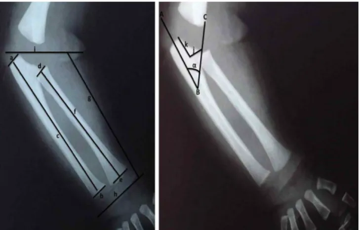
Later on, the mammographies and forearm radiographies of the fetuses were shot in the way that the forearms would remain in a prone position (Fig. 1). The radiographies that were shot were placed on the negatoscope, after which morphometric measurement were taken from the structures belonging to the forearm through the use of a digital compass.
Morphometric Measurements Taken:
Proximal width of Ulna (a): The largest transverse distance pertaining to the proximal region of the ulnar bone (Fig. 1). Distal width of Ulna (b): The largest transverse distance pertaining
to the distal region of the ulnar bone (Fig. 1).
Ulnar length (c): The longest vertical distance between the transverse axes that pass through the starting point and end-point of the ulnar bone (Fig. 1).
Proximal width of Radius (d): The largest transverse distance pertaining to the proximal region of the radial bone (Fig. 1). Distal width of Radius (e): The largest transverse distance pertaining
to the distal region of the radial bone (Fig. 1).
Radius length (f): The longest vertical distance between the transverse axes that pass through the starting point and end-point of the radial bone (Fig. 1).
Forearm length (g): The longest vertical distance between the transverse axes that pass through the middle region of the wrist and the elbow joint (Fig. 1).
Wrist width (h): The longest transverse distance between the inner and outer sides of the wrist (Fig. 1).
Elbow width (i): The longest transverse distance between the inner and outer sides of the elbow (Fig. 1).
Width of trochlear notch (j): The largest transverse distance between the inner and outer sides of trochlear notch (Fig. 1).
Depth of trochlear notch (k): The longest vertical distance between the transverse axes that pass through the upper and lower margins of trochlear notch (Fig. 1).
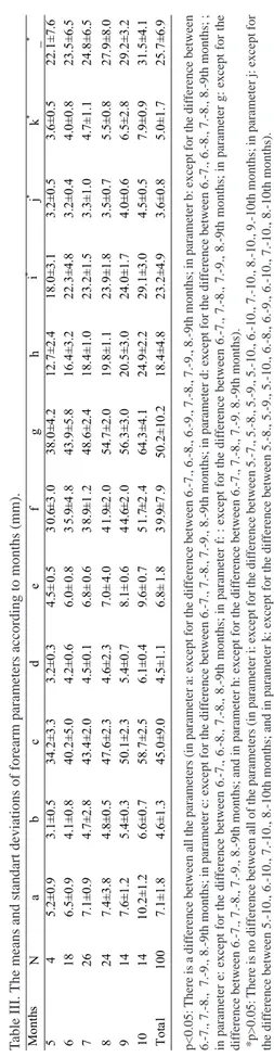
Coronoidal angle (a): The angle between the BC line that passes through coronoid process and the AB line that passes through the truncus of the ulnar bone (Fig. 1).
By utilizing the SPSS statistical program, the averages and standard deviations of the parameters according to sexs, gestational age and groups were ascertained. The significance level in the statistical analysis was taken as p<0.05. The parametric values given in accordance with the
groups were shown with the average ± standard deviation.
In the comparison of the groups, non-parametric tests were used due to the scarcity of the number of cases in some groups. Firstly, the Kruskall-Wallis variance analysis was performed. As the result of this analysis, the groups regarded as significant were compared in groups of twos by means of Mann-Whitney U test. The levels of significance were assessed through the Bonferroni Correction. The relationships between the parameters taken and the gestational age (week) were determined through the use of Pearson correlation test. In the inter-sex comparison of the
Fig. 1 The forearm radiography taken from 26 week old male fetus. The picture pertaining to the measurement areas of the forearm paramaters (a: proximal width of ulna, b: distal width of ulna, c: ulnar length, d: proximal width of radius, e: distal width of radius, f: radial length, g: forearm length, h: wrist width, i: elbow width, j: width of trochlear notch, k: depth of trochlear notch, a: coronoidal angle).
parametric data, the Student- T test (in total for all the ca-ses) and Mann-Whitney U test (within each group while comparing separately) were utilized. The values, p obtained were given in the findings section and under the tables involved.
RESULTS
The general external measurements of the fetuses were performed in the first place. During the measurement practice, it was determined that there was no difference between sexs (p>0.05). Later on, the mammographies and forearm radiographies of the fetuses were shot in the way that the forearms would remain in a prone position. The radiographies that were shot were placed on the negatoscope, and morphometric measurements of the forearm were taken (Fig. 1). The mean values and the standard deviations of the morphometric measurements in question were determined according to weeks, trimesters and months (Tables I, II, III). In the comparison of these taken parameters between trimesters, it was observed that there were some differences between the other parameters with respect to trochlear notch width and trochlear notch depth parameters, except for the difference between 2nd and 3rd trimester groups, and some differences between the other parameters with respect to coronoidal angle parameter, except for the difference between 3rd and 4th trimester groups (p<0.05, Table II). In
Weeks N a b c d e f g h i j k _ 20 4 5.53 2.61 31.87 3.02 3.98 29.19 35.32 10.90 14.70 2.80 3.66 19.00 21 2 6.39 2.92 33.96 3.17 4.20 29.95 36.71 11.66 16.64 3.00 4.00 19.75 22 8 6.89 3.32 35.05 3.49 4.58 31.53 37.11 12.29 18.74 3.25 4.42 20.00 23 2 7.01 3.75 37.62 3.71 4.80 33.81 38.39 13.51 19.81 3.33 4.68 20.00 24 6 7.39 3.91 38.53 3.87 5.12 34.97 39.21 14.46 20.47 3.45 4.80 20.50 25 6 7.51 4.18 40.47 4.06 5.34 35.53 41.29 15.51 21.70 3.50 4.93 21.16 26 10 7.66 4.58 41.87 4.28 5.67 36.47 43.65 16.30 22.02 3.60 5.02 23.00 27 6 7.75 4.71 42.75 4.33 5.90 37.53 45.62 16.75 22.93 3.68 5.24 23.30 28 4 7.92 4.95 43.31 4.53 6.14 38.25 47.40 17.25 23.05 3.75 5.40 24.00 29 6 8.39 5.04 44.62 4.61 6.55 41.16 49.18 18.64 23.80 3.90 5.66 25.00 30 10 8.42 5.31 45.02 4.76 6.79 42.39 51.80 19.68 24.14 4.00 5.85 25.71 31 4 8.48 5.56 46.62 4.90 6.93 43.57 53.42 20.12 25.22 4.12 6.00 27.83 32 4 8.99 5.71 47.00 5.08 7.16 44.28 54.42 20.80 25.89 4.25 6.33 28.50 33 2 9.23 6.00 48.39 5.15 7.47 45.85 55.83 21.47 26.24 4.33 6.66 28.50 34 4 9.52 6.21 49.73 5.30 7.75 46.72 57.20 22.28 27.54 4.42 6.80 29.00 35 2 9.76 6.58 51.13 5.55 7.93 47.28 58.83 23.68 27.91 4.55 7.10 29,50 36 6 9.92 7.22 53.29 5.69 8.22 48.16 60.48 24.26 28.65 4.70 7.40 30.00 37 2 10.26 7.46 54.92 5.87 8.59 49.39 61.52 25.47 29.17 4.78 7.65 31.50 38 4 10.56 7.71 56.70 6.03 8.88 51.85 62.75 26.85 29.62 4.85 7.90 33.00 39 2 10.88 7.89 58.52 6.27 9.14 52.97 63.51 27.96 29.81 4.92 8.20 35.00 40 6 11.24 7.97 60.44 6.42 9.36 53.69 65.12 28.22 30.28 5.00 8.50 35.00
the comparison made between months, on the other hand, it was determined that there were some differences between the other parameters with respect to the ulnar proximal width parameter, except for the difference between 7., 8., 6.-9., 7.-8., 7.-6.-9., 8.-9th months; and with respect to the ulnar distal width parameter, except for the difference between 6.-7., 7.-8., 7.-9., 8.-9th months; and with respect to the ulnar length parameter, except for the difference between 6.-7., 7.-8., 7.-9., 8.-9th months; and with respect to the radial proximal width parameter, except for the difference between 6.-7., 6.-8., 7.-8., 8.-9th months; and with respect to the ra-dial distal width parameter, except for the difference between 6.-7., 6.-8., 7.-8., 8.-9th months; and with respect to the ra-dial length parameter, except for the difference between 6.-7., 7.-8., 7.-9., 8.-9th months; and with respect to the forearm length parameter, except for the difference between 6.-7., 7.-8., 7.-9., 8.-9th months; and with respect to the wrist width parameter, except for the difference between 6.-7., 8., 7.-9. 8.-9th months; and also with respect to the coronoidal angle parameter, except for the difference between all the months (p<0.05, Table III). Separately, it was ascertained that there was no difference between the other months, except for the difference between 5.-7., 5.-8., 5.-9., 5.-10., 6.-10., 7.-10., 8.-10., 9.-10th months in elbow width parameter; and except for the difference between 5.-10., 6.-10., 7.-10., 8.-10th months in trochlear notch width parameter; and except for the difference between 5.-8., 5.-9., 5.-10., 8., 9., 6.-10., 7.-6.-10., 8.-10th months in trochlear notch depth parameter (p>0.05, Table III). On the other hand; in the comparison of Table I The means forearm parameters according to weeks (mm).
the parameters in question between sexs and right-left forearms, it was observed that there was no statistically significant difference (p>0.05, Tables I, II, III). It was determined that there was a significant correlation between the measured parameters and the gestational age (p<0.001).
M onth s N a bc de f gh i * j * k * _ * 5 4 5. 2 ± 0.9 3 .1 ± 0.5 3 4 .2± 3 .3 3 .2 ± 0.3 4 .5 ± 0.5 3 0 .6 ± 3 .0 38 .0 ± 4 .2 1 2 .7± 2 .4 1 8 .0± 3 .1 3 .2 ± 0.5 3 .6 ± 0.5 2 2 .1± 7 .6 6 1 8 6 .5±0 .9 4 .1±0 .8 4 0 .2 ± 5 .0 4 .2±0 .6 6 .0± 0 .8 3 5 .9 ± 4 .8 4 3 .9 ± 5 .8 1 6 .4 ± 3 .2 2 2 .3 ± 4 .8 3 .2±0 .4 4 .0±0 .8 2 3 .5 ± 6 .5 7 2 6 7 .1±0 .9 4 .7±2 .8 4 3 .4 ± 2 .0 4 .5±0 .1 6 .8± 0 .6 3 8 .9 ± 1 .2 4 8 .6 ± 2 .4 1 8 .4 ± 1 .0 2 3 .2 ± 1 .5 3 .3±1 .0 4 .7±1 .1 2 4 .8 ± 6 .5 8 2 4 7 .4±3 .8 4 .8±0 .5 4 7 .6 ± 2 .3 4 .6±2 .3 7 .0± 4 .0 4 1 .9 ± 2 .0 5 4 .7 ± 2 .0 1 9 .8 ± 1 .1 2 3 .9 ± 1 .8 3 .5±0 .7 5 .5±0 .8 2 7 .9 ± 8 .0 9 1 4 7 .6±1 .2 5 .4±0 .3 5 0 .1 ± 2 .3 5 .4±0 .7 8 .1± 0 .6 4 4 .6 ± 2 .0 5 6 .3 ± 3 .0 2 0 .5 ± 3 .0 2 4 .0 ± 1 .7 4 .0±0 .6 6 .5±2 .8 2 9 .2 ± 3 .2 10 14 10 .2 ± 1 .2 6 .6 ± 0.7 5 8 .7± 2 .5 6 .1 ± 0.4 9 .6 ± 0.7 5 1 .7 ± 2 .4 64 .3 ± 4 .1 2 4 .9± 2 .2 2 9 .1± 3 .0 4 .5 ± 0.5 7 .9 ± 0.9 3 1 .5± 4 .1 To ta l 100 7 .1±1 .8 4 .6±1 .3 4 5 .0 ± 9 .0 4 .5±1 .1 6 .8± 1 .8 3 9 .9 ± 7 .9 5 0 .2 ±10 .2 1 8 .4 ±4. 8 2 3. 2 ±4. 9 3 .6 ± 0 .8 5 .0 ± 1 .7 2 5. 7 ±6. 9 p<0.05: The dif
ference between all the parameters (except for the dif
ference between 2nd and 3rd trimester groups pertaining to
j and k parameters, and except for the dif
ference between 3rd and 4th
trimester groups pertaining to the a parameter).
p>0.05: no dif
ferences between sexs.
T
able II.
The means and standart deviations of forearm parameters according to trimester groups (mm).
T ri m e st e r N a b cd ef g h i j k _ I. gr ou p 2 8 5 .7 ± 1 .6 3.4± 1.2 36.4 ± 8 .0 3.5± 0.9 5 .2± 1 .6 3 2.5± 6.9 40.7 ± 8 .9 1 4.2± 4 .6 18.9 ± 4 .9 3.2± 0.5 4 .1 ± 1.5 23.7 ± 6 .6 II. g ro u p 60 7 .2 ± 1 .1 4.8± 0.8 46.3 ± 5 .8 4.7± 0.7 7 .0± 1 .1 4 1.1± 5.0 51.9 ± 7 .2 19.22 ± 3.4 24.0 ± 3 .3 3.6± 0.8 4 .8 ± 1.3 28.0 ± 7 .6 II I. g ro u p 1 2 1 0.2± 1.3 6.5± 0.8 58.5 ± 2 .7 6.1± 0.4 9 .6± 0 .8 5 1.7± 2.6 64.1 ± 4 .5 2 4.5± 2 .2 28.9 ± 3 .3 4.5± 0.5 7 .8 ± 0.9 29.4 ± 3 .5 T ota l 100 7 .1± 1 .8 4.6± 1.3 45.0 ± 9 .0 4.5± 1.1 6 .8± 1 .8 3 9.9± 7.9 5 0.2± 10.2 1 8.4± 4 .8 23.2 ± 4 .9 3.6± 0.8 5 .0 ± 1.7 25.7 ± 6 .9 T able III.
The means and standart deviations of forearm parameters according to months (mm).
p<0.05:
There is a dif
ference between all the parameters (in parameter a: except for the dif
ference between 6.-7., 6.-8., 6.-9.
, 7.-8., 7.-9., 8.-9th months; in parameter b: except for the dif
ference between
6.-7., 7.-8.,
7.-9., 8.-9th months; in parameter c: except for the dif
ference between 6.-7., 7.-8., 7.-9., 8.-9th months; in pa
rameter d: except for the dif
ference between 6.-7., 6.-8., 7.-8., 8.-9th months; ;
in parameter e: except for the dif
ference between 6.-7., 6.-8., 7.-8., 8.-9th months; in parameter f: : except for the dif
feren
ce between 6.-7., 7.-8., 7.-9., 8.-9th months; in parameter g: except for the
dif
ference between 6.-7., 7.-8., 7.-9., 8.-9th months; and in parameter h: except for the dif
ference between 6.-7., 7.-8., 7.-9
. 8.-9th months).
*p>0.05:
There is no dif
ference between all of the parameters (in parameter i: except for the dif
ference between 5.-7., 5.-8.,
5.-9., 5.-10., 6.-10., 7.-10., 8.-10., 9.-10th months; in parameter j: except for
the dif
ference between 5.-10., 6.-10., 7.-10., 8.-10th months; and in parameter k: except for the dif
ference between 5.-8., 5.-9., 5.-10., 6.-8., 6.-9., 6.-10., 7.-10., 8.-10th months).
Fig. 3. The relationship of the width of trochlear notch (j), the depth of trochlear notch (k) and the coronoidal angle (a) with the gestational age.
Fig. 2. The relationship of the ulnar length (c) /radial length (f), forearm length (g) /ulnar length (c), forearm length (g) / radial length (f) and elbow width (i) /wrist width (h) rates with thegestational age.
DISCUSSION
The development of the skeletal system during the intrauterine period takes place in an orderly manner (Moore et al.). There have been several ultrasonographic studies conducted on the development of fetal limb (Brons et al., 1990; Ma-las et al., 2000; Parilla et al.; Huang et al.; Chen et al.; Song & Wang), since the prenatal and postnatal analyses of fetal structure and fetal limbs provide information as to fetal growth, growth retardation, gestational age, and congenital malformations (Sadler; Collins; Malas et al., 2006).
1.4 1,2 0,8 0,6 0,4 0,2 0+ ~ -0 JO 30 40 50 I 2 3 4 S 6 7 B 9 10 U 12 13 14 lS 16 17 18 19 20 2l - c/f - -,Jc - ,Jr - Vh - r - k - -a
When we reviewed the former studies as regards fetal forearm development, we did not come across any study that investigated the development of the forearm during the fetal period through radiological methods. The studies in this matter were conducted on dead fetuses and were concerned with the morphometric measurements of upper and lower limbs on dead fetuses (Malas et al., 2000; Malas et al., 2005; Uluutku et al.). Separately, they involved ultrasonographic studies regarding the measurements of limb that were performed during the fe-tal period (Huang et al.; Chen et al.; Song & Wang). In our study, different from the others, we aimed to investigate the morphometric development of the forearm on dead human fetuses pertaining to the fetal period between 20-40 weeks over radiographic images and also to evaluate its importance in terms of clinics.
In our study, morphometric measurements pertaining to the ulna were taken from the forearm bones in the first pla-ce (Tables I, II, III). It was determined that there was a significant correlation between the measured parameters and the gestational age (p<0.001). In addition, it was ascertained that the morphometric measurements pertaining to the ulna proved to be higher in male fetuses throughout the fetal period; yet, there was no statistically significant difference between sexs (p>0.05, Tablo 2, 3). As in adults, the ulnar proximal part was determined to be wider than the distal part throughout the fetal period (Tables I, II, III). Separately, the total length of the ulnar bone was measured as 45.8 mm (Tables II, III). Matsushita et al. measured the ulnar length on 122 Japanese fetuses, which varied between 18-40 weeks. As the result of the study, they report the total length of the ulnar bone as 25.47 mm and also state that there is no difference between sexs. In addition to this, they emphasize the fact that the ulnar length is correlated with the gestational age. Our study results are in accordance with other study results, except for the total ulnar length found as the result of the study conducted by Matsushita et al. We identified the difference in the total ulnar length with ethnic reasons and with the factors that affected the fetal bone development.
In our study, morphometric measurements pertaining to the radial bone were taken from the forearm bones afterwards (Tables I, II, III). It was determined that there was a significant correlation between the measured parameters and the gestational age (p<0.001). Separately, it was ascertained that the morphometric measurements pertaining to the radial bone proved to be higher in male fetuses throughout the fetal period; yet, there was no statistically significant difference between sexs (p>0.05, Tables II, III). As in adults, the radial distal part was determined to be wider than the radial proximal part throughout the fetal period (Tables I, II, III). Separately, the total length of the radial bone was measured as 41.64 mm (Tables II, III). Matsushita et al. measured the radial length
on 122 Japanese fetuses, which varied between 18-40 weeks. As the result of the study, they report the total length of the radial bone as 28.99 mm and also state that there is no difference between sexs. Additionally, they report that the ra-dial length is correlated with the gestational age. Our study results are in accordance with other study results, except for the total radial length found as the result of the study conducted by Matsushita et al. We identified the difference in the total radial length with ethnic reasons and with the factors that affected the fetal bone development.
In our study, morphometric measurements pertaining to the forearm length, wrist width and elbow width were taken, as well (Tables I, II, III). It was determined that there was a correlation between the measured parameters and the gestational age (p<0.001). It was also ascertained that the measurements taken proved to be higher in male fetuses throughout the fetal period; yet, there was no statistically significant difference between sexs (p>0.05, Tables II, III). As the result of the study, we identified the total length of the forearm as 50.2 mm, the total width of the wrist as 18.4 mm, and the total width of the elbow as 23.2 mm (Tables II, III). Malas et al. (2000), in their study, report the total length of the forearm as 44.1 mm. In another study they conducted (Malas et al., 2006), they report the total width of the wrist as 14 mm. On the other hand, as the result of the study conducted by Uluutku et al. on 21 fetuses involving 17-35.8 gestational weeks, the total width of the right wrist is said to be 15.25 mm, whereas the total width of the left wrist is said to be 15.27 mm; on the other hand, the total width of the right elbow is stated to be 18.85 mm, while the total width of the left elbow is stated to be 18.87 mm. Separately, just as the result seen in our study, it is reported in each of these three studies that there is no difference between sexs, nor between right-left parts of the forearm (Malas et al., 2000; Uluutku et al.). There are differences between the results of our study and those of the other studies in terms of the mean values pertaining to the parametric measurements. We identified this difference with the fact that the other studies mentioned above were fetal studies covering earlier gestational weeks and also with the factors that affected the development of the fetal skeletal system.
In our study, we also examined the relationship of the proportion of ulnar length (c) to radial length (f), forearm length (g) /ulnar length (c), forearm length (g) /radial length (f) and elbow width (i) /wrist width (h) with the gestational age (Fig. 2). We have not come across any study like this that had been conducted on adults and on fetal period. As the result of the study, we determined that the ulna had developed more rapidly
until 29th and 30th gestational weeks, and that after this period,
the radius had developed more rapidly (Fig. 2). In the same way, we also ascertained that depending on the rapid development of the ulna and the slow development of the
radius, the forearm had developed more slowly until 29th and 30th gestational weeks, and that after this period, depending on the rapid development of the radius, the forearm had developed more rapidly (Fig. 2). Separately, it was also determined that the wrist had developed more rapidly until 29th and 30th gestational weeks, and following this period, the elbow was seen to have developed more rapidly.
In our study, we, later on, examined the width and depth of trochlear notch, one of the structures belonging to the proximal part of the ulnar bone, as well as the coronoidal angle. We have not come across such a study that had been conducted during the fetal period. We could only find the study conducted by Kumar et al. on the hand radiography of 53 adults. As the result of our study, it was determined that there was a significant correlation between the measured parameters and the gestational age (p<0.001, Fig. 2). It was also ascertained that the measurements taken proved to be higher in male fetuses throughout the fetal period; yet, there was no statistically significant difference between sexs (p>0.05, Tables II, III). Separately, we determined that the depth of trochlear notch had increased in proportion to the coronoidal angle (Fig. 2). According to the results of the study conducted by Kumar et al. it is expressed that there is no difference in terms of right-left parts and sexs in the parameter results pertaining to the width and depth of trochlear notch as well as coronoidal angle. Our study result, on the other hand, is in accordance with the results found in the study by Kumar et al.. We interpreted this outcome as the fact that these parameters had begun to develop during the fetal period and that they continued developing in the same way during adulthood, as well. We are
of the opinion that the data we obtained as the result of our study will be of use in determining the prostheses to be performed on the elbow area, the pathologies regarding the skeletal system in this region as well as the fetal age and sex. Our study involves radiologically detailed information as to the morphometric development of the forearm, which has not been included in the formerly studies. We regard our study as a precursor one that is useful in terms of evaluating the development of the fetal skeletal system and determining the pathologies and anomalies regarding the fetal skeletal system in the early stage.
In conclusion, we are of the opinion that the morphometric data we obtained as the result of this study will be beneficial for the involved clinicians, such as those in charge of gynecology, radiology, pathology, forensic medicine and perinatology, in terms of evaluating the studies related to the development of the forearm and the skeletal system throughout the fetal period, in identifying the pathologies and variations regarding the development of the fetal skeletal system, and also in determining fetal age and sex.
ACKNOWLEDGEMENTS
We would like to express our sincere thanks to the families who have donated the fetuses to our study as well as Isparta Maternity and Paediatric Hospital that has been a mediator for the fetus donations.
DESDICIOGLU, R.; UGUZ, C.; DESDICIOGLU, K.; SULAK, O. & MALAS, M. A. Investigación radiológica y evaluación clínica del desarrollo morfométrico del antebrazo en fetos humanos. Int. J. Morphol., 35(2):629-636, 2017.
RESUMEN: El período fetal es el tiempo en el cual el feto crece rápidamente y se forman los órganos. Los análisis prenatal y postnatal de la estructura fetal proporcionan información sobre el crecimiento fetal, el retraso de crecimiento, la edad gestacional y las malformaciones congénitas. El desarrollo del sistema esquelético, como también el de otros sistemas durante el período intrauterino, avanza de manera ordenada. Se investigó radiológicamente el desarrollo morfométrico del antebrazo en fetos humanos durante el perío-do comprendiperío-do entre 20-40 semanas gestacionales y se evaluó su importancia clínica. Un total de 100 antebrazos fetales (50 fetos: 23 de sexo masculino, 27 de sexo femenino), cuya edad varió entre 20-40 semanas de gestación, sin patología externa o anomalía, fueron incluidos en el estudio. Los fetos fueron separados en grupos de semanas, trimestres y meses. Después de realizar las mediciones externas generales de los fetos, las mamografías y las radiografías fueron realizadas de tal manera que los antebrazos permanecieran en pronación. Las radiografías de las medidas morfométricas correspondientes a las estructuras del antebrazo se tomaron con apoyo de una compás digital; posteriormente, las medidas fueron tratadas estadísticamente. Los valores medios y las desviaciones estándar de los parámetros medidos se determinaron de acuerdo con las semanas de gestación, los trimestres y los meses. Hubo una correlación signifi-cativa entre los parámetros medidos y la edad gestacional (p <0,001). En la comparación de los parámetros medidos entre los trimestres y los meses, se observó una diferencia estadísticamente significativa entre los grupos (p <0,05). Se determinó también que no hubo diferencias estadísticamente significativas en la comparación de los parámetros, que se realizó entre los sexos y los antebrazos derecho-izquierdo (p> 0,05). En cuanto a los resultados de nuestro estudio, los datos obtenidos durante este período de estudio serán beneficiosos para los clínicos, como también para profesionales de las áreas de ginecología, radiología, medicina forense y perinatología, en la evaluación de estudios clínicos relacionados con el desarrollo morfométrico del antebrazo durante todo el período fetal, determinación de la edad y el sexo fetal, así como en la determinación de variaciones en el desarrollo del sistema esquelético fetal.
REFERENCES
Brons, J. T.; van Geijn, H. P.; Bezemer, P. D.; Nauta, J. P. & Arts, N. F. The fetal skeleton; ultrasonographic evaluation of the normal growth. Eur. J. Obstet. Gynecol. Reprod. Biol., 34(1-2):21-36, 1990.
Chen, M.; Lee, C. P.; Lam, Y. H.; Ou, C. Q. & Tang, M. H. First-trimester fetal limb biometry in Chinese population. Prenat. Diagn., 27(2):133-8, 2007.
Collins, P. Neonatal Anatomy and Growth. In: Williams, P. L.; Warwich, R.; Dyson, M. & Bannister, L. H. Gray’s Anatomy. 38th ed. London, Churchill Livingstone, 1995. pp.343-75.
Huang, L. H.; Fang, Q.; Xie, H. N.; Yang, Z. Y.; Yang, Y. Z.; Shi, H. J. & Huang, X. Clinical study and prenatal diagnosis of fetus with shortened long bones. Zhonghua Yi Xue Za Zhi, 87(45):3178-82, 2007. Jacquemyn, Y.; Sys, S. U. & Verdonk, P. Fetal biometry in different ethnic
groups. Early Hum. Dev., 57(1):1-13, 2000.
Johnson, M. P.; Barr, M. Jr.; Treadwell, M. C.; Michaelson, J.; Isada, N. B.; Pryde, P. G.; Dombrowski, M. P.; Cotton, D. B. & Evans, M. I. Fetal leg and femur/foot length ratio: a marker for trisomy 21. Am. J. Obstet. Gynecol., 169(3):557-63, 1993.
Kulkarni, M. L. & Rajendran, N. K. Values for foot length in newborns. Indian Pediatr., 29(4):507-9, 1992.
Kumar, B.; Pai, S.; Ray, B.; Mishra, S.; K. S. S.; K.S.; Pandey, A. K. & S. B. Radiographic study of carrying angle and morphometry of skeletal elements of human elbow. Rom. J. Morphol. Embryol., 51(3):521-6, 2010.
Malas, M. A.; Dogan, S.; Evcil, E. H. & Desdicioglu, K. Fetal development of the hand, digits and digit ratio (2D:4D). Early Hum. Dev., 82(7):469-75, 2006.
Malas, M. A.; Dogan, S.; Evcil, E. H.; Desdicioglu, K.; Tagil, S. M. & Sulak, O. The rate of growth between the upper and lower extremity in the fetal period. Med. J. Suleyman Demirel Univ., 12(2):1-8, 2005. Malas, M. A.; Salbacak, A. & Sulak, O. The growth of the upper and lower
extremities of Turkish fetuses during the fetal period. Surg. Radiol. Anat., 22(5-6):249-54, 2000.
Matsushita, K.; Shinoda, K.; Akiyoshi, T. & Watanabe, H. Multivariate analysis of limb long bone growth during the human prenatal period. Tohoku J. Exp. Med., 176(2):109-20, 1995.
Moore, K. L.; Persaud, T. V. N. The Developing Human. Clinically Oriented Embryology. 8th ed. Philadelphia, Saunders, 2004.
Munsick, R. A. Similarities of Negro and Caucasian fetal extremity lengths in the interval from 9 to 20 weeks of pregnancy. Am. J. Obstet. Gynecol., 156(1):183-5, 1987.
O’Brien, G. D.; Rodeck, C. & Queenan, J. T. Early prenatal diagnosis of diastrophic dwarfism by ultrasound. Br. Med. J., 280(6227):1300, 1980. Parilla, B. V.; Leeth, E. A.; Kambich, M. P.; Chilis, P. & MacGregor, S. N. Antenatal detection of skeletal dysplasias. J. Ultrasound Med., 22(3):255-8, 2003.
Sadler, T. W. Langman’s Medical Embryology. 6th ed. Baltimore, Williams & Wilkins, 1990. pp.134-40.
Song, J. & Wang, X. Prenatal diagnose of abnormalities of fetal limb bone. Zhonghua Fu Chan Ke Za Zhi, 45(10):745-9, 2010.
Taeusch, H. W. Initial Evaluations: History and Physical Examination of the Newborn. In: Taeusch, H. W.; Ballord, R. A. & Avery, M. E. Diseases of the Newborn. Philadelphia, W. B. Saunders Company, 1991. pp.207-24.
Uluutku, M. H.; Akbaytürk, N.; Çan, M. A. & Özyasar, A. F. Gestational age and its relationship with elbow width, wrist width and forearm length. Turk. Klin. J. Med. Sci., 30(6):1993-8, 2010.
Witters, I.; Moerman, P. & Fryns, J. P. Skeletal dysplasias: 38 prenatal cases. Genet. Couns., 19(3):267-75, 2008.
Corresponding author: Dr. Raziye Desdicioglu Yıldırım Beyazıt University Medical Faculty
Department of Obstetrics and Gynecology Ankara
TURKEY
E-mail: [email protected]
Received: 19-10-2016 Accepted: 20-03-2017